R E S U M O
Introdução: A Pressão Venosa Central (PVC),
é um importante parâmetro hemodinâmico, reflectindo o estado de hidratação. Embora existam estudos de cálculo não invasivo da PVC por ecocardiografia, não está descrito um método de quantificação exacta da mesma.
Objectivos: Analisando diversos parâmetros
ecocardiográficos, os autores propõem uma fórmula de cálculo da PVC que mostra boa correlação estatística com a PVC medida de forma invasiva.
Material e Métodos: O estudo foi efectuado
em 45 doentes internados na UCI, 32 em ritmo sinusal e 13 em fibrilhação auricular, 32 dos quais submetidos a ventilação mecânica. Fizeram-se simultaneamente medições invasivas da PVC e o estudo ecocardiográfico transtorácico com Doppler. Os métodos estatísticos utilizados foram a correlação bivariada e análise de variâncias.
Resultados: Foram encontrados diversos
parâmetros ecocardiográficos que mostram boa correlação com o valor da PVC medida. A estes foram aplicados coeficientes estandardizados e obteve-se a seguinte fórmula de cálculo da PVC: (desaceleração E tricúspide) x 0,11 + (gradiente VD/AD) x 0,16 - (variação da VCI).
Conclusão: Acrescenta-se mais um elemento
de avaliação hemodinâmica não invasiva quantificando um parâmetro até agora só avaliado de forma aproximada por esta abordagem.
A
RTIGOS
O
RIGINAIS
Avaliação Não Invasiva da Pressão Venosa
Central por Ecocardiografia
[10]
PAULOMARCELINO, ANAPAULAFERNANDES, SUSANMARUM, JOAQUIMPALMEIRORIBEIRO
Unidade de Cuidados Intensivos – Hospital de Curry Cabral, Lisboa
125
A B S T R A C T
Non-Invasive Evaluation of Central Venous Pressure by Echocardiography
Introduction: Central venous pressure (CVP)
is an important hemodynamic parameter, reflecting the patient’s volume status. Although some studies of non-invasive evaluation of CVP by echocardiography exist, an exact quantitative method has not been described.
Objectives: Analyzing several
echocardiographic variables, the authors present an equation to calculate CVP with good statistical correlation with CVP measured invasively.
Material and Methods: The study included
45 patients, 32 undergoing mechanical ventilation, 32 in sinus rhythm and 13 with atrial fibrillation. Patients underwent simultaneous CVP measurement and transthoracic Doppler echocardiography. Statistical analysis was performed using bivariate correlation and analysis of variance.
Results: Several echocardiographic
parameters measured showed a good correlation with the measured CVP. Standardized coefficients were applied to them and an equation to calculate CVP was obtained (tricuspid E decelaration) x 0.11 + (RU/RA gradient) x 0.16 - (IVC variation).
Conclusion: This new method seems reliable
to evaluate non-invasively a hemodynamic parameter that until now has only been measured approximately by this approach.
Recebido para publicação: Agosto 2001 • Aceite para publicação: Novembro 2001 Received for publication: August 2001 • Accepted for publication: November 2001
Rev Port Cardiol 2002; 21 (2) : 125-133
Palavras-Chave
Pressão venosa central; Ecocardiografia
Key words
INTRODUÇÃO
É
hoje possível fazer-se uma análise por-menorizada do estado hemodinâmico dos doentes por exame não invasivo, utilizando a ecocardiografia.Se uma primeira abordagem ecocardiográ-fica pode dar a noção do estado de repleção hídrica ou identificar a natureza de uma hipo-tensão, utilizando o estudo Doppler é possível quantificar importantes parâmetros hemodinâ-micos tais como o débito cardíaco(1), a pressão
telediastólica do ventrículo esquerdo(2, 3) e as
pressões do coração direito(4, 5).
A avaliação da pressão venosa central pode ser feita pela análise da veia cava inferior (VCI) e respectivo colapso inspiratório, calculando-se assim o índex da VCI. Obtém-se um valor apro-ximado e não um valor exacto, mas que mostra boa correlação com os valores obtidos de forma invasiva(6, 7, 8). Encontram-se descritos outros
mé-todos de cálculo da PVC através da análise do fluxo das veias hepáticas(9), bem como de outras
veias de grande calibre acessíveis às técnicas de
Doppler(10, 11). Descrevem-se igualmente
altera-ções do Doppler transvalvular tricúspide perante alterações da pré-carga(12).
Porém, não se encontra descrito nenhum método não invasivo de quantificação exacta da PVC. O estudo apresentado pelos autores teve como objectivo encontrar um método de cálculo quantificado da PVC, aplicável a um número alargado de doentes, especialmente nos submetidos a ventilação mecânica numa unidade de Cuidados Intensivos Polivalente.
MATERIAL E MÉTODOS
O estudo foi efectuado em 45 doentes inter-nados numa UCI polivalente, aleatoriamente seleccionados. Destes, 30 eram do sexo mascu-lino e 15 do sexo feminino, com uma média etária de 61 anos, trinta e dois dos quais sob suporte ventilatório. Trinta e dois doentes en-contravam-se em ritmo sinusal, e 13 em fibri-lhação auricular. Em relação às patologias de base, 12 doentes estavam internados por agra-vamento da insuficiência respiratória crónica, 10 por sépsis (sem choque hemodinâmico, de-finido por pressão arterial média inferior a 60 mmHg) ou outras patologias infecciosas, 6 por intoxicações diversas, e os restantes apresenta-vam outros diagnósticos. Excluíram-se os doen-tes com valvulopatias e os doendoen-tes em choque hemodinâmico (pressão arterial média inferior a 60 mmHg), e com frequência cardíaca supe-rior a 120 bpm.
126
INTRODUCTION
I
t is now possible to carry out a detailed analysis of a patient’s hemodynamic status by non-invasive means using echocardio-graphy.While an initial echocardiographic assess-ment can give an idea of hydration status or identify the nature of hypotension, Doppler imaging can quantify important hemodynamic parameters such as cardiac output(1), left
ven-tricular end-diastolic pressure(2, 3) and right
heart pressures(4, 5).
Central venous pressure can be evaluated by analyzing the inferior vena cava (IVC) and its inspiratory collapse, and thence calculating the IVC index. An approximate value is obtain-ed rather than a precise figure, but this shows good correlation with values obtained using in-vasive methods(6, 7, 8). Other methods of
calculat-ing CVP have been described by analyzcalculat-ing he-patic vein flow(9), as well as other large veins
accessible to Doppler techniques(10, 11). Changes
in transvalvular tricuspid Doppler, caused by changes in preload, are also described(12).
However, no non-invasive method for pre-cise quantification of CVP has been described. The aim of the study presented here was to find a method for quantified calculation of CVP that can be applied to a wider range of patients, especially those undergoing mechani-cal ventilation in an intensive care unit (ICU).
METHODS
The study was performed in 45 randomly selected patients admitted to an ICU. Thirty of these patients were male and 15 female, with a mean age of 61 years, and 32 were on ventila-tory support. Thirty-two patients were in sinus rhythm and 13 with atrial fibrillation. With re-gard to underlying pathologies, 12 patients had been admitted with worsening chronic respira-tory failure, 10 with sepsis (without hemodyna-mic shock, defined as mean blood pressure of less than 60 mmHg) or other infections, 6 with various types of poisoning, and the remainder with other diagnoses. Patients with heart valve disease, those in hemodynamic shock (mean blood pressure less than 60 mmHg), and those with heart rate of over 120 bpm, were excluded. In patients undergoing positive pressure mechanical ventilation, PEEP (positive end-ex-piration pressure) was 4 cmH2O, the same in
Nos doentes submetidos a ventilação mecâ-nica com pressão positiva, a PEEP foi de 4 cm H2O, igual em todos os doentes.
Em relação à avaliação ecocardiográfica ge-ral, 7 doentes apresentavam dilatação das ca-vidades direitas e 3 tinham um ventrículo es-querdo dilatado com disfunção sistólica. Os restantes não apresentaram alterações a assi-nalar.
A pressão venosa central foi medida através de catéter venoso central colocado em veia ju-gular ou subclávia por método da régua de água, após correcto posicionamento do doente e verificação da localização do catéter venoso central por radiografia do tórax. O método da régua de água é o mais utilizado na avaliação da PVC, tendo sido por isso o método de com-paração escolhido.
Simultaneamente, realizou-se estudo diográfico transtorácico utilizando um ecocar-diógrafo ALOKA-SSD 2200 equipado com sonda de 2,5 MHZ com Doppler contínuo, pulsado e codificado em côr. Por Doppler pulsado na câ-mara de entrada do ventrículo direito analisa-ram-se o fluxo de entrada neste, a relação E/A do fluxo transvalvular tricúspide dos doentes em ritmo sinusal, o tempo de desaceleração da onda E do fluxo transvalvular tricúspide, o gra-diente entre o ventrículo direito e a aurícula di-reita por Doppler contínuo, a veia cava inferior com a respectiva dimensão expiratória máxima (medida a 2 cm da emergência da aurícula di-reita e em decúbito dorsal) e dimensão durante a inspiração. Não foi possível obter o fluxo das veias pulmonares em todos os doentes, princi-palmente devido à ventilação mecânica, pelo que este parâmetro não foi considerado na ava-liação final. Todos estes parâmetros foram obti-dos no final da expiração, com o intuito de ho-mogeneizar as amostras. Nos treze doentes em fibrilhação auricular, a desaceleração da onda E tricúspide e o gradiente VD/AD foram calcula-dos como a média de 5 medições. Neste grupo não foi possível determinar a relação E/A do fluxo transvalvular tricúspide.
Seguidamente, através do cálculo de uma re-gressão linear, determinou-se a correlação entre as diversas variáveis obtidas por eco Doppler cardíaco e a PVC medida, de modo a encontrar uma forma de cálculo não invasivo da PVC.
RESULTADOS
A análise estatística dos resultados dos pa-râmetros analisados (Quadro I) mostrou o
se-guinte: 127
On general echocardiographic evaluation, 7 patients had dilatation of the right chambers and 3 had dilated left ventricles with systolic dysfunction. The others had no relevant abnor-malities.
Central venous pressure was measured with a central venous catheter placed in the jugular or subclavian vein using a fluid-filled U-tube manometer, after the patient had been appro-priately positioned and the location of the ca-theter had been verified by chest X-ray. The U-tube manometer is the most commonly used method to assess CVP, and was accordingly chosen as the method for comparison.
Simultaneously, a transthoracic echocardio-graphic study was performed using an Aloka SSD 2200 echocardiograph fitted with a 2.5 MHz probe with continuous-wave, pulsed and color Doppler. Using pulsed Doppler in the right ventricular inflow tract, the flow was mea-sured here, together with the E/A ratio of tri-cuspid inflow in patients in sinus rhythm, dece-leration time of the E wave of tricuspid inflow, the gradient between right ventricle and right atrium (RV/RA) by continuous Doppler, the in-ferior vena cava with the corresponding maxi-mum expiratory diameter (measured at 2 cm from its emergence from the right atrium and in dorsal decubitus), and its diameter during ins-piration. It was not possible to measure pulmo-nary vein flow in all patients, mainly because of mechanical ventilation, and so this parame-ter was not included in the final assessment. All measurements were taken at end-expiration in order to standardize the samples. In the thir-teen patients with atrial fibrillation, the decele-ration of the tricuspid E wave and the RV/RA gradient were calculated as the mean of 5 mea-surements. In this group it was not possible to determine the E/A ratio of tricuspid inflow.
Subsequently, by calculating a linear re-gression, the correlation between the different variables obtained by Doppler echocardio-graphy and measured CVP was determined, in order to find a non-invasive way of calculating CVP.
RESULTS
Statistical analysis of the results of the pa-rameters measured (Table I) shows the follow-ing:
1. CVP was more significantly related to the inspiratory collapse of the vena cava, with a correlation of 67.4 %, giving p<0.001. These findings are in
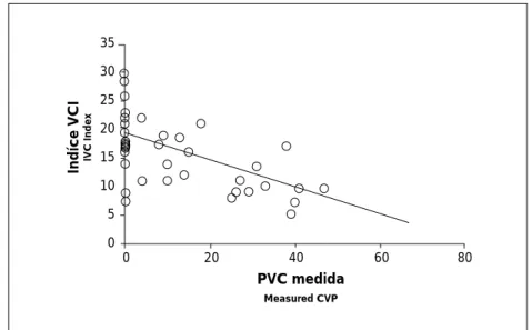
agree-1. A PVC foi mais significativamente rela-cionável com o colapso inspiratório da VCI, com uma correlação de 67,4 % para um p<0,001. Estes dados são concordan-tes com o descrito na literatura. O cali-bre expiratório da VCI não mostrou uma relação tão forte com a PVC, embora ainda estatisticamente significativa (43,1 % para p<0,005). O índice da VCI, calculado pela diferença relativa entre as
128
ment with those described in the litera-ture. The expiratory diameter of the vena cava did not show such a strong relation with CVP, although it was statistically significant (43.1 %, p < 0.005). The IVC index, calculated as the relative dif-ference between the diameter of the ve-na cava at expiration and inspiration, showed a relation with measured CVP (p<0.0001) – Fig. 1. Its mean value was
Quadro I
Correlação bivariada entre as variáveis analisadas, utilizando o método de Pearson a duas caudas
Desacel. E Gradiente VD/AD VCI 1 VCI 2 Calculada
Desaceleração P 1.000 .048 .298 .303* .738** p .771 .047 .043 .000 Gradiente VD/AD P .048 1.000 .142 .087 .468** p .771 .388 .598 .003 VCI 1 P .298 .142 1.000 .843 .418** p .047 .388 .000 .004 45 39 45 45 45 VCI 2 P .303 .087 .843** 1.000 .632** p .043 .598 .000 .000 Calculada P .738** .468 .418** .632** 1.000 p .000 .003 .004 .000 Medida P .670** 403* .431** .666** .959** p .000 .012 .003 .000 .000 E/A P .276 .373 .273 .457** .502** p .126 .051 .130 .009 .003 Delta VCI P -.186 .021 -.318* -.778** -.626** p .222 .901 .033 .000 .000 P - correlação de Pearson; * - p<0,001; ** - p<0,01 p - valor de significância do teste de Pearson
Table I
Bivariate correlation between the variables analyzed, using the two-tailed Pearson’s test
E-Decel. Gradient RV/RA IVC 1 IVC 2 Calculated
Deceleration P 1.000 .048 .298 .303* .738** p .771 .047 .043 .000 RV/RA Gradient P .048 1.000 .142 .087 .468** p .771 .388 .598 .003 IVC 1 P .298 .142 1.000 .843 .418** p .047 .388 .000 .004 45 39 45 45 45 IVC 2 P .303 .087 .843** 1.000 .632** p .043 .598 .000 .000 Calculated P .738** .468 .418** .632** 1.000 p .000 .003 .004 .000 Measured P .670** 403* .431** .666** .959** p .000 .012 .003 .000 .000 E/A P .276 .373 .273 .457** .502** p .126 .051 .130 .009 .003 Delta IVC P -.186 .021 -.318* -.778** -.626** p .222 .901 .033 .000 .000 P - Pearson’s correlation; * - p<0.001; ** - p<0.01 p - value of significance of Pearson’s test
dimensões da veia cava em expiração e inspiração, mostrou-se relacionável com a PVC medida (p<0.0001) – Fig. 1. O seu valor médio foi de 14,9 %, inferior ao valor normal descrito(13), o que pode estar
associado ao valor médio elevado das PVCs medidas (14,8 cm H2O) e à
venti-lação mecânica.
2. Foi obtida uma relação igualmente signi-ficativa entre a desaceleração da onda E do fluxo transvalvular tricúspide e o lor da PVC (67,0 % para p<0,001). O va-lor médio deste parâmetro foi de 110 ms (52-220ms).
3. O gradiente entre o ventrículo direito e a aurícula direita mostrou uma correlação com a PVC de 40,3 % para p<0,05. O valor médio deste gradiente no grupo de doentes estudado foi de 18,7 mmHg (0-102 mmHg).
4. Assistiu-se igualmente a uma boa corres-pondência entre a relação E/A do fluxo transvalvular tricúspide e a PVC nos doentes em ritmo sinusal, embora a consi-deração deste parâmetro exclua os doen-tes em fibrilhação auricular.
5. Quando comparadas entre si, as variáveis independentes não mostraram correlação estatisticamente significativa, à excepção da desaceleração da onda E tricúspide e da relação E/A do fluxo transvalvular tri-cúspide.
Assim, os preditores do valor da PVC foram a desaceleração da onda E tricúspide, o co-lapso inspiratório da VCI e o gradiente entre o ventrículo direito e a aurícula direita. A estes
129
14.9 %, lower than the figure usually re-ported(13), which may be related to the
high mean value of the CVPs measured (14.8 cmH2O) and to mechanical
ventila-tion.
2. An equally significant relation was obtain-ed between the deceleration of the E wave of tricuspid inflow and the value of CVP (67.0 %, p<0.001). The mean value of this parameter was 110 ms (52-220 ms). 3. The gradient between the right ventricle and the right atrium showed a correlation with CVP of 40.3 %, giving p<0.05. The mean value of this parameter in the group of patients studied was 18.7 mmHg (0-102 mmHg).
4. A good correspondence was also seen between the E/A ratio of tricuspid inflow and CVP in patients in sinus rhythm, al-though inclusion of this parameter exclu-des patients with atrial fibrillation. 5. When compared between each other, the
independent variables do not show statis-tically significant correlations, with the exception of the tricuspid E wave and the E/A ratio of tricuspid inflow.
Thus, the predictors of the value of CVP were the deceleration of the tricuspid E wave, the inspiratory collapse of the vena cava, and the gradient between the right ventricle and right atrium. Standardized coefficients were applied to these parameters in order to obtain a equation to calculate CVP.
Equation:
(tricuspid E deceleration) x 0.11 + (RV/RA gradient) x 0.16 - (variation in IVC)
Indíce VCI IVC Index
35 30 25 20 15 10 5 0 PVC medida Measured CVP
0 20 40 60 80 Fig. 1 Relação entre o índice da VCI e a PVC medida.
Fig. 1 Relation between IVC
foram aplicados coeficientes estandardizados para obtenção de uma fórmula de cálculo da PVC.
Fórmula de cálculo:
(desaceleração E tricúspide) x 0,11 + (gradiente VD/AD) x 0,16 - (variação da VCI)
Foram de seguida comparados os resultados obtidos de forma invasiva com os obtidos com a aplicação desta fórmula. A PVC assim calcu-lada ajusta-se numa relação linear com um coe-ficiente de 97,8% à PVC medida, pelo que se pode dizer que são significativamente seme-lhantes (p<0.001) – Fig. 2.
DISCUSSÃO
O objectivo inicial de encontrar um método não invasivo de quantificação da PVC foi ple-namente conseguido. A fórmula aplicada mos-tra uma correlação estatisticamente significa-tiva com a PVC medida de forma invasiva. Nela conjugam-se diversas variáveis influencia-doras do resultado final da PVC e não um único parâmetro.
Existem diversas publicações que estudam as características do Doppler do ventrículo di-reito perante diversas alterações hemodinâmi-cas. A relação E/A do fluxo transvalvular tri-cúspide é influenciada pelas alterações induzidas pela respiração(13), bem como pela
função diastólica e sistólica do ventrículo di-reito(14, 15), à semelhança do fluxo transvalvular
mitral. No estudo hemodinâmico não invasivo o gradiente de pressão entre o ventrículo di-reito e a aurícula direita é utilizado para
deter-130
The results from invasive testing were then compared with those obtained by applying this formula. CVP calculated in this way has a li-near relationship with measured CVP by a coefficient of 97.8%, and so they can be said to be significantly similar (p<0.001) – Fig. 2.
DISCUSSION
The initial objective of finding a non-invas-ive method for quantifying CVP was fully achieved. The formula applied shows a statistic-ally significant correlation with CVP measured invasively. The formula brings together several variables that influence the final result of CVP, rather than a single parameter.
Various studies have been published des-cribing the Doppler characteristics of the right ventricle in the presence of different hemody-namic changes. The E/A ratio of tricuspid in-flow is influenced by changes brought on by respiration(13), as well as by the diastolic and
systolic function of the right ventricle(14, 15), in a
similar way as for mitral inflow. In non-invas-ive hemodynamic study the pressure gradient between the right ventricle and the right atrium is used to determine systolic pressure in the pulmonary artery, and mitral regurgita-tion is used to ascertain the pressure in the left atrium(1). Recent studies suggest that results of
Doppler studies of the left and the right heart are similar(16, 17). The E/A ratio of tricuspid
in-flow has not yet been clearly related to CVP, although in the present study it was signifi-cant, which may result from differences in vol-ume status. This aspect may merit more tho-rough study. On the other hand, inclusion of
0 10 20 30 40 40 30 20 10 0 PVC medida Measured CVP
Indíce VCI IVC Index
Fig. 2 Relação entre a PVC
me-dida com régua de água e a cal-culada pela fórmula:
(desaceleração E tricúspide) x 0,11 + (gradiente VD/AD) x 0,16 – (variação da VCI).
Fig. 2 Relation between CVP
measured by U-tube manometer and CVP calculated by the equa-tion:
(tricuspid E deceleration) x 0.11 + (RV/RA gradient) x 0.16 - (va-riation in IVC).
minar a pressão sistólica da artéria pulmonar, e a regurgitação mitral para o cálculo da pres-são da aurícula esquerda(1). Os estudos
recen-tes apontam para uma semelhança de resulta-dos entre o estudo Doppler do coração esquerdo e direito(16, 17). A relação E/A do fluxo
transvalvular tricúspide não foi ainda clara-mente relacionada com a PVC, embora no pre-sente estudo tenha sido significativa, o que pode estar associado a diferentes estados de volémia. Possivelmente, este aspecto é merece-dor de um estudo mais aprofundado. Por outro lado, a consideração deste parâmetro excluiria os doentes em fibrilhação auricular.
A desaceleração da onda E tricúspide não é influenciada pelas variações respiratórias e os resultados obtidos com a sua análise são sobre-poníveis à caracterização recente do Doppler transvalvular tricúspide(13). A sua relação com
as variações de volémia e consequentemente com a PVC, já se encontra referida(13).
O gradiente entre o ventrículo e a aurícula direita, não é referido na literatura como possí-vel influenciador do valor da PVC, mas a sua relação com a sobrecarga de pressão é sobeja-mente conhecida(1, 14).
Os dados obtidos com os valores derivados da VCI estão plenamente de acordo com o que já se encontra descrito, embora para a fórmula de cálculo se tenha utilizado o valor absoluto do colapso e não o índice da VCI. De notar ainda que o calibre inspiratório da VCI não se mostrou correlacionável com a PVC, sendo neste aspecto fundamental a avaliação do co-lapso inspiratório da mesma.
Nenhum método descrito foi aplicado a doentes submetidos a ventilação mecânica. A avaliação ecocardiográfica da PVC, é frequen-temente estabelecida pelo colapso inspiratório da VCI. A correlação entre estes dois parâme-tros foi aqui confirmada, mas revela-se insufi-ciente para a obtenção de um valor exacto da mesma. A avaliação do fluxo das veias hepáti-cas revelou-se difícil no grupo de doentes por nós estudado, mas obtiveram-se outros parâme-tros de eco-Doppler, com boa relação com a PVC e independentes entre si. As avaliações feitas pelos diversos autores revelaram-se mui-to sobreponíveis embora seja desejável a con-firmação da reprodutibilidade dos resultados por outros ecocardiografistas.
As limitações do estudo prendem-se funda-mentalmente com a aquisição de imagem e a execução do estudo Doppler das cavidades
di-reitas, principalmente nos doentes sob ventila- 131
this parameter would exclude patients with atrial fibrillation.
The deceleration of the tricuspid E wave is not influenced by respiratory variations and the results obtained on analysis are compar-able to the recent characterization of tricuspid transvalvular Doppler (13). Its relation to varia-tions in volume and therefore to CVP have al-ready been described(13).
The gradient between the right ventricle and atrium is not mentioned in the literature as a factor influencing the value of CVP, but its relation with pressure overload is well known(1, 14).
The data obtained with the values derived from the IVC are in full agreement with what has been described, although the absolute va-lue of collapse rather than the IVC index was used in the formula. It should also be noted that the inspiratory diameter of the IVC has not been shown to correlate with CVP, and evaluation of its inspiratory collapse is there-fore essential.
None of the methods described in the lite-rature has been applied to patients undergoing mechanical ventilation. Echocardiographic as-sessment of CVP is frequently determined by the inspiratory collapse of the IVC. The corre-lation between these two parameters has been confirmed here, but it is insufficient to obtain an exact value of CVP. Assessment of flow in the hepatic veins proved difficult in the group of patients studied by us, but other Doppler echo parameters were obtained that showed a good relation with CVP and that were mutually independent. Assessments made by the various authors were very similar, although confirma-tion of the reproducibility of the results by other echocardiographers is desirable.
The limitations of the study are essentially related to acquiring the image and carrying out the Doppler study of the right chambers, parti-cularly in patients undergoing mechanical ven-tilation. In patients who have undergone abdo-minal surgery, abdoabdo-minal dressings can make it impossible to study the IVC.
We did not include patients with heart valve disease or in hemodynamic shock, and so the application of the equation to these pa-tients remains to be studied. This would be of particular relevance for patients in hemodyna-mic shock in an intensive care unit. In patients with elevated heart rate, non-invasive hemody-namic studies are limited and unreliable.
We consider that the equation presented could be useful when a quantified estimate of
ção mecânica. Nos doentes submetidos a cirur-gia abdominal, a presença da pensos abdomi-nais pode inviabilizar o estudo da VCI.
Não incluímos doentes com valvulopatias ou em choque hemodinâmico, pelo que a apli-cação desta fórmula nestes doentes deverá ser ainda estudada. Principalmente estes últimos terão particular interesse numa UCI Poliva-lente. Nos doentes com frequência cardíaca elevada, o estudo hemodinâmico não invasivo é limitado e pouco fidedigno.
Pensamos que a fórmula encontrada poderá ser útil quando uma estimativa quantificada da PVC é necessária e não pode ser obtida de forma invasiva. Pode ser aplicada em larga es-cala(18), pelo menos em doentes internados em
UCIs polivalentes e submetidos a ventilação mecânica.
AGRADECIMENTOS
Agradecemos à Dra. Maria João Marques pela realização do tratamento estatístico pre-sente neste trabalho.
132
CVP is required and cannot be obtained by in-vasive means. It can be widely applied(18), not
least with patients hospitalized in intensive care units and undergoing mechanical ventila-tion.
ACKNOWLEDGEMENTS
We are grateful to Dr. Maria João Marques for the statistical processing presented in this work.
Pedido de separatas para: Address for reprints: PAULO MARCELINO
Unidade de Cuidados Intensivos Hospital de Curry Cabral Rua da Beneficência, 8 1069-166 LISBOA
1. Feigenbaum H. Hemodynamic Information Derived from Echocardiography. Echocardiography. 5th Edition 1994;181-215.
2. Gonzalez-Vilchez F, Ares M, Ayuela J, Alonso L. Combin-ed Use of PulsCombin-ed and Color M-Mode Doppler Echocardio-graphy for the Estimation of Pulmonary Capillary Wedge Pressure: An Empirical Approach Based on an Analytical Relation. J Am Coll Cardiol 1999;34:515-23.
3. Omnen SR, Nishimura RA, Appleton CP, et al. Clinical utility of Doppler echocardiography and tissue Doppler ima-ging in the estimation of left ventricular filling pressures: A comparative simultaneous Doppler-catheterization study. Cir-culation 2000;102:1788-94.
4. Masuyama T, Kodama K, Kitabake A, et al. Continuous-wave Doppler echocardiographic detection of pulmonary re-gurgitation and its application to non-invasive estimation of pulmonary artery pressure. Circulation 1986;74: 484-92. 5. Ensing G, Seward J, Rarragh R, Caldwell R. Feasibility of generating hemodynamic pressure curves from non-invasive Doppler echocardiographic signals. J Am Coll Cardiol 1994; 23:434-42.
6. Minutiello L. Value of the vena cava index in healthy young subjects. Echocardiographic study. Minerva Cardio-angiol 1994;42:229-32.
7. Mandelbaum A, Link A, Wambach G, Ritz E. Vena cava ultrasonography for the assessment of hydration status in kidney insufficiency. Dtsch Med Wochenschr 1993;118: 1309-15.
8. Minutiello L. Non-invasive evaluation of central venous pressure derived from respiratory variations in the diameter of the inferior vena cava. Minerva Cardioangiol 1993;41: 433-7.
9. Reeves WC, Leaman DM, Buonocore E, et al. Detection of tricuspid regurgitation and estimation of central venous
pres-133
sure by two-dimensional contrast echocardiography of the right superior hepatic vein. Am Heart J 1981; 102:374-7. 10. Cozcolluela MR, Sarria L, Sanz L, et al. Correlation of central venous pressure with Doppler waveform of the com-mon femoral veins. J Ultrasound Med 2000;19:587-92. 11. Nahum E, Dagan O, Sulkes J, Schoefeld T. A compa-rison between continuous central venous pressure measure-ment from right atrium and abdominal vena cava or common iliac vein. Intensive Care Med 1996;22:571-4.
12. Kyriakides ZS, Kremastinos DT, Paraskevaides IA, et al. Effects of a preload increase on ventricular filling in coronary artery disease. Cardiology 1993;82:229-37.
13. Klein AL, Dominic YL, Murray RD, et al. Effects of Age and Physiologic Variables on Right Ventricular Filling Dynamics in Normal Subjects. Am J Cardiol 1999; 84:440-8. 14. Tominaga T, Oki T, Okushi H, et al. Diastolic right ven-tricular hemodynamics in right venven-tricular overloads asses-sed by pulasses-sed Doppler echocardiography. J Cardiol 1988;18:1115-26.
15. Seibold H, Henze E, Kohler J, et al. Right ventricular function in patients with chronic obstructive pulmonary di-sease. Klin Wochenschr 1985;63:1041-7.
16. Zoghbi WA, Habib GB, Quinones MA. Doppler assess-ment of right ventricular filling in a normal population: com-parison with left filling dynamics. Circulation 1990;82: 1316-24.
17. Iwase M, Nagata K, Izawa H, et al. Age-related changes in left and right ventricular filling velocity profiles and their relationship in normal subjects. Am Heart J 1993;126:419-26. 18. Randazzo MR, Snoey ER. Accuracy of emergency physi-cian assessment of left ventricular ejection fraction and cen-tral venous pressure using echocardiography. Acad Emerg Med 2001;8:550-1.
À ATENÇÃO DOS AUTORES
Lembra-se que a reprodução de figuras de outras publicações (livros ou revistas) está sujeita às normas de copyright pelo qual deverá ser solicitadas pelos autores,
com antecedência aos editores.