The Myocardium in Tetralogy of Fallot: a Histological and
Morphometric Study
Maria Cecília Knoll Farah
2, Cláudia Regina Pinheiro de Castro
1, Valéria Mello Moreira
1, Arlindo de Almeida Riso
1,
Antonio Augusto Barbosa Lopes
1, Vera Demarchi Aiello
1Instituto do Coração (InCor) Hospital das Clínicas-FMUSP, São Paulo, SP1; Faculdade de Medicina - Centro de Ciências Biológicas da Saúde - Universidade Federal de Mato Grosso, Cuiabá, MT2 - Brazil
Summary
Background: Patients with Tetralogy of Fallot frequently develop ventricular dysfunction in the postoperative period. The histological basis of this functional alteration has been scarcely studied.
Objective: To evaluate myocardial remodeling in anatomical specimens, comparing the subepicardial and subendocardial regions, especially because the subendocardial region is easily approached by means of endomyocardial biopsy.
Methods: Transmural sections of myocardium from the right ventricular (RV) inflow tract, anterior wall and infundibulum, and from the left ventricular (LV) free wall were evaluated regarding the degree of cardiomyocyte hypertrophy, vascularization and interstitial fibrosis were analyzed.
Results: The mean diameter of subendocardial cardiomyocytes is similar to that of subepicardial cardiomyocytes in all regions, except for the RV infundibulum, in which subendocardial cardiomyocytes are significantly larger in relation to those of the subepicardium (p=0.007). The amount of interstitial collagen is in the upper limits of normal and was similar in the subendocardial layers in comparison with the subpericardial layer of each region; however, it was greater in the inflow tract and RV anterior wall than in the LV lateral wall. The numerical density of subendocardial capillaries was similar to that of the subepicardium and was lower than the mean minus two standard deviations of normal in all regions and layers, except for the infundibulum, in which the subepicardium showed normal values and the subendocardium showed values lower than the mean minus two standard deviations.
Conclusion: The postnatal myocardial changes in Tetralogy of Fallot are homogeneously distributed in the subepicardial and subendocardial halves of the ventricular walls, except for the infundibulum, which has peculiar remodeling characteristics and, therefore, is not representative of the other ventricular regions and layers for morphometric studies. (Arq Bras Cardiol 2009;92(3): 160-167)
Key words: Tetralogy of Fallot; heart defects, congenital; myocardium/anatomy and histology; cardiomegaly.
Mailing address: Vera Demarchi Aiello •
Av. Dr. Enéas Carvalho Aguiar, 44 - 05406-000 - Cerqueira César - São Paulo, SP - Brazil
E-mail: [email protected], [email protected]
Manuscript received November 30, 2007; revised manuscript received January 21, 2008; accepted February 26, 2008.
Introduction
Histological studies of the myocardium in Tetralogy of Fallot demonstrated the existence of cardiomyocyte hypertrophy and disarray in addition to varying degrees and types of interstitial fibrosis and edema1. On gross examination, ventricular
hypertrophy is particularly evident in the infundibular region, precisely where the basic embryologic defect of this anomaly is manifested, leading to the anterior deviation of the infundibular septum. In adults with acquired heart diseases it has been demonstrated that the different patterns of fibrosis and remodeling in the different ventricular regions
and muscle layers (subendocardial and subepicardial) have varying functional consequences2,3.
It is important to know the distribution and intensity of the histological changes in the different ventricular regions in Tetralogy of Fallot, including cardiomyocyte hypertrophy, percentage of fibrosis and capillary network, since this anomaly is frequently accompanied by ventricular dysfunction in the postoperative period and its histological basis has been scarcely studied.
Original Article
Farah et al Myocardial histology in tetralogy of fallot
Objective
To morphometrically study the histological characteristics (degree of myocardial hypertrophy, interstitial fibrosis, and capillary density) of samples from three different right ventricular (RV) regions – inflow tract, anterior wall and outflow tract, and to compare them with each other and with left ventricular (LV) samples, in addition to verifying the transmural distribution of these samples.
Methods
A total of 192 histological sections prepared from eight anatomical pieces of hearts with congenital defects that characterize Tetralogy of Fallot were analyzed. These specimens are part of the collection of the Laboratory of Pathology from the Heart Institute (InCor), University of São Paulo Medical School, São Paulo, and were obtained from autopsies performed in the period between 1999 and 2005.
The cases were selected according to the integrity of the specimens filed, considering the possibility of their representing all walls, and also according to the quality of the histological sections. Sections with signs of poor fixation were excluded. Table 1 shows the selected cases.
Transmural myocardial slices from four different ventricular regions were removed – RV inflow tract, anterior wall and infundibulum, and LV free wall. The samples underwent conventional histological processing, and then histological sections with 5-micrometer (µm) width were obtained. These were stained with hematoxylin-eosin; picrosirius (Sirius Red) for quantitative analysis of collagen in the myocardial interstitium; and also immunohistochemical staining for Von Willebrand factor to label the myocardial capillaries. All sections were divided into two halves for analysis: subendocardial sample and subepicardial sample.
Morphometric measurements were taken with the help of an interactive computerized system of image analysis
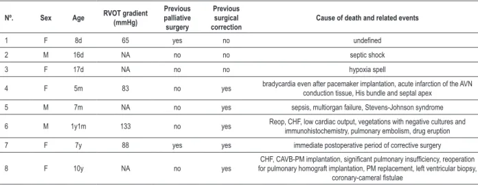
Table 1 – Patients’ clinical data regarding the specimens studied
Nº. Sex Age RVOT gradient
(mmHg) Previous palliative surgery Previous surgical correction
Cause of death and related events
1 F 8d 65 yes no undeined
2 M 16d NA no no septic shock
3 F 17d NA no no hypoxia spell
4 F 5m 83 no yes bradycardia even after pacemaker implantation, acute infarction of the AVN
conduction tissue, His bundle and septal apex
5 M 7m NA no yes sepsis, multiorgan failure, Stevens-Johnson syndrome
6 M 1y1m 133 no yes Reop, CHF, low cardiac output, vegetations with negative cultures and
immunohistochemistry, pulmonary embolism, drug eruption
7 F 7y 88 yes yes immediate postoperative period of corrective surgery
8 F 10y NA no yes
CHF, CAVB-PM implantation, signiicant pulmonary insuficiency, reoperation for pulmonary homograft implantation, PM replacement, left ventricular biopsy,
coronary-cameral istulae
(Leica Qwin version 2.2 – Leica Imaging Systems Inc, 1997). A video camera (JVC model TK-1280U) connected to the light microscope (Leica DMLS) transmits each microscopic field to the system. The fields are then turned into a binary digital image. A sequence of mathematical and morphologic operations allows the identification and quantification of all structures of interest. The technique of histological analysis proposed was tested and showed a small interobserver variation4. A 400X magnification was used for linear
measurements (diameters).
After study of the evolution of the mean and variance, 60 cells per region/case were measured in at least 10 different fields. The smallest diameters of cardiomyocytes were measured along a line that intercepted the respective nucleus. The smallest diameter of the nucleus was also measured (Figure 1). The results were compared with normal indexes for age as established in the literature1,5,,6. For collagen quantification,
the area fraction occupied by interstitial collagen (% of the myocardial area) was determined using a 200X magnification and adopting color as the parameter to discriminate collagen in 40 fields of each sample (Figure 2).
Healing areas characterized by stellate fibrosis with newly formed vessels and collagen around the arterioles with diameter greater than 30µm, measured through the edition functionality of the Quantimet software, were excluded from this analysis. Evaluation of the capillaries was feasible in histological sections of five out of eight specimens, whose results of immunohistochemical reaction enabled the morphometric analysis. The unsatisfactory results of the slides of the three remaining specimens were probably related to the period of time in which they had been stored in formaldehyde, and therefore they were excluded from the analysis.
Figure 1 -Histological section of the myocardium showing measurement of the cardiomyocyte diameter (arrows). Hematoxylin-eosin staining. 40X magniication.
Figure 2 -Histological section of Picrosirius-stained myocardium for morphometric collagen quantiication – in red. 20X magniication.
(number of capillaries/mm2). In this phase, five fields of each
sample were analyzed, corresponding to an area of 72384µm2,
using an 80-point grid with 400X magnification to count the points falling upon the cardiomyocytes and capillaries.
In these fields, the shortest distance from each point of the test system to the nearest capillary was evaluated and used as a parameter of homogeneity of the distribution of capillaries in the fields (capillary diffusion distance – Figure 3). For each field, the total number of capillaries (vessels with diameter smaller than or equal to 7µm) inserted into the grid was also counted.
Statistical analysis
Data were analyzed and computed using the SigmaStat software version 2.03. Continuous variables were descriptively expressed as mean, median and standard deviation. The Student’s t test or Mann-Whitney test were used for the comparison between the groups, and the test of analysis of variance was used for repeated measures with one or two factors. The chi square test was used for the analysis of the association between variables. Pearson’s or Spearman’s correlation tests were used to test the correlation between variables. The results were analyzed considering a significance level of 5%.
Results
Cardiomyocyte diameter
Original Article
Farah et al Myocardial histology in tetralogy of fallot
Figure 3 -Histological section of myocardium immunohistochemically stained for Factor VIII for determination of numerical density of capillaries and measurement of capillary diffusion distance, showing the 80-point grid. 40X magniication.
wall (p=0.487) as well as from the left ventricular lateral wall (p=0.959). However, subendocardial cardiomyocytes of the right ventricular infundibular wall were significantly larger than those of the subepicardium (p=0.007).
The comparative analysis of the mean diameter of cardiomyocytes from the different ventricular walls – inflow tract, anterior wall, right ventricular infundibular region and left ventricular lateral wall-did not show any significant difference among them, both for the subepicardial samples (p=0.415) and for the subendocardial samples (p=0.5222).
A statistically significant correlation was observed between age and cardiomyocyte diameter in the subendocardial and subepicardial layers of the inflow tract (r=0.94, p<0.001; r=0.89, p<0.001), anterior wall (r=0.76, p<0.05; r=0.84 p<0.001) and right ventricular infundibulum (r=0.96, p<0.001; r=0.94 p<0.001); and in the subepicardial layer of the left ventricular lateral wall (r=0.75, p<0.05), but not in the subendocardial layer of this wall (r=0.41, p=0.311).
When the mean values of cardiomyocyte diameters of the specimens studied were plotted in the graph with the values established for normal hearts for the same age range5, we could
observe that in the specimens studied the values obtained from the right ventricular cardiomyocytes in general are higher than the normal values for the same age range, by more than one standard deviation above normal most of the times. A similar analysis for left ventricular cardiomyocytes showed that this difference is not evident in all cases (Graph 1).
Interstitial ibrosis
The area fraction occupied by collagen in the
of the same region in the different ventricular walls studied. Comparing only the subendocardium of the different ventricular walls, we observed that in the right ventricular inflow tract the area fraction occupied by collagen had values significantly higher than those of the left ventricle (p=0.02). A similar analysis performed for the subepicardium showed that in the right ventricular inflow tract and anterior wall the area fraction of collagen is also higher in comparison with the left ventricle (p=0.002 and p=0.03, respectively) and that in the right ventricular inflow tract the values are higher in comparison with the right ventricular infundibular region (p=0.04) (Table 2).
Comparing the normal values of myocardial fibrosis published in the literature for normal hearts (up to 3%)7, we observed that
the mean values obtained from the right ventricular samples were in the upper limit or slightly above the mean, except for the subepicardial sample of the infundibular region which presented a mean fibrosis level below the maximum normal value. Values obtained from the left ventricular samples, in turn, were below the maximum normal value. In the histological analysis of these specimens, several patterns of interstitial fibrosis were found, including samples with perimysial fibrosis and samples with endomysial fibrosis (Figure 4).
Capillaries
Capillary density (number of capillaries/mm2 of cardiomyocytes)
Graph 1 - Mean cardiomyocyte diameter (µm) of the specimens with tetralogy of Fallot plotted in a graph with the normal values for age according to Nishikawa (1990) in
Original Article
Farah et al Myocardial histology in tetralogy of fallot
Figure 4 -Histological section of Picrosirius-stained myocardium showing an example of endomysial ibrosis, where each cardiomyocyte is surrounded by ibrosis (in red). 20X magniication.
Table 2 – Descriptive analysis of the area fraction occupied by collagen (%) in each region and muscle layer
Region RV-inlow tract RV-anterior wall RV-infundibulum LV-Lateral wall
Layer Subendo Subepi Subendo Subepi Subendo Subepi Subendo Subepi
Maximum 6.9 4.9 6.5 6.3 10.9 3.2 3.4 2.4
Minimum 2.5 1.6 1.2 1.0 0.6 0.9 1.1 0.9
Median 3.4 3.4 3.5 2.3 1.6 1.4 1.8 1.3
Mean 3.9 3.1 3.6 3.0 3.0 1.8 2.0 1.5
Standard deviation 1.5 1.2 1.5 2.0 3.4 0.8 0.8 0.6
RV - right ventricle; LV - left ventricle; Endo - endocardium; epi - epicardium.
(p>0.11). However, the subendocardium of the subpulmonary infundibulum showed a lower capillary density in relation to the anterior wall (p=0.031).
Comparing the data obtained with the normal values of capillary density published in the literature for normal hearts of children younger than nine years of age (3315±85 capillaries/ mm2)8, we observed that the mean values from the four patients
in this age range were lower than the mean minus two standard deviations, except for the subepicardial layer of the infundibular region (Table 3).The spacing of capillary diffusion (µm) in the subendocardium and subepicardium was similar in each of the different ventricular regions studied for the left ventricle (p=0.693) and for the right ventricle in the infundibular region (p=0.208), inflow tract (p=0.595) and anterior wall (p=0.687).
Discussion
promote myocardial adaptation and may occasionally induce ventricular dysfunction. Previous studies showed that patients with Tetralogy of Fallot present considerable cardiomyocyte hypertrophy and disarray and varying degrees and types of fibrosis, edema, mononuclear cell infiltration and degenerative alterations such as vacuolar degeneration of cardiomyocytes1.
It was observed that there is no difference between the diameter of cardiomyocytes of patients with Tetralogy of Fallot and those of normal individuals at birth9. After birth, this
diameter increases progressively and gradually, and connective tissue proliferation occurs proportionally to hypertrophy and age9. These findings corroborate the hypothesis that
during fetal life the ventricular septal defect and the low left ventricular afterload relieve the pressure overload imposed by the obstruction of the right ventricular outflow tract, thus decreasing the stimulus for right ventricular remodeling.
Table 3 – Descriptive analysis of the values of capillary density (number of capillaries/mm2 of cardiomyocytes) in each region and muscle layer of the specimens from patients younger than nine years of age
Region RV –inlow tract RV- anterior wall RV- infundibulum LV- lateral wall
Layer Subendo Subepi Subendo Subepi Subendo Subepi Subendo Subepi
Mean 2334.34 3127.30 3031.69 2729.18 2244.21 3224.96 2553.12 3105.44
Standard deviation 378.65 742.16 608.26 331.62 773.89 365.98 783.35 1496.83
RV - right ventricle; LV - left ventricle; Endo - endocardium; epi - epicardium.
by the obstruction of the right ventricular outflow tract, thus generating a strong stimulus to right ventricular remodeling. Thus, we can consider that the right ventricular remodeling possibly occurs mainly in the postnatal period.
Although a previous study in anatomical specimens had demonstrated that the pulmonary infundibulum is of greater length in Tetralogy of Fallot10, a more recent prospective
echocardiographic study demonstrated that the pulmonary infundibulum is smaller and shorter and the infundibular septum is thicker and deviated antero-superiorly in Tetralogy of Fallot in comparison with normal infants11. Although
infundibular obstruction is progressive with age, the absolute dimension of the pulmonary infundibular volume did not change significantly over time, nor did it increase in proportion with the increase in body surface.
Possibly, the reason for this finding would be an abnormal infundibular growth intrinsic to the congenital anomaly, which makes it relatively smaller in relation to the somatic growth, thus collaborating for the worsening of the obstruction of the right ventricular outflow tract. Therefore, the images confirm the concept that, in the case of congenital heart defects, and provided that these conditions of volume and/ or pressure overload are present since the morphogenesis process, the remodeling process occurs concomitantly with the morphogenesis process and heart growth, both before and after birth.
In the case of Tetralogy of Fallot in particular we should consider that the infundibular hypertrophy present from birth is the result of the intrinsic abnormality of the infundibular morphogenesis that can become more accentuated due to the postnatal remodeling effect. Some authors have already given their opinions regarding the characteristics of myocardial remodeling in congenital heart diseases12 in the different
ventricular wall regions, and in the subendocardial and subepicardial layers.
Salih et al13 demonstrated that in the hypoplastic left heart
syndrome (HLHS) a lower percentage of collagen per field in the left ventricle is observed in comparison with normal controls as well as with the right ventricle. They showed a higher collagen percentage in the subendocardium in comparison with the subepicardium, and in the subepicardium in comparison with the mesocardium. The authors concluded that these alterations represent an intrinsic abnormality and suggested that this disproportionality in the amount of fibrous tissue may have an important implication in the ventricular function over time.
Binotto et al4 studied anatomic specimens with tricuspid
atresia and showed a higher proportion of fibrosis in the left
ventricle of specimens with tricuspid atresia in comparison with normal specimens. They also observed that the proportion of fibrosis was not homogeneous in the entire left ventricle, with higher values in the inflow tract and apex in comparison with the outflow tract. A higher proportion was also observed in the subendocardial layer. Increased interstitial myocardial fibrosis was also documented in Ebstein’s anomaly14 and pulmonary
atresia with intact ventricular septum15. Collagen deposit is
believed to result in increased stiffness that may impair the ventricular filling, thus causing a restrictive physiology.
In addition to proportionally following the physiological growth of the heart, the capillaries are involved in all mechanisms of adaptation under adverse conditions16. In some
situations that lead to myocardial hypertrophy, the inadequate number of capillaries is believed to increase the potential of the ischemic damage. Although hypoxia is considered an effective stimulus to endothelial cell growth, experimental data on adaptation of capillary supply in response to hypoxia are controversial. The number of capillaries was considered inadequate in hearts with hypoplastic left heart13 and in
tricuspid atresia4.
The findings of the present study showed that in the case of Tetralogy of Fallot the changes verified in the subendocardial samples are representative of the whole wall for the different characteristics studied (cardiomyocyte diameter, percentage of interstitial fibrosis, and capillary density and diffusion) in most of the ventricular regions, with two exceptions that occur exactly in the region that most stands out in the anatomy of Tetralogy of Fallot: the pulmonary infundibulum.
We verified that, in this region, the cardiomyocytes have a greater diameter in the subendocardial layer in comparison with the subepicardium, and that capillary density is higher in the subepicardial layer in comparison with the subendocardial layer. This is consistent with the assertion that hypertrophy in response to pressure overload is greater in the subendocardium and is not accompanied by an adequate proportional capillarization16. In relation to fibrosis, the
result shows that although this is increased in the entire right ventricle, this increase is more significant in the inflow tract and anterior wall, which are precisely the most important regions for the right ventricular function.
Contraction of these walls toward the septum has an important role both in the systolic function and in the diastolic relaxation. Rushmer17, cited by Nakasato et al18, reports
Original Article
Farah et al Myocardial histology in tetralogy of fallot
References
1. Kawai S, Okada R, Kitamura K, Suzuki A, Saito S. A morphometrical study of myocardial disarray associated with right ventricular outflow tract obstruction. Jpn Circ J. 1984; 48 (5): 445-56.
2. Matsubara BB, Ferreira ALA, Matsubara LS. Aspectos anatomopatológicos da disfunção ventricular. Rev Soc Cardiol Estado São Paulo. 2002; 12 (3): 361-70.
3. Perdigão C. Fibrose miocárdica: fundamentos teóricos, aspectos clínicos e implicações terapêuticas. Rev Port Cardiol. 1993; 12: 675-85.
4. Binotto MA, Higuchi ML, Aiello VD. Left ventricular remodeling in hearts with tricuspid atresia: morphological observations and possible basis for ventricular dysfunction afther surgery. J Thorac Cardiovasc Surg. 2003; 126 (4): 1026-32.
5. Nishikawa T, Sekiguchi M, Takao A, Ando M, Hiroe M, Morimoto S, et al. Histopathological assessment of endomyocardial biopsy in children: semiquantitative study on the hypertrophy of cardiac myocytes. Am J Cardiovasc Pathol. 1990; 3 (1): 5-11.
6. Noma M, Sekiguchi A, Chikada M, Ishizawa A, Miyauchi J, Okada R. Quantitative analysis of hypertrophy in cardiac chambers in cyanotic tetralogy of Fallot. Jpn Heart J. 2001; 42 (2): 173-84.
7. Schwartz SM, Gordon D, Mosca RS, Bove EL, Heidelberger KP, Kulik TJ. Collagen content in normal, pressure, and pressure-volume overloaded developing human hearts. Am J Cardiol. 1996; 77 (9): 734-8.
8. Rakusan K, Flanagan MF, Geva T, Southern J, Van Praagh R. Morphometry of human coronary capillaries during normal growth and the effect of age in left ventricular pressure-overload hypertrophy. Circulation. 1992; 86: 38-46.
9. Kato M, Kawashima Y, Fugita T, Mori T, Manabe H. Right ventricular hypertrophy in the tetralogy of Fallot. Recent Adv Stud Cardiac Struct Metab. 1976; 53 (3): 555-61.
geometric study. Am J Cardiol. 1975; 35 (3): 402-12.
11. Geva T, Ayres NA, Pac FA, Pignatelli R. Quantitative morphometric analysis of progressive infundibular obstruction in tetralogy of Fallot: a prospective longitudinal echocardiographic study. Circulation. 1995; 92 (4): 886-92.
12. Aiello VD, Binotto MA. Myocardial remodeling in congenital heart disease. Arq Bras Cardiol. 2007; 88 (6): e185-6.
13. Salih C, Sheppard MN, Ho SY. Morphometry of coronary capillaries in hypoplastic left heart syndrome. Ann Thorac Surg. 2004; 77: 903-7.
14. Celermajer DS, Dodd SM, Greenwald SE, Wyse RK, Deanfeld JE. Morbid anatomy in neonates with Ebstein’s anomaly of the tricuspid valve: pathophysiologic and clinical implications. J Am Coll Cardiol. 1992; 19: 1049-53.
15. Akiba T, Nakasato M, Sato S, Ishikawa A, Sato T. Left and right ventricular volume characteristics in tetralogy of Fallot and their relationship to arterial oxygen saturation and age. Acta Paediatric Jpn. 1996; 38 (6): 657-60.
16. Anversa P, Rici R, Olivetti G. Coronary capillaries during normal and pathological growth. Can J Cardiol. 1986; 2 (2): 104-13.
17. Rushmer RF. Cardiovascular dynamics. Philadephia: WB Saunders, 1976. p. 92-3 apud Nakasato M, Akiba T, Sato S, Suzuki H, Hayasaka K. Right and left ventricular function assessed by regional wall motion analysis in patients with Tetralogy of Fallot. Int J Cardiol. 1997; 58 (2): 127-34.
18. Nakasato M, Akiba T, Sato S, Suzuki H, Hayasaka K. Right and left ventricular function assessed by regional wall motion analysis in patients with tetralogy of Fallot. Int J Cardiol. 1997; 58 (2): 127-34.
19. Slesnick T C, Chang AC. Right ventricular dysfunction in congenital heart disease.
ventricular “twist” effect thus ending the systole19. Therefore,
the aspect of the remodeling described above may be related to the existence of right ventricular dysfunction. We should also consider that the different patterns of remodeling have different functional consequences3.
The higher percentage of myocardial fibrosis is known to result in greater stiffness. Impairment of the diastolic function is more related to increased collagen concentration than to cardiomyocyte hypertrophy2. Our findings make us believe
that hypertrophy in response to myocardial remodeling in Tetralogy of Fallot occurs in all right ventricular regions, but becomes significant in the subendocardial infundibular region, where a proportional increase of capillaries does not occur. This phenomenon is possibly related to the degree of abnormality in the infundibular septum and to the obstruction of the right ventricular outflow tract.
As we verified in this study, the alterations seen in the postnatal myocardium in tetralogy of Fallot are homogeneously distributed in the subepicardial and subendocardial halves of the ventricular wall, except for the infundibulum, which presents peculiar myocardial remodeling characteristics. Therefore, we conclude that the subendocardial myocardial samples of the infundibular region are not representative of the remaining ventricular regions for the purpose of morphometric studies.
Further studies are essential to define whether some pattern or intensity of histological damage is correlated with the
ventricular dysfunction clinically detected in the postoperative period of some patients with this congenital anomaly. In this context, and as a prospect of the present study, endocardial biopsy performed in the intraoperative period would be useful to predict the behavior of the ventricular function after correction. We have to recognize that the number of specimens analyzed in our study was small and that the interpretation of the amount of capillaries in the different age ranges requires more extensive studies, including a wider age range.
Acknowledgement
This study was supported by Fapesp (project no. 05/01476-2).
Potential Conflict of Interest
No potential conflict of interest relevant to this article was reported.
Sources of Funding
This study was partially funded by FAPESP.
Study Association