Prophylactic removal of unerupted asymptomatic third
molars: is it justifiable?
Marlus da Silva Pedrosa,1 Evelyn Bianca Soares Silva,1 Thais Oliveira Cordeiro,1 Luiz Gustavo Fernandes Lima Oliveira,2 Rodrigo Richard da Silveira,3 Cláudio Heliomar Vicente da Silva,4 José Guilherme Férrer Pompeu5
1Department of Dentistry, DeVry Facid – DeVry Education Group, Terezina, PI, Brazil 2Private Practice, Luiz Gustavo Aesthetic Dentistry and Oral Implantology, Terezina, PI, Brazil
3Department of Restorative Dentistry, Federal University of Minas Gerais – UFMG, Belo Horizonte, MG, Brazil
4Department of Prosthetic Dentistry and Maxillofacial Surgery, Federal University of Pernambuco – UFPE, Recife, PE, Brazil 5Department of Restorative Dentistry, Federal University of Piauí – UFPI, Terezina, PI, Brazil
•Conflicts of interest: none declared.
AbstrAct
Objective: to review the literature currently available on the evidence that does or does not justify the prophylactic extraction of unerupted asymptomatic third molars. Material and Methods: the electronic databases PubMed, Capes Periodicals, Web of Science and Scopus were searched from November to December 2016 by two
au-thors, simultaneously, using as search terms: Terceiro Molar/Molar, Third AND Extração Profilática/Prophylatic Removal OR Prophylatic Extraction. We included articles from original research and clinical trials published in English and Portuguese. No limits were applied to the date of publication. Review articles and clinical case reports were removed.
Results: we identified 13 studies that addressed, at some aspect, the prophylactic removal of unerupted asymptomatic third molars. The results of this literature review which
alluded to the potential for the formation of pathological alterations in asymptomatic third molars are conflicting; While some justifies the prophylactic procedure based on the possible formation of associated lesions, other scientific evidence does not support such practice. Conclusion: in view of the conflicting viewpoints found in the literature, the prophylactic removal of asymptomatic third molars requires case-by-case evaluation of each patient, and the decision-making process, regarding the retention versus the prophylactic removal of these teeth should be based on scientific evidence combined with the clinical experience of the professional.
Keywords: Oral surgery; Third molar; Unerupted tooth; Disease prevention.
Introduction
T
he extraction of third molars is one of the mostcom-mon procedures in the clinical practice of dentists worldwide. It is estimated that, in the United States, ap-proximately 10 million impacted teeth are extracted annually from approximately 5 million individuals, generating a reve-nue of 3 billion dollars.1,2 In England and Wales, extractions of
third molars between 1995 and 1996 totaled approximately 5.2 million pounds.3
Prophylactic extraction, the most common reason for ex-traction of third molars,4 is widely recognized by a
consider-able number of dental surgeons.2,5 This fact is based on the
as-sociation of these teeth with oral pathological changes such as pericoronitis, periodontal problems, caries in third or second molars, different types of odontogenic cysts and tumors as well as crowding of the lower incisors.6-14
The scientific literature also mentions other reasons to jus-tify this procedure, considering the fact that these teeth do not always play a functional role in the oral cavity as well as an increased risk for postoperative complications, pain and dis-comfort when their surgical removal is performed in more ad-vanced aged patients.15-21 However, other studies suggest that
the lower third molars should not be removed prophylactically in some cases2,14,22-23 and vigilant monitoring of these teeth is
more appropriate strategy.24
Debate about indications for prophylactic removal of
im-pacted third molars remains heated.11 The decision-making
process regarding retention versus prophylactic removal of these teeth should be based on the available scientific evi-dence.25 However, the literature is lacking in studies to
sup-port adequate clinical decision-making regarding prophylactic extraction of third molars.14 This study aimed to review the
literature currently available on scientific evidence that does or does not justify the prophylactic extraction of unerupted asymptomatic third molars.
Material and Methods
For the purpose of this study, we followed guidelines pro-vided by Moher et al.26 in Preferred Reporting Items for
System-atic Reviews and Meta-Analyses: The PRISMA Statement.
Identification and Selection of Relevant
Research
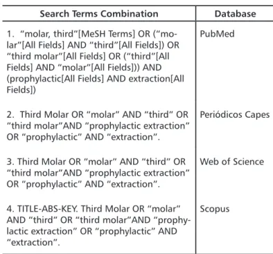
An exploratory bibliographic search was conducted from January to February of 2017 using the electronic databases: Public Medline (PubMed), Periódicos da Capes, Web of Sci-ence and Scopus. Two authors performed the search employ-ing the term Third Molar in combination with Prophylactic removal OR Prophylactic Extraction (Table 1). Inclusion crite-ria were: original research articles and clinical tcrite-rials published in Portuguese and English. No limits were applied to the year of publication.
Table 1. Search strategy for all databases
Search Terms Combination Database
1. “molar, third”[MeSH Terms] OR (“mo-lar”[All Fields] AND “third”[All Fields]) OR “third molar”[All Fields] OR (“third”[All Fields] AND “molar”[All Fields])) AND (prophylactic[All Fields] AND extraction[All Fields])
PubMed
2. Third Molar OR “molar” AND “third” OR “third molar”AND “prophylactic extraction” OR “prophylactic” AND “extraction”.
Periódicos Capes
3. Third Molar OR “molar” AND “third” OR “third molar”AND “prophylactic extraction” OR “prophylactic” AND “extraction”.
Web of Science
4. TITLE-ABS-KEY. Third Molar OR “molar” AND “third” OR “third molar”AND “prophy-lactic extraction” OR “prophy“prophy-lactic” AND “extraction”.
Scopus
Studies were identified and duplicates were removed. Subse-quently, titles and abstracts were screened for relevance, conside-ring the exclusion criteria. Next, the remaining studies were ob-tained in full-text and were screened using the self-same criteria, the eligible the ones being included in this review. We removed review studies, clinical case reports, articles not available in full-text, and publications that did not address the prophylactic
remo-val of unerupted asymptomatic third molars. Eligible studies also had their reference lists screened following the specified criteria for the eligible ones.
Data Collection and Analyses
All the selected articles addressed the relationship between third molars and pathological changes and included the following parameters: authorship, year of publication, country of publica-tion, type of study, sample size (and age), outcomes measured, re-levant data and results, and study considerations.
Based on the findings of the studies, we determined the following themes for critical analysis of the results: characteris-tics of the studies, prophylactic removal of third molars and im-plications for practice. Yet, considering our study design and its findings, we presented the section “study limitations”.
Results
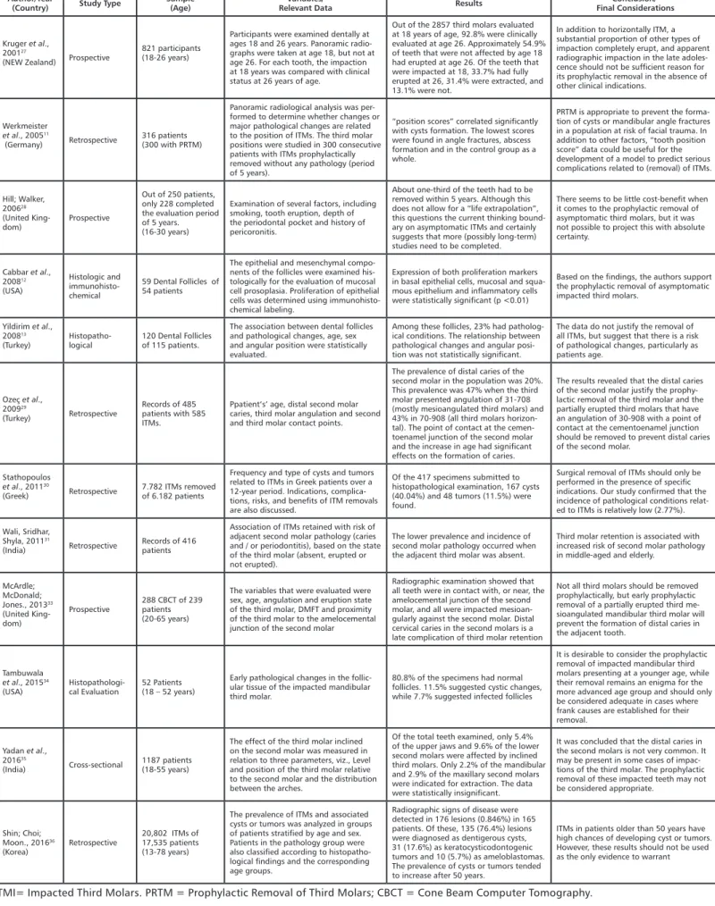
For this systematic review, the initial electronic search yielded a total of 540 titles found in the databases: PubMed, Periódicos da Capes, Web of Science and Scopus. 433 studies were excluded for duplication and the remaining 107 unique papers were scre-ened for relevance to this study. 55 publications were excluded after reading their titles and abstracts. Then, the 52 remaining documents were obtained in full-text and assessed for eligibility in consideration of the prophylactic removal of unerupted asymp-tomatic third molars. After reading the documents in full text, 13 studies were included (Figure 1 and Table 2). No studies from reference lists were added due to their either not being eligible or not having come up on the database search.
Author/Year
(Country) Study Type Sample(Age) Relevant DataVariables/ Results Final ConsiderationsConclusion/
Kruger et al., 200127
(NEW Zealand) Prospective
821 participants (18-26 years)
Participants were examined dentally at ages 18 and 26 years. Panoramic radio-graphs were taken at age 18, but not at age 26. For each tooth, the impaction at 18 years was compared with clinical status at 26 years of age.
Out of the 2857 third molars evaluated at 18 years of age, 92.8% were clinically evaluated at age 26. Approximately 54.9% of teeth that were not affected by age 18 had erupted at age 26. Of the teeth that were impacted at 18, 33.7% had fully erupted at 26, 31.4% were extracted, and 13.1% were not.
In addition to horizontally ITM, a substantial proportion of other types of impaction completely erupt, and apparent radiographic impaction in the late adoles-cence should not be sufficient reason for its prophylactic removal in the absence of other clinical indications.
Werkmeister
et al., 200511
(Germany) Retrospective 316 patients(300 with PRTM)
Panoramic radiological analysis was per-formed to determine whether changes or major pathological changes are related to the position of ITMs. The third molar positions were studied in 300 consecutive patients with ITMs prophylactically removed without any pathology (period of 5 years).
“position scores” correlated significantly with cysts formation. The lowest scores were found in angle fractures, abscess formation and in the control group as a whole.
PRTM is appropriate to prevent the forma-tion of cysts or mandibular angle fractures in a population at risk of facial trauma. In addition to other factors, “tooth position score” data could be useful for the development of a model to predict serious complications related to (removal) of ITMs. Hill; Walker, 200628 (United King-dom) Prospective Out of 250 patients, only 228 completed the evaluation period of 5 years. (16-30 years)
Examination of several factors, including smoking, tooth eruption, depth of the periodontal pocket and history of pericoronitis.
About one-third of the teeth had to be removed within 5 years. Although this does not allow for a “life extrapolation”, this questions the current thinking bound-ary on asymptomatic ITMs and certainly suggests that more (possibly long-term) studies need to be completed.
There seems to be little cost-benefit when it comes to the prophylactic removal of asymptomatic third molars, but it was not possible to project this with absolute certainty. Cabbar et al., 200812 (USA) Histologic and immunohisto-chemical 59 Dental Follicles of 54 patients
The epithelial and mesenchymal compo-nents of the follicles were examined his-tologically for the evaluation of mucosal cell prosoplasia. Proliferation of epithelial cells was determined using immunohisto-chemical labeling.
Expression of both proliferation markers in basal epithelial cells, mucosal and squa-mous epithelium and inflammatory cells were statistically significant (p <0.01)
Based on the findings, the authors support the prophylactic removal of asymptomatic impacted third molars.
Yildirim et al., 200813
(Turkey) Histopatho-logical 120 Dental Follicles of 115 patients.
The association between dental follicles and pathological changes, age, sex and angular position were statistically evaluated.
Among these follicles, 23% had patholog-ical conditions. The relationship between pathological changes and angular posi-tion was not statistically significant.
The data do not justify the removal of all ITMs, but suggest that there is a risk of pathological changes, particularly as patients age. Ozeç et al., 200929 (Turkey) Retrospective Records of 485 patients with 585 ITMs.
Ppatient’s’ age, distal second molar caries, third molar angulation and second and third molar contact points.
The prevalence of distal caries of the second molar in the population was 20%. This prevalence was 47% when the third molar presented angulation of 31-708 (mostly mesioangulated third molars) and 43% in 70-908 (all third molars horizon-tal). The point of contact at the cemen-toenamel junction of the second molar and the increase in age had significant effects on the formation of caries.
The results revealed that the distal caries of the second molar justify the prophy-lactic removal of the third molar and the partially erupted third molars that have an angulation of 30-908 with a point of contact at the cementoenamel junction should be removed to prevent distal caries of the second molar.
Stathopoulos
et al., 201130
(Greek) Retrospective 7.782 ITMs removed of 6.182 patients
Frequency and type of cysts and tumors related to ITMs in Greek patients over a 12-year period. Indications, complica-tions, risks, and benefits of ITM removals are also discussed.
Of the 417 specimens submitted to histopathological examination, 167 cysts (40.04%) and 48 tumors (11.5%) were found.
Surgical removal of ITMs should only be performed in the presence of specific indications. Our study confirmed that the incidence of pathological conditions relat-ed to ITMs is relatively low (2.77%). Wali, Sridhar,
Shyla, 201131
(India) Retrospective Records of 416 patients
Association of ITMs retained with risk of adjacent second molar pathology (caries and / or periodontitis), based on the state of the third molar (absent, erupted or not erupted).
The lower prevalence and incidence of second molar pathology occurred when the adjacent third molar was absent.
Third molar retention is associated with increased risk of second molar pathology in middle-aged and elderly.
McArdle; McDonald; Jones., 201333 (United King-dom) Prospective 288 CBCT of 239 patients (20-65 years)
The variables that were evaluated were sex, age, angulation and eruption state of the third molar, DMFT and proximity of the third molar to the amelocemental junction of the second molar
Radiographic examination showed that all teeth were in contact with, or near, the amelocemental junction of the second molar, and all were impacted mesioan-gularly against the second molar. Distal cervical caries in the second molars is a late complication of third molar retention
Not all third molars should be removed prophylactically, but early prophylactic removal of a partially erupted third me-sioangulated mandibular third molar will prevent the formation of distal caries in the adjacent tooth.
Tambuwala
et al., 201534
(USA) Histopathologi-cal Evaluation 52 Patients(18 – 52 years)
Early pathological changes in the follic-ular tissue of the impacted mandibfollic-ular third molar.
80.8% of the specimens had normal follicles. 11.5% suggested cystic changes, while 7.7% suggested infected follicles
It is desirable to consider the prophylactic removal of impacted mandibular third molars presenting at a younger age, while their removal remains an enigma for the more advanced age group and should only be considered adequate in cases where frank causes are established for their removal.
Yadan et al., 201635
(India) Cross-sectional 1187 patients(18-55 years)
The effect of the third molar inclined on the second molar was measured in relation to three parameters, viz., Level and position of the third molar relative to the second molar and the distribution between the arches.
Of the total teeth examined, only 5.4% of the upper jaws and 9.6% of the lower second molars were affected by inclined third molars. Only 2.2% of the mandibular and 2.9% of the maxillary second molars were indicated for extraction. The data were statistically insignificant.
It was concluded that the distal caries in the second molars is not very common. It may be present in some cases of impac-tions of the third molar. The prophylactic removal of these impacted teeth may not be considered appropriate. Shin; Choi; Moon., 201636 (Korea) Retrospective 20,802 ITMs of 17,535 patients (13-78 years)
The prevalence of ITMs and associated cysts or tumors was analyzed in groups of patients stratified by age and sex. Patients in the pathology group were also classified according to histopatho-logical findings and the corresponding age groups.
Radiographic signs of disease were detected in 176 lesions (0.846%) in 165 patients. Of these, 135 (76.4%) lesions were diagnosed as dentigerous cysts, 31 (17.6%) as keratocysticodontogenic tumors and 10 (5.7%) as ameloblastomas. The prevalence of cysts or tumors tended to increase after 50 years.
ITMs in patients older than 50 years have high chances of developing cyst or tumors. However, these results should not be used as the only evidence to warrant
Table 2. Results of literature review displayed in chronological order
Characteristics of the Studies
We identified 13 studies that addressed, in some respect, the prophylactic removal of asymptomatic third molars. The 13 doc-uments fell into several different categories: retrospective (n = 5), prospective (n = 4), histopathological (n = 2), cross-sectional (n = 1), and histologic and immunohistochemical (n = 2).
According to our findings, we identified only six studies pub-lished at sporadic intervals within a ten-year time frame: 2001 (n = 1), 2005 (n = 1), 2006 (n = 1), 2008 (n = 2), e 2009 (n = 1). In the last six years, seven studies were published: 2011 (n = 2), 2013 (n = 2), 2015 (n = 1) and 2016 (n = 2) The results indicated the need for future research on the prophylactic extraction of unerupted asymptomatic third molars.
Prophylactic Removal of Unerupted
Asymptomatic Third Molars
Table 2 presents the main findings of all the 13 selected articles that addressed the prophylactic removal of unerupted asymptom-atic third molars and included the following parameters: author-ship, year of publication, country of publication, type of study, sample size (and age), outcomes measured, relevant data and re-sults, and study considerations.
Implications for Practice
In recent years, the shift in emphasis to nonintervention in pa-tients with asymptomatic impacted third molars has been accom-panied by a considerable debate.37 The supporters of prophylactic
removal argue that the benefits outweigh the risks. Nonetheless, the scientific evidence is too inconclusive to support prophylactic removal. Unfortunately, most of the clinical research has failed, leading to contradictory interpretations that have not completely clarified the relative risks and benefits of early intervention. 38
Conflicting reports persists surrounding to the incidence of pathological conditions associated with impacted third molars, and the subsequent need for prophylactic removal. The data re-main limited regarding the long-term effects of unerupted third molars on adjacent teeth.32 According to Hicks,38 unreliable data
would serve only to fuel this debate and the controversy over proper protocols.
It is likely that disagreements persists on which clinical rec-ommendations should be followed when considering the pro-phylactic removal of asymptomatic third molars.39 Therefore, the
decision to perform prophylactic removal of these teeth should be based on the probability of retained third molars causing future problems.30
Thus, the prophylactic removal of asymptomatic third molars requires individual care and case-by-case evaluation of each pa-tient and the decision-making process regarding the retention versus prophylactic removal of these teeth should be based on the available scientific evidence combined with the professional’s clinical experience.
Study Limitations
This literature review has some limitations given that the
lit-Other limitation found by the authors was the lacking in some full texts publications. However, available scientific evidence were included in order to better work on the subject. Sample sizes found in most studies were acceptable.
Discussion
Prophylactic removal of unerupted asymptomatic third molars is defined as a surgical procedure in which the patient does not present or has not presented any symptoms or pathologies associ-ated with unerupted third molars.29 Currently, there is no general
agreement as far as the necessity of surgical removal of asymp-tomatic third molars is concerned.
In order to minimize the risk of disease associated with these teeth30 or to avoid complications at more advanced ages, due to
the risk of trauma or mandibular fractures,11,40 development of
cysts and tumors,36 patient’s recovery and prognosis,41 some
au-thors believe that all unerupted third molars should be removed. Nevertheless, in this sense, there is still a need to compare the morbidity rates of tooth removal in people of several age groups.37
Occasionally, orthodontists propose the removal of asymp-tomatic third molars to complete orthodontic therapy.30 Despite
the fact that the role of third molars has been the subject of re-search, clinical interest, and debate for years, there is still a lack of scientific evidence from high-quality clinical studies on this subject.42 However, Normando et al.43 suggest that, in general, the
best clinical conduct is not to proceed with the prophylactic ex-traction of third molars, except in situations where removal of a third molar is mandatory from the beginning of treatment.
The studies addressed in this literature review alluded to the potential for development of pathological alterations in unerupted asymptomatic third molars.11-13,27-36 Some evidence shows a greater
risk in the occurrence of mandibular fractures11 or associated
le-sions such as cysts,11,12 especially dentigerous ones,31,36 suggesting
the prophylactic extraction of unerupted third asymptomatic mo-lars might be a treatment option worth considering.
Other studies do not support such clinical conduct,17
consider-ing that, even with the risk of occurrence of lesions13, which was
relatively low,30 the relationship between pathological changes and
dental position was not statistically significant, and that it was not possible to come up with a significant cost-benefit relationship.28
According to some authors,28,32 caries in the distal region of
the second molar seem to be a factor that justifies the extraction of asymptomatic third molars, especially if the tooth is mesiongu-lated.32 However, given that distal caries in the second molars is
not very common in cases of third molar impactions, the pro-phylactic removal of these impacted teeth may not be considered appropriate.35
In this sense, in the absence of any other indication, the pres-ence of radiologically diagnosable retention is not sufficient in-dication for the prophylactic removal of an asymptomatic third molar.27 This is specially true given the lack of evidence from
ran-domized clinical trials that this procedure would avoid painful or infectious pathological complications due to its retention.25
molar removal: a prospective cohort study. J Oral Maxillofac Surg. 2008 Nov;66(11):2276-83.
21. Chuang Sk, Perrott DH, Susarla SM, Dodson TB. Risk Factors for Inflam-matory Complications Following Third Molar Surgery in Adults. Journal of Oral and Maxillofacial Surgery. 2008;66(11):2213-8.
22. Brickley M, Kay E, Shepherd JP, Armstrong RA. Decision Analysis for lower-third-molar Surgery. Medical Decision Making. 1995;15(2):143-51. 23. Weyant R. No Evidence to Support Removal of Asymptomatic Impact-ed Third Molars in Adolescents or Adults. Journal of Evidence-BasImpact-ed Dental Practice. 2007;7(3):108-9.
24. Mettes TG, Nienhuijs ME, van der Sanden WJ, Verdonschot EH, Plass-chaert AJ. Interventions for treating asymptomatic impacted wisdom teeth in adolescents and adults. Cochrane Database Syst Rev. 2005;18:(2). 25. Mettes TG, Ghaeminia H, Nienhuijs ME, Perry J, van der Sanden WJ, Plass-chaert AJ. Surgical removal versus retention for the management of as-ymptomatic impacted wisdom teeth. Cochrane Database Syst Rev. 2012;13(6). 26. Moher D, Liberati A, Tetzlaff J, et al. Preferred reporting items for sys-tematic reviews and meta-analyses: the PRISMA statement. PLos Med. 2009;6(7):264-9.
27. Kruger E, Thomson WM, Konthasinghe P. Third molar outcomes from age 18 to 26: findings from a population-based New Zealand longitudinal study. Oral Surg Oral Med Oral Pathol Oral Radiol Endod. 2001;92(2):150-5. 28. Hill CM, Walker RV. Conservative, non-surgical management of patients presenting with impacted lower third molars: a 5-year study. Br J Oral Maxil-lofac Surg. 2006;44(5):347-50.
29. Ozeç I, Hergüner Siso S, Taşdemir U, Ezirganli S, Göktolga G. Prevalence and factors affecting the formation of second molar distal caries in a Turkish population. Int J Oral Maxillofac Surg. 2009;38(12):1279-82.
30. Stathopoulos P, Mezitis M, Kappatos C, Titsinides S, Stylogianni E. Cysts and tumors associated with impacted third molars: is prophylactic removal justified? J Oral Maxillofac Surg. 2011;69(2):405-8.
31. Wali GG, Sridhar V, Shyla HN. A study on dentigerous cystic changes with radiographically normal impacted mandibular third molars. J Maxillo-fac Oral Surg. 2012;11(4):458-65.
32. Nunn ME, Fish MD, Garcia RI, Kaye EK, Figueroa R, Gohel A, et al. Re-tained asymptomatic third molars and risk for second molar pathology. J Dent Res. 2013;92(12):1095-9.
33. Mcardle LW, Mcdonald F, Jones J. Distal cervical caries in the mandibu-lar second momandibu-lar: an indication for the prophylactic removal of third momandibu-lar teeth? Update. Br J Oral Maxillofac Surg. 2014;52(2):185-9.
34. Tambuwala AA, Oswal RG, Desale RS, Oswal NP, Mall PE, Sayed AR, et al. An evaluation of pathologic changes in the follicle of impacted mandibular third molars. J Int Oral Health. 2015;7(4):58-62.
35. Yadav P, Pruthi PJ, Nawal RR, Talwar S, Verma M. Saving the 2nd Molar from the 3rd Is it Really the Guilt of the Tilt? Journal of Clinical and Diag-nostic Research. 2016;10(5).
36. Shin SM, Choi EJ, Moon S-Y. Prevalence of pathologies related to impact-ed mandibular third molars. SpringerPlus. 2016;5(1):915.
37. Hill CM, Walker RV. Conservative, non-surgical management of patients presenting with impacted lower third molars: a 5-year study. Br J Oral Maxil-lofac Surg. 2006;44(5):347-50.
38. Hicks EP. Third molar management: a case against routine removal in adolescent and young adult orthodontic patients. J Oral Maxillofac Surg. 1999;57(7):831-6.
39. Godfrey K. Prophylactic removal of asymptomatic third molars: a review. Aust Dent J. 1999;44(4):233-7.
Referências
1. American Dental Association. 1999 survey of dental services rendered. ADA Catalog SDSR-1999. American Dental Association. 1999.
2. Friedman JW. The prophylactic extraction of third molars: a public health hazard. Am J Public Health. 2007;97(9):1554-9.
3. Song F, O’Meara S, Wilson P, Golder S, Kleijnen J. The effectiveness and cost-effectiveness of prophylactic removal of wisdom teeth. Health Technol Assess. 2000;4(15):1-55.
4. Torres MAF, Albiol JG, Aytés LB, Escoda CG. Evaluation of the indication for surgical extraction of third molars according to the oral surgeon and the primary care dentist. Experience in the Master of Oral Surgery and Implan-tology at Barcelona University Dental School. Med Oral Patol Oral Cir Bucal. 2008;13(8):E499-504.
5. American Association of Oral and Maxillofacial Surgeons (AAOMS). State-ments by the AAOMS concerning the management of selected clinical condi-tions and associated clinical procedures: The management of impacted third molar teeth. American Association of Oral and Maxillofacial Surgeons, 2007. 6. Laskin DM. Evaluation of the third molar problem. J Am Dent Assoc. 1971;82(4):824-8.
7. Schulhof RJ. Third molars and orthodontic diagnosis. J Clin Orthod 1976;10:272-81.
8. Lysell L, Rohlin M. A study of indications used for removal of the mandib-ular third molar. Int J Oral Maxillofac Surg. 1988;17(3):161-4.
9. Stanley HR, Alattar M, Collett WK, Stringfellow HR Jr, Spiegel EH. Pathological sequelae of “neglected” impacted third molars. J Oral Pathol. 1988;17(3):113-7.
10. Al-Khateeb TL, El-Marsafi AI, Butler NP. The relationship between the indications for the surgical removal of impacted third molars and the inci-dence of alveolar osteitis. J Oral Maxillofac Surg. 1991;49(2):141-5.
11. Werkmeister R, Fillies T, Joos U, Smolka K. Relationship between lower wisdom tooth position and cyst development, deep abscess formation and mandibular angle fracture. J Craniomaxillofac Surg. 2005;33(3):164-8. 12. Cabbar F, Güler N, Comunoğlu N, Sençift K, Cöloğlu S. Determination of potential cellular proliferation in the odontogenic epithelia of the dental follicle of the asymptomatic impacted third molars. J Oral Maxillofac Surg. 2008;66(10):2004-11.
13. Yildirim G, Ataoğlu H, Mihmanli A, Kiziloğlu D, Avunduk MC. Patho-logic changes in soft tissues associated with asymptomatic impacted third molars. Oral Surg Oral Med Oral Pathol Oral Radiol Endod. 2008;106(1):14-8.
14. Costa MG, Pazzini CA, Pantuzo MC, Jorge ML, Marques LS. Is there jus-tification for prophylactic extraction of third molars? A systematic review. Braz Oral Res. 2013;27(2):183-8.
15. Brokaw WC. The third molar question: when and why should we recom-mend removal?. Virginia Dental Journal; 1991;68(4):18-21.
16. Mercier P, Precious D. Risks and benefits of removal of impacted third mo-lars. International Journal of Oral and Maxillofacial Surgery. 1992;21(1):17-27.
17. Chuang SK, Perrott DH, Susarla SM, Dodson TB. Age as a risk factor for third molar surgery complications. Journal of Oral and Maxillofacial Sur-gery. 2007;65(9):1685-92.
18. Stavisky E. Clinical justification for the prophylactic removal of impacted third molars. Pennsylvania Dental Journal. 1989;56(3):8-9.
19. Tate TE. Impactions: observe or treat?. Journal of Californian Dental As-sociation. 1994;22(6):59-64.
20. Baqain ZH, Karaky AA, Sawair F, Khraisat A, Duaibis R, Rajab LD. Fre-quency estimates and risk factors for postoperative morbidity after third justify the prophylactic removal based on the potential of devel-opment of pathological changes while other available scientific evidence does not support such conduct.
The routine removal of unerupted asymptomatic or dis-ease-free third molars will require individual care and assess-ment. A case-by-case management protocol is needed. The close monitoring of these teeth may be an acceptable option.
The decision-making process regarding the retention versus
prophylactic removal of unerupted asymptomatic third molars should be based on the available scientific evidences combined with the professional’s clinical experience.
Acknowledgment
We thank Kevan Self from the Center for English as a Second Language at Southern Illinois University Carbondale for editing and proofreading this paper.
Submitted: 01/26/2017 / Accepted for publication: 03/06/2017
Corresponding Author Marlus da Silva Pedrosa
E-mail: [email protected]
Mini Curriculum and Author’s Contribution
1. Marlus da Silva Pedrosa - dental student. Contribution: scientific and intellectual participation for the study; study design; critical review; data acquisition; data interpretation; technical procedures; manuscript writing; final review.
2. Evelyn Bianca Soares Silva - dental student. Contribution: scientific and intellectual participation for the study; data acquisition; data interpretation; manuscript writing; final review.
3. Thais Oliveira Cordeiro - dental student. Contribution: data acquisition; data interpretation; manuscript writing; final review.
4. Luiz Gustavo Fernandes Lima Oliveira - DDS. Contribution: scientific and intellectual participation for the study; data acquisition; data interpretation; manuscript writing; final review.
5. Rodrigo Richard da Silveira - DDS, Msc and PhD. Contribution: scientific and intellectual participation for the study; data interpretation; technical procedures; final review.
6. Cláudio Heliomar Vicente da Silva - DDS, Msc and PhD. Contribution: scientific and intellectual participation for the study; study design; critical review; data interpretation; technical procedures; final review.
7. José Guilherme Férrer Pompeu - DDS, Msc and PhD. Contribution: scientific and intellectual participation for the study; study design; critical review; data acqui-sition; data interpretation; technical procedures; manuscript writing; final review.
40. Duarte BG, Assis D, Ribeiro-Júnior P, Gonçales ES. Does the Relationship between Retained Mandibular Third Molar and Mandibular Angle Fracture Exist? An Assessment of Three Possible Causes. Craniomaxillofacial Trauma & Reconstruction. 2012;5(3):127-36.
41. Zhang QB, Zhang ZQ. Early extraction: a silver bullet to avoid nerve injury
in lower third molar removal? Int J Oral Maxillofac Surg 2012;18;41(10):1280-3. 42. Almpani K, Kolokitha O-E. Role of third molars in orthodontics. World Journal of Clinical Cases. 2015;3(2):132-40.
43. Normando D. Third molars: To extract or not to extract? Dental Press Journal of Orthodontics. 2015;20(4):17-8.