2016
UNIVERSIDADE DE LISBOA FACULDADE DE CIÊNCIAS
DEPARTAMENTO DE BIOLOGIA VEGETAL
TRIMETHOPRIM-SULFAMETHOXAZOLE RESISTANCE IN
STAPHYLOCOCCUS AUREUS IN AFRICA: DISTRIBUTION OF
RESISTANCE GENES AND EVALUATION OF THE SUCCESS OF
MAJOR MRSA CLONES
CÉLINE CATHERINE BOTTINEAU COELHO
MESTRADO EM MICROBIOLOGIA APLICADA
Dissertação orientada por:
Dr. Marta Aires-de-Sousa Dr. Francisco Dionísio
ii
TRIMETHOPRIM-SULFAMETHOXAZOLE RESISTANCE IN
STAPHYLOCOCCUS AUREUS IN AFRICA: DISTRIBUTION OF
RESISTANCE GENES AND EVALUATION OF THE SUCCESS OF
MAJOR MRSA CLONES
CÉLINE CATHERINE BOTTINEAU COELHO
2016
This thesis was fully performed at Instituto de Tecnologia Química e Biológica António Xavier da Universidade NOVA de Lisboa (ITQB-NOVA), under direct supervision of Dr. Marta Aires-de-Sousa in the scope of the Master in Applied Microbiology of the Faculty of Sciences of the University of Lisbon.
iii Aos meus Pais, Por tudo o que sempre foram.
iv
ACKNOWLEDGEMENTS
First of all, I would like to thank Dr. Marta Aires-de-Sousa, my external supervisor, for accepting me as a master student, for always being available, and for all her support. I must also thank her for all her supervision and valuable guidance during this master thesis that enabled the conclusion of this study.
To Professor Hermínia de Lencastre, head of the Laboratory of Molecular Genetics, at Instituto de Tecnologia Química e Biológica António Xavier da Universidade Nova de Lisboa, for accepting me as a master student at her laboratory, where I performed all the experimental work presented in this thesis.
To Dr. Francisco Dionísio, for being my internal supervisor of this master thesis and for all knowledge shared during his classes.
To all members of Microbiology of Human Pathogens Unit, for all the technical assistance at the laboratory and the happy moments shared together.
To the Coordination and to all teachers of the Master in Applied Microbiology, for always being available and from whom I learned so much.
To D. Manuela and D. Eugénia for their friendship and all the help during this thesis.
To Faculty of Science of University of Lisbon, for providing excellent conditions during the first year of this master.
To Instituto de Tecnologia Química e Biológica António Xavier da Universidade
Nova de Lisboa, for providing excellent research facilities for the development of this thesis.
À Bandeira, por estar sempre ao meu lado e por me ter ajudado nos momentos mais difíceis.
v Aos meus amigos “Oh filho, um Pastis”, pela nossa amizade e por todos os momentos de descompressão quando mais precisava.
Aos meus irmãos, Dada et Ma Crêpe, por tudo, por serem quem são e pela nossa cumplicidade.
vi
ABSTRACT
Staphylococcus aureus is a major pathogen worldwide due to its remarkable ability to develop resistance to antibiotics coupled with the emergence of highly virulent strains. Trimethoprim-sulfamethoxazole (SXT) is a fixed-dose combination of the antifolate compounds trimethoprim and sulfamethoxazole, which act synergistically by inhibiting distinct steps in the synthesis of bacterial folic acid. SXT is recommended as a first-line treatment of uncomplicated urinary tract infections and skin and soft tissue infections. It is also prescribed for respiratory infections and is widely used in resource-constrained areas as a prophylaxy among HIV-infected or HIV-exposed children. Very high SXT resistance rates have been reported among S. aureus isolates from the African continent, namely among major methicillin-resistant S. aureus (MRSA) clonal types circulating in Portuguese-speaking African countries, such as Angola, São Tomé and Príncipe and Cape Verde.
The aim of the present study was to provide new insights on the high rates of SXT-resistant S. aureus isolates in the African continent. Furthermore, we also evaluated the success of three major MRSA clones in Portuguese-speaking African countries by comparing with the Brazilian MRSA-ST239-III (or variant ST241-III), also resistant to SXT, which is widely spread all over the word but not detected so far in Portuguese-speaking African countries.
Our results demonstrated for the first time an SXT-hetero-resistance phenotype, highly frequent in S. aureus isolates from Africa. We also evidenced a wide prevalence of dfrG gene in Portuguese-speaking African countries, reinforcing its potential origin in the African continent. Since these countries present important demographic and economic exchanges with Portugal, future spread of dfrG within MRSA in populations where antifolate resistance is currently considered to be low should be monitored. Moreover, we showed that the epidemiological success of the major MRSA clones in Portuguese-speaking African countries may be due to their capacity to survive under different stress situations including a better adaptation to alkaline conditions, and to their potential to outgrow other epidemic SXT-resistant MRSA lineages such as the Brazilian clone.
Keywords: Staphylococcus aureus, MRSA, antibiotic resistance, sulfonamides, trimethoprim,
viii
RESUMO
Staphylococcus aureus é uma bactéria gram-positiva, imóvel, anaeróbia facultativa, pertencente à família Micrococcaceae. É um agente patogénico de maior relevância e mundialmente disseminado devido à sua notável capacidade em desenvolver resistência aos antibióticos, juntamente com a emergência de estirpes altamente virulentas. A bactéria tem adquirido sucessivamente resistência aos antimicrobianos introduzidos na prática clínica, nomeadamente aos β-lactâmicos, levando à emergência de isolados resistentes à meticilina (MRSA – do inglês, “methicillin-resistant Staphylococcus aureus”). Além da resistência a este β-lactâmico, os isolados de S. aureus podem igualmente adquirir resistência a múltiplas outras classes de antibióticos, reduzindo as opções de tratamento no caso de infeções causadas por esta bactéria. A bactéria é responsável por uma grande variedade de infeções de gravidade variável, desde infeções de pele menos graves a infeções sistémicas e fatais.
A propagação mundial de isolados MRSA deve-se sobretudo à disseminação de determinadas linhagens clonais epidémicas. Os clones MRSA são geralmente definidos pela combinação do perfil de MLST (do inglês “multilocus sequence type”) e da cassete SCCmec (do inglês, “staphylococcal chromossome cassette mec”) (i.e. ST239-III). Os principais clones MRSA associados ao ambiente hospitalar (HA-MRSA – do inglês “hospital-associated MRSA), incluem o clone Brasileiro (ST239-III), Nova Iorque/Japão II), Pediátrico (ST5-VI e ST5-IVa), EMRSA-15 (ST22-IVh) e EMRSA-16 (ST36-II). Em relação às principais
linhagens clonais MRSA associadas à comunidade (CA-MRSA – do inglês, “community-associated MRSA”), destacam-se os clones USA300 (ST8-IVa), Europeu (ST80-IVNT e
ST80-IVc), Sudoeste/Pacífico (ST30-IV), Queensland (ST93-IV) e ST59-IV.
No continente Africano verifica-se uma predominância de isolados MRSA associados ao complexo clonal (CC) 5, tais como o clone Pediátrico (ST5-IVa), nomeadamente nos Países
Africanos de Língua Oficial Portuguesa (PALOP). Além disso, vários clones CA-MRSA, incluindo o clone “West Australia MRSA-2” (ST88-IVa) e o clone ST8-IVa, são também muito
frequentes, tanto no ambiente hospitalar como na comunidade. Os factores que contribuem para o sucesso destes tipos clonais MRSA no continente Africano são ainda desconhecidos.
ix O trimetoprim-sulfametoxazol (SXT) é uma combinação, de dose fixa, dos compostos antifolatos trimetoprim e sulfametoxazol, que atuam sinergicamente ao inibir várias etapas da síntese do ácido fólico bacteriano. O SXT está recomendado como primeira linha de tratamento de infeções não complicadas do trato urinário, e infeções da pele e tecidos moles. Este composto de amplo espectro é igualmente utilizado para o tratamento de infeções respiratórias, bem como para a profilaxia de pacientes infetados ou expostos ao Vírus da Imunodeficiência Humana (VIH). Tendo em conta a sua ampla utilização, e uma vez que o SXT é um antimicrobiano de baixo custo, bem tolerado, e com boa absorção pelos tecidos celulares, tem-se verificado um aumento significativo da prescrição médica deste antibiótico nos últimos anos, nomeadamente nos países em desenvolvimento.
A resistência ao sulfametoxazol em isolados de S. aureus é geralmente mediada por mutações não-sinónimas no gene folP, que codifica a proteína dihidropteroato sintetase (DHPS). Em relação ao trimetoprim, são conhecidos dois mecanismos de resistência: (i) a aquisição de genes de resistência (dfrA, dfrG and dfrK) transportados em plasmídeos, que codificam variantes da proteína DHFR e conferem uma resistência elevada ao antibiótico (CMI ≥ 512 μg/mL); e (ii) mutações não sinónimas no gene cromossómico dfrB que codifica a proteína intrínseca S. aureus DHFR (SaDHFR), conferindo uma resistência intermédia ao trimetoprim (CMI ≤ 256 μg/mL). Estudos quanto à contribuição relativa dos diferentes marcadores de resistência ao trimetoprim em isolados clínicos de S. aureus são escassos, e representam implicações de maior importância para o desenvolvimento de novos antimicrobianos antifolatos.
As taxas de resistência ao SXT variam consideravelmente consoante a zona geográfica e o período de isolamento dos isolados S. aureus. Embora a resistência a este antimicrobiano em isolados clínicos de S. aureus não seja muito comum na Europa, elevadas taxas de resistência têm sido descritas nos PALOP, nomeadamente em Angola, São Tomé e Príncipe e Cabo Verde, provavelmente devido ao uso frequente do antibiótico. Por outro lado, o clone Brasileiro (MRSA-ST239-III) ou variante (ST241-III), especialmente disseminado hoje em dia na Ásia e América do Sul mas nunca detetado nos PALOP, tem sido normalmente associado à resistência ao SXT.
x O presente estudo teve como principal objetivo fornecer informação quanto às elevadas taxas de resistência ao SXT em isolados de S. aureus do continente Africano, (i) determinando pela primeira vez a prevalência dos diferentes marcadores de resistência ao trimetoprim em isolados recolhidos nos PALOP, nomeadamente em São Tomé e Príncipe, Angola e Cabo Verde e (ii) avaliando o “fitness” e a resistência a diferentes condições de “stress” químico em representativos dos três principais clones MRSA nos PALOP – ST5-IVa, ST88-IVa e ST8-IVa
– todos resistentes ao SXT. Por outro lado, pretendemos perceber quais os fatores que contribuem para o sucesso dos principais clones MRSA disseminados nos PALOP e comparar com o clone MRSA Brasileiro (ST239-III) ou variante (ST241-III), igualmente resistentes ao SXT.
Os nossos resultados demonstraram pela primeira vez um fenótipo de hetero-resistência para o SXT em isolados de S. aureus, fenómeno este muito frequente em isolados de S. aureus recolhidos nos países Africanos e que por conseguinte necessita de estudos adicionais. Foi evidenciada uma elevada prevalência (81,1%) do gene dfrG nos PALOP, reforçando a sua possível origem no continente Africano. Cerca de 20% dos isolados apresentaram o gene dfrA, maioritariamente associado a isolados MRSA pertencentes ao clone ST88-IVa. No entanto o
gene dfrK não foi detetado em nenhum isolado. Uma vez que o gene dfrG é facilmente transferido através de elementos genéticos móveis e que Portugal detém importantes trocas demográficas e económicas com os PALOP, a eventual propagação do gene dfrG em isolados MRSA em populações onde a resistência aos antifolatos é atualmente considerada ainda baixa deve ser monitorizado. Sequenciámos ainda o gene dfrB em 10 dos 15 isolados resistentes ao trimetoprim pertencentes ao clone Brasileiro e que não apresentaram dfrG, dfrA nem dfrK, verificando-se três mutações sinónimas e duas não sinónimas (F99Y e R150H) em todos os isolados. Os restantes cinco isolados, do Brasil (n = 2), Argentina (n = 1), Taiwan (n = 1) e Portugal (n = 1), não amplificaram nenhum dos determinantes de resistência ao trimetoprim descritos até à data.
Por outro lado, demonstrámos que o sucesso epidemiológico dos principais clones MRSA que circulam atualmente nos PALOP poderá ser devido à sua capacidade de sobreviver em diferentes situações de stress, incluindo uma melhor adaptação a ambientes alcalinos e a uma elevada resistência à salinidade e à dissecação. Verificámos igualmente que os principais tipos clonais MRSA do continente Africano superaram outros clones MRSA epidémicos
xi resistentes ao SXT, tais como o clone Brasileiro, tanto no crescimento independente como em condições de competição explicando provavelmente a ausência deste clone pandémico em África. Embora não tenhamos esgotados todas as condições de crescimento encontradas nos hospitais e em instituições de saúde, o facto dos isolados MRSA recolhidos no continente Africano serem dominantes na grande maioria dos ensaios, incluindo em condições sub-óptimas de crescimento, sugere que poderá ser uma vantagem significativa de “fitness” e explica o sucesso destes clones nos PALOP.
Palavras-chaves: Staphylococcus aureus, MRSA, resistência aos antibióticos, sulfametoxazol,
xii
TABLE OF CONTENTS
ACKNOWLEDGMENTS ……… iv
ABSTRACT ………... vi
RESUMO ………... viii
TABLE OF CONTENTS ………... xii
FIGURES INDEX ………. xiv
TABLES INDEX ………... xv
ABBREVIATIONS ………... xvi
CHAPTER I – INTRODUCTION ………. 1
1. Staphylococcus aureus – general features ………...1
2. Evolution of resistance in Staphylococcus aureus ………... 2
3. Molecular evolution of MRSA: from hospital to the community ……….. 3
4. MRSA clonal distribution ……….. 4
5. Antifolate antibiotics: sulfonamides and trimethoprim ………. 5
5.1. Use of trimethoprim and sulfamethoxazole ……… 6
5.2. Mechanisms of action of SMZ and TMP………... 7
5.3. Resistance to sulfonamides ………. 9
5.4. Resistance to trimethoprim ………. 9
5.5. Resistance to SXT and its global distribution ………. 10
6. Hetero-resistance in S. aureus ……… 11
7. Aim of the study ……… 12
CHAPTER II – MATERIALS AND METHODS ………. 13
1. Bacterial collection ……… 13
2. Antimicrobial susceptibility testing and minimum inhibitory concentration (MIC) ………. 15
3. DNA extraction ………... 15
xiii
5. Fitness experiments ……… 18
5.1. Growth curves ………. 18
5.2. Competition assays ………. 18
5.3. Survival assays ……… 19
6. Resistance to chemical stresses ………... 19
6.1. Growth under stress conditions ………... 19
6.2. Whole-cell autolysis assays ………... 20
CHAPTER III – RESULTS ………... 21
1. Antibiotic resistance and screening of TMP resistance genes ………... 21
1.1. Antimicrobial susceptibility testing and MIC ………. 21
1.2. Detection of TMP resistance genes ………. 23
2. Comparison of major SXT-resistant MRSA clonal types recovered from São Tomé and Príncipe against major SXT-resistant MRSA ST239/241 international clones ………... 24
2.1. Fitness experiments ………. 24
2.2. Resistance to chemical stresses ………... 27
CHAPTER IV – DISCUSSION AND CONCLUSIONS ……… 30
CHAPTER V – REFERENCES ……… 35
CHAPTER VI – ANNEXES ………. 45
xiv
FIGURES INDEX
Figure 1 – Chemical structure of Sulfamethoxazole (SMZ) ………. 6
Figure 2 – Chemical structure of Trimethoprim (TMP) ………... 6
Figure 3 – Staphylococcal folic acid biosynthesis pathway and locals of inhibition of sulfonamides and trimethoprim antibiotics ……… 8
Figure 4 – Antibiomicrobial susceptibility testing of an isolate full resistant to sulfonamides and SXT by A) disk diffusion test; B) MIC determination for SMZ determined with the Tecan spectrophotometer ……….. 21
Figure 5 – Antibiomicrobial susceptibility testing of an isolate hetero-resistant to sulfonamides and SXT by A) disk diffusion test; B) MIC determination for SMZ determined with the Tecan spectrophotometer ……….. 22
Figure 6 – Antibiomicrobial susceptibility testing of an isolate hetero-resistant to SXT and susceptible to sulfonamides by A) disk diffusion test; B) MIC determination for SMZ determined with the Tecan spectrophotometer ………. 23
Figure 7 – Independent growth curves of selected MRSA strains resistant and hetero-resistant to SXT: STP33, STP46A, STP151, TAW10 and HSJ216 ………...…... 25
Figure 8 – Co-culture growth of the strain pairs STP33/HSJ216, STP46A/HSJ216, STP151/HSJ216 and STP151/TAW10 ……….. 26
Figure 9 – Survival experiment ………. 27
Figure 10 – Growth in the presence of several chemical stresses ………. 28
xv
TABLE INDEX
Table 1 – Genotypic properties of SXT-resistant MRSA isolates from Angola, São Tomé
and Príncipe, and Cape Verde ………. 13
Table 2 – Genotypic properties of SXT-resistant MSSA isolates from Angola, São Tomé
and Príncipe, and Cape Verde ………. 14
Table 3 – Genotypic properties and origin of SXT-resistant MRSA isolates from other
continents belonging to the Brazilian clone or variant ……….. 14
Table 4 – Primers nucleotide sequences, products size and PCR conditions of the genes
amplified in this study ……….... 17
Table 5 – Viable cell ratio and relative fitness of each strain pairs………... 26 Table 6 – Growth measure from survival experiments ………. 27 Table 7 – OD values after 24 hours of growth in TSB and in the presence of several
xvi
ABBREVIATIONS
aa - aminoacid agr – Arginine
ATCC – American Type Culture Collection (Manassas, USA)
bp - Base pairs
C – Cytosine
CA-MRSA – Community-associated methicillin-resistant Staphylococcus aureus CC – Clonal complex
CD4 – Cluster of differentiation 4
CDC – Centers for Disease Control and Prevention (Atlanta, USA) cfu – Colony forming units
CLSI – Clinical and Laboratory Standards Institute (Wayne, USA)
DHFR – Dihydrofolate reductase DHPS - Dihydropteroate synthase DNA – Deoxyribonucleic acid DNase – Deoxyribonuclease
dNTP – Deoxynucleoside triphosphate
EDTA – Ethylenediamine tetraacetic acid
F - Phenylalanine
G – Guanine
H2O2 – Hydrogen peroxide
H - Histidine
HA-MRSA – hospital-associated methicillin-resistant Staphylococcus aureus HCl – Hydrochloric acid
xvii HIV – Human immunodeficiency virus
M – Molar
MH – Mueller-Hinton
MIC – Minimal inhibitory concentration mg - Milligram
mL – Milliliter mM – milimole
MLST – Multilocus sequence typing
MRSA – Methicillin-resistant Staphylococcus aureus MSSA – Methicillin-susceptible Staphylococcus aureus
NaCl – Sodium chloride nm – Nanometer
OD – Optical density
PBP – Penicillin-binding protein PCR – Polymerase chain reaction PFGE – Pulsed-field gel electrophoresis pH – Potential of hydrogen
PVL – Panton-Valentine leukocidin
R - Arginine
rpm – Rotation per minute
SCCmec – Staphylococcal Cassette Chromosome mec spa – Staphylococcus protein A
SMZ – Sulfamethoxazole
SXT – Trimethoprim-sulfamethoxazole ST – Sequence type
xviii TE – Tris-EDTA
TAE – Tris-acetate-EDTA TMP – Trimethoprim TSA – Tryptic soy agar TSB – Tryptic soy broth U - Units
USA – United States of America
V – Volt
VISA – Vancomycin-intermediate Staphylococcus aureus VRSA – Vancomycin-resistant Staphylococcus aureus
Y - Tyrosine
µg - Microgram µL – Microliter µM - Micromole
1
CHAPTER I – INTRODUCTION
1. Staphylococcus aureus – general features
Staphylococcus was firstly identified in 1880 by the surgeon Sir Alexander Ogston, who isolated the bacterium in pus from a surgical abscess in a knee joint, and demonstrated its capacity to produce inflammation and suppuration. However, the binomial nomenclature Staphylococcus aureus was only introduced in 1884 by Friedrich Julius Rosenbach. The name of the genus derived from the Greek term “staphyle”, which means brunch of grapes and the species epithet “aureus” that comes from the Latin word for gold, due to its golden pigmentation (72, 101).
S. aureus is a Gram-positive coccus nonmotile, non-spore forming, facultative anaerobic, and member of the Micrococcaceae family. When grown on agar plates, S. aureus appears as large round, golden yellow colonies and as grape-like cluster when viewed under the microscope (84). It is catalase, coagulase and DNase positive, ferments mannitol and presents a low G + C content (30-38%). This microorganism is extremely versatile, tolerating high salt concentration (up to 1.7 M), extreme temperatures (up to 50ºC), different pH (4.8 to 9.4) and drying conditions (72).
S. aureus is an ubiquitous bacterium, part of the human microbiota, with up to one third of normal healthy population being carriers (124). This microorganism can be found in different parts of the body, however anterior nares are the primary ecological niche in humans (124). S. aureus is also known as an opportunistic pathogen when the immune system becomes compromised, and it is a major cause of infections worldwide. It is responsible for a wide array of diseases ranging from pyogenic skin infections to complicated life-threatening diseases and toxinoses (115, 118).
2
2. Evolution of resistance in Staphylococcus aureus
In the pre-antibiotic era, the mortality of patients with S. aureus bacteremia exceeded 80% and over 70% developed metastatic infections (109). The introduction of penicillin in 1941 into clinical practice dramatically improved the prognosis of patients infected with pathogenic S. aureus. However, one year later, in 1942, the first penicillin-resistant staphylococci were recognized in the hospital and subsequently in the community (9, 94). At the end of 1960, more than 80% of staphylococcal isolates were resistant to penicillin due to the acquisition of a plasmid borne-β-lactamase capable of hydrolyzing and destroying the penicillin molecule before it reaches the intracelular target. Nowadays, the great majority of S. aureus strains are β-lactamase producers and penicillin has almost become useless (15, 24).
Methicillin, initially called celbenin, was the first semisynthetic penicillinase-stable β -lactam antimicrobial, which was introduced into clinical practice in 1960. However, in 1961, one year after its introduction, the first methicillin-resistant S. aureus (MRSA) isolates were reported in England (66). Its resistance was due to the introduction of a large piece of foreign DNA, called Staphyloccocal cassette chromosome mec (SCCmec), which carries the central element of methicillin resistance – the mecA gene (64). The mecA gene encodes an extra penicillin-binding protein (PBP), the PBP2a, characterized by low affinity to virtually all β-lactam antibiotics, including cephalosporins and carbapenems.
During the following decades, S. aureus has sequentially acquired and developed resistance to a wide variety of antimicrobial agents, either by mutations in genetic determinants encoding target proteins, or through horizontal acquisition of antibiotic resistance genes. Nowadays, the increasing rates of multidrug resistance pose a serious challenge for the treatment of MRSA infections, limiting the therapeutic to the “last-resort” antimicrobials such as vancomycin, linezolid, quinupristin-dalfopristin and daptomycin (103). Once again, the emergence of resistance to linezolid, quinupristin-dalfopristin and daptomycin was reported shortly after their clinical introduction (78, 120, 123). Furthermore, in 1997, the first S. aureus clinical isolate with low-level resistance to vancomycin (VISA) was reported in Japan (59), followed by the emergence of vancomycin-resistant S. aureus (VRSA) isolates in 2002 (22). Several recent reports of infections caused by VRSA (8, 50, 75) led to concerns that, in the near future, staphylococcal infections may no longer be treatable (25). Although antimicrobial
3 resistance is not imperative for survival of S. aureus, it undoubtedly contributes to the success of the bacterium under the selective pressure of antimicrobial chemotherapy (25).
3. Molecular evolution of MRSA: from the hospital to the community
After the first report of MRSA in England, these resistant isolates began to spread, reaching epidemic proportions in several European countries in the 1960s and in other parts of the world, such as Australia, Japan and the USA in the late 1970s (41).
From 1961 until 1993, MRSA isolates were restricted to healthcare facilities, being considered hospital-associated MRSA (HA-MRSA). However, since the late 1990s, there was a significantly increase in the prevalence of MRSA isolates in children and adults with no contact with hospital settings, leading to the emergence of community-associated MRSA (CA-MRSA) (41). The first CA-MRSA emerged among aboriginal communities in Australia in 1993 (121) and nowadays CA-MRSA has been described worldwide (80, 99). CA-MRSA strains tend to be more virulent due to the presence of various virulence factors, namely the Panton-Valentine leukocidin, show a faster growth, are frequently susceptible to non-β-lactams and have a lower degree of β-lactam resistance (89). Nowadays, the difference between HA-MRSA and CA-HA-MRSA is beginning to fade; major HA-HA-MRSA clones have been described in the community and CA-MRSA clones are being the cause of nosocomial outbreaks (37).
MRSA has been considered as a globally important pathogen and it remains a major cause of healthcare associated infections not only in the developed world, but in developing regions as well where the human immunodeficiency virus (HIV), malaria, malnutrition, crowded living conditions, high temperatures and humidity additionally contribute to the increased risk of bacterial infections (12, 122).
4
4. MRSA clonal distribution
The worldwide spread of MRSA is driven by the dissemination of a number of clones with a specific genetic background (41). MRSA clones are usually defined by the combination of their multilocus sequence type (MLST) and the SCCmec type they carry (e.g. ST239-SCCmec III, abbreviated as ST239-III) (44).
Some of the major international HA-MRSA clones include the Brazilian/Hungarian (ST239-III), New York/Japan (ST5-II), Pediatric (ST5-VI and ST5-IVa), EMRSA-15
(ST22-IVh) and EMRSA-16 (ST36-II).
ST239-III is one of the most successful and persistent HA-MRSA clones; it is multidrug-resistant and accounts for 90% of HA-MRSA in the Asiatic continent (58). This clone currently represents one of the major lineages in South America (5, 98), the major clone in Asia (6, 28), represents 43% of the MRSA isolates in Australia (34), and is also present in Europe (55) and Africa (1). Likewise, the New York/Japan (ST5-II) and the Pediatric (ST5-VI and ST5-IVa) clones, belonging to Clonal Complex (CC) 5, are globally disseminated in North
(97) and South America (98), Europe (55) and Asia (74), causing serious infections in healthcare settings and in the community. The EMRSA-15 (ST22-IVh) and EMRSA-16
(ST36-II) clones, firstly identified in England (82, 87), are nowadays predominant clones in many European countries in the hospital (2, 46, 55) and community settings (114), but only sporadically found in the United States (79, 125).
Regarding CA-MRSA clonal lineages, the major lineages are the USA300 (ST8-IVa),
European (ST80-IVNT and ST80-IVc), Southwest/Pacific (ST30-IV), Queensland (ST93-IV)
and ST59-IV clones (40). ST80-IV is widely spread in Europe, Northern Africa, Singapore and the Middle-East (37). USA300 clone (ST8-IVa) is predominately disseminated in the United
States (127), while the Southwest/Pacific clone (ST30-IV) is mainly found in Oceania together with ST93-IV and in Asia together with ST59-IV (60).
On the other side, the scenario in the African continent appears to be quite different. It was shown that the predominant hospital-associated epidemic clones, such as Brazilian/Hungarian, EMRSA-15 and EMRSA 16 clones, were reported only sporadically in the African continent (81). However, MRSA assigned to CC5, such as the Pediatric clone
(ST5-5 IVa), are widely spread across Africa, namely in Portuguese speaking African countries (1, 33).
In addition, CA-MRSA clones, such as the West Australia MRSA-2 clone (ST88-IVa) and
ST8-IVa clone, are highly prevalent in the African continent, in both hospital and community
settings (1, 33).
5. Antifolate antibiotics: sulfonamides and trimethoprim
Antifolate antimicrobials are the compounds that act on the bacterial folic acid biosynthesis pathway, inhibiting its final production. Folic acid derivatives, such as tetrahydrofolate, is essential for bacterial DNA synthesis and thus for its replication. The two most known antifolate compounds with antibacterial properties are sulfonamides and trimethoprim (13).
Sulfonamides were the first class of antimicrobial agents with a selective effect on bacteria that could be used systemically against bacterial infections. In 1932 Gerhard Domagk showed that mice infected intraperitoneally with Streptococcus pyogenes could be protected from peritonitis by the chemically synthetized Prontosil (4-sulfonamide-2’, 4’-diaminoazobenzene) (108). Sulfonamides were introduced into clinical practice in 1935 and, since then, they have been used extensively for different clinical indications. The medium long-acting sulfonamide, sulfamethoxazole (SMZ) – Figure 1, remains the most useful member of this class of antimicrobial agents. Nevertheless, sulfonamides can cause serious side effects, including significant hypersensitivity or toxic reactions, and are not used very frequently as a single drug. In addition, sulfonamides are the most important drugs to be considered causes of blood dyscrasias. With the introduction of new and safer antibacterial agents, these side effects have reduced the attractiveness of sulfonamides in most developed countries (62).
6 Trimethoprim (TMP), a synthetic antimicrobial agent (Figure 2), was initially used for the treatment of infections in 1962 (63). Ten years later, it was firstly introduced as prophylaxis for urinary tract infections, in Finland, and then for the treatment of patients with acute urinary tract infections in 1979. TMP presents fewer side effects than sulfonamides, however rashes and other hypersensitivity reactions have been reported, especially among patients with HIV (62).
5.1. Use of trimethoprim and sulfamethoxazole
Use of TMP in combination with sulfonamides was registered for clinical use in 1968 (19, 63). Currently, the combination of trimethoprim-sulfamethoxazole (SXT), also called co-trimoxazole, is used as a 5:1 fixed-dose that produces a 20:1 plasma concentration due to the greater volume distribution of TMP compared with that of SMZ. SXT is effective for empirical treatment of skin and soft tissue infections caused by CA-MRSA in Africa, but also in Europe and North America (38, 112).
Figure 1 – Chemical structure of Sulfamethoxazole (SMZ).
Figure 2 – Chemical structure of Trimethoprim (TMP).
7 The drastic stagnation in the development of novel antibacterial chemotherapies increasingly forces infectious diseases practitioners to rediscover “old antibiotics”, such as antifolate antagonists (17, 21). The recent observation that SXT, in combination with rifampicin, is a non-inferior treatment to linezolid for the treatment of severe staphylococcal infections, illustrates the renewed interest in, and potential of antifolate compounds (57).
5.2. Mechanism of action of SMZ and TMP
The combination of the two antifolate compounds, SXT, acts synergistically by inhibiting two essential enzymes in the bacterial folic acid biosynthesis pathway: SMZ inhibits dihydropteroate synthetase (DHPS), which catalyzes the formation of dihydropteroic acid from para-aminobenzoic acid and pteridine; in the subsequent step of the pathway, TMP inhibits dihydrofolate reductase (DHFR), essential for the formation of tetrahydrofolate from dihydrofolate – Figure 3 (62).
8 Figure 3 – Staphylococcal folic acid biosynthesis pathway and locals of inhibition of sulfonamides and trimethoprim antibiotics. p-aminobenzoic acid (PABA)
+
Pteridine Dihydropteroic acid Dihydropteroate synthetase (DHPS) Glutamate Dihydrofolate Sulfonamides Tetrahydrofolate Dihydrofolate reductase (DHFR) Trimethoprim9
5.3. Resistance to sulfonamides
Sulfonamides resistance is usually conferred by non-synonymous mutations in the gene encoding the dihydropteroate synthase (DHPS), which alters the amino acid sequence of this protein (62). In 1997, the DHPS gene from S. aureus, designated folP or dpsA, was cloned for the first time, sequenced and the protein expressed in Escherichia coli. Southern-blot analysis of S. aureus chromosomal DNA indicated that there is only one gene encoding DHPS, the S. aureus DHPS (SaDHPS) (56).
Several clinical MRSA isolates were studied showing that resistance to sulfonamides in S. aureus is only due to mutations in DHPS gene, and not to transferable markers, since the strains remained resistant when cured of their plasmids (116). In this context, the analysis of mutations in the chromosomal folP gene could discern four different groups, based upon the amino acid changes, and implicate as many as 14 residues scattered over the surface of the protein (56, 108).
5.4. Resistance to trimethoprim
Resistance of S. aureus to TMP was first reported in 1980s (77). Two genetic mechanisms are known: (i) resistance genes that encode variant DHFRs that are less sensitive to TMP than intrinsic DFHR [S. aureus DHFR (SaDHFR)], located on exchangeable genetic elements; and (ii) mutations on the chromosomal DHFR gene (dfrB) (36, 68, 100, 104). The acquired dfr gene variants mediate high-level resistance to TMP (MIC > 512 μg/mL). To date, three of such genes have been identified in S. aureus: dfrA, dfrG and dfrK, all of them being carried in plasmids (68, 100, 104). In contrast, the mutation of the instrinsic dfrB gene in S. aureus, a single functional mutation at position 98 (F99Y), confers intermediate-level TMP resistance (MIC ≤ 256 μg/mL) (36).
Until recently, the single dfrB F99Y mutation and the acquired dfrA gene are considered to be the key determinants of TMP resistance in S. aureus isolated from humans (48, 49, 65,
10 88). In contrast, dfrG gene was perceived as being rare among S. aureus isolates from human origin (88), but plays an important role in particular clones of Staphylococcus pseudointermedius (93) and S. aureus of animal origin (10). dfrG gene was reported for the first time among MRSA isolates in Thailand in 2005 (104), and five years later, an outbreak of MRSA carrying this gene occurred in a hospital in London (61). More recently, a widespread prevalence of dfrG in S. aureus isolates causing human infections in the sub-Saharan Africa and its abundance in imported S. aureus from ill returning travelers from Africa to Europe, suggest an African origin of this gene (85). The third DHFR variant, dfrK gene, has been identified predominantly in livestock-associated MRSA belonging to Sequence Type (ST) 398 from Europe and only sporadically in humans (10, 35, 47, 67).
Comprehensive studies on the relative contribution of the different TMP resistance genes in human S. aureus isolates are scarce, namely from low resource countries where it is widely used, and represent major implications for the development of new antifolate antibiotics.
5.5. Resistance to SXT and its global distribution
S. aureus isolates fully resistant to SXT were firstly identified in the 1980s (63). SXT resistance rates among S. aureus vary considerably depending on the location, as well as the time period. In North America, very low resistance rates have been reported in clinical S. aureus isolates, ranging from 0 to 7% (69, 96, 113). In addition, a recent report showed a decreased of SXT-resistant MRSA strains in the United States over the years (16). In Europe, low-to-medium SXT resistance levels have been generally observed in most of the countries (111); 1% in Spain (76), 1.1% in Turkey (42) and Greece (110), and 14.5% in Italy (31). Portugal is one of the exceptions, where the reported rate of SXT resistance reached 67% of the staphylococcal isolates (7). On the other hand, high levels of SXT resistance among MRSA isolates have been observed in South America (up to 100%) (5, 106), as well as in the Asiatic continent, namely in Taiwan (89%) and China (21%) (6).
11 Clonal outbreaks of MRSA resistant to SXT have been reported; of these, the globally disseminated hospital-associated Brazilian/Hungarian clone and variants (ST239/241) has been described to be usually associated with SXT resistance (53). The high prevalence of ST239 in Asia, South America and Portugal (58) explains the high resistance rate of SXT in these geographical locations.
Although the prevalence of ST239 is less frequent in the African continent (1) and completely absent in Portuguese-speaking African countries (33), high levels of SXT resistance were also reported in this continent associated to ST5-IVa, ST88-IVa and ST8-IVa clones. In
fact, up to 55% of colonizing and 72% of clinical S. aureus isolates from Africa, are resistant to SXT (18, 33, 85, 90, 91). The frequent use of this inexpensive antifolate compound as first-line option for the treatment and prevention of infections in the African continent, might explain the high resistance rates (90). Moreover, prophylactic use of SXT in resource-constrained areas is widely recommended for HIV-infected or HIV-exposed children, to everyone with CD4 cell counts bellow 350, and to those with stage III and IV disease (126), which also partly explains the increased SXT resistance in Africa. Therefore, monitoring the genetic basis of SXT resistance in this continent is essential to anticipate its further spread.
6. Hetero-resistance in S. aureus
In board terms, hetero-resistance is defined as resistance to certain antibiotics expressed by a subset of a microbial population that is generally considered to be susceptible to these antibiotics, according to traditional in vitro susceptibility testing. It could be a tool for natural evolution to drug resistance since it provides an opportunity to the bacteria to explore the possibility of growth in the presence of antibiotics before acquisition of resistance by the major proportion of the microbial population (45).
This phenomenon has been described in a wide variety of microorganisms, but much attention has been directed towards its expression in S. aureus. Firstly described in 1994 in MRSA isolates with heterogeneous resistance to methicillin (102), reports of this phenomenon have been restricted to β-lactams and glycopeptides until now (39, 45, 71, 102).
12
7. Aim of the study
The aim of the present study is to provide new insights on the high rates of SXT-resistant S. aureus isolates in the African continent (i) by determining for the first time the prevalence of the different TMP resistance markers in Portuguese-speaking African countries, namely São Tomé and Príncipe, Angola and Cape Verde and (ii) by evaluating the fitness and resistance to chemical stresses of the three major MRSA clones circulating in these African countries, all SXT-resistant. Furthermore, we will evaluate the success of these clones by comparing to the Brazilian MRSA (ST239 or variant ST241), also resistant to SXT, which is widely spread all over the word and prevalent in some other countries in Africa but not detected so far in these three Portuguese-speaking African countries.
13
CHAPTER II – MATERIALS AND METHODS
1. Bacterial collection
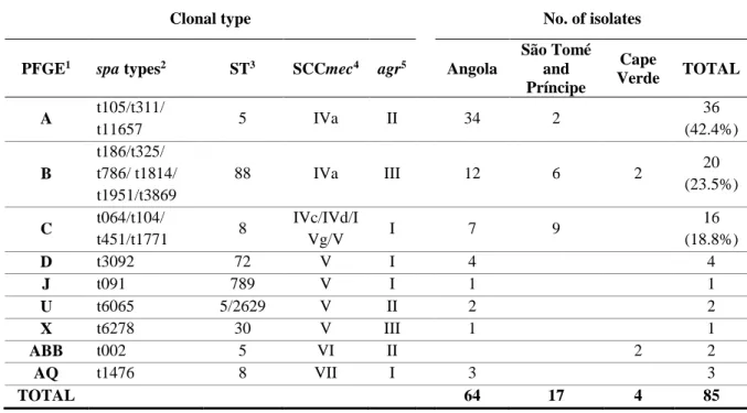
For this study, we selected a total of 122 S. aureus isolates previously found resistant to SXT (85 MRSA and 37 MSSA), from three Portuguese-speaking African countries (Angola, São Tomé and Príncipe, and Cape Verde), recovered from nasal swabs between 2010 and 2014 (33) – Tables 1 and 2. The isolates were chosen from an initial collection of 507 isolates, in order to represent the proportion of each clonal type in the three countries included in this study.
Table 1 – Genotypic properties of SXT-resistant MRSA isolates from Angola, São Tomé and Príncipe, and Cape Verde (33).
Clonal type
No. of isolates PFGE1 spa types2 ST3 SCCmec4 agr5 Angola
São Tomé and Príncipe Cape Verde TOTAL A t105/t311/ t11657 5 IVa II 34 2 36 (42.4%) B t186/t325/ t786/ t1814/ t1951/t3869 88 IVa III 12 6 2 20 (23.5%) C t064/t104/ t451/t1771 8 IVc/IVd/I Vg/V I 7 9 16 (18.8%) D t3092 72 V I 4 4 J t091 789 V I 1 1 U t6065 5/2629 V II 2 2 X t6278 30 V III 1 1 ABB t002 5 VI II 2 2 AQ t1476 8 VII I 3 3 TOTAL 64 17 4 85
1 PFGE – Pulsed-field gel electrophoresis 2 spa – Staphylococcus protein A 3 ST – Sequence type
4 SCCmec – Staphylococcal cassette chromosome mec 5 arg - Arginine
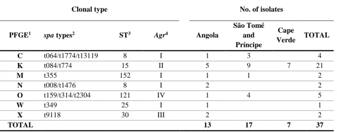
14 Table 2 – Genotypic properties of SXT-resistant MSSA isolates from Angola, São Tomé and Príncipe, and Cape Verde (33).
Clonal type No. of isolates
PFGE1 spa types2 ST3 Agr4 Angola
São Tomé and Príncipe Cape Verde TOTAL C t064/t1774/t13119 8 I 1 3 4 K t084/t774 15 II 5 9 7 21 M t355 152 I 1 1 2 N t008/t1476 8 I 2 2 O t159/t314/t2304 121 IV 1 4 5 W t349 25 I 1 1 X t9118 30 III 2 2 TOTAL 13 17 7 37
1 PFGE – Pulsed-field gel electrophoresis 2 spa – Staphylococcus protein A 3 ST – Sequence type
4 arg – Arginine
In addition, 18 SXT-resistant MRSA isolates representative of major clones widely spread in South America (Argentina, Brazil, Chile and Uruguay), Portugal and Taiwan, were also included (Table 3), reaching a total of 140 S. aureus isolates (4-6).
Table 3 – Genotypic properties and origin of SXT-resistant MRSA isolates from other continents belonging to the Brazilian clone or variant (4-6).
ST1 Origin Period of isolation SCCmec 2 No. of isolates TOTAL ST239 Brazil 1996-1998 III 3 18 Argentina 1995-1996 III 2 Uruguay 1996-1998 III 3 Chile 1997-1998 III 3 Portugal 1991-1997 III 4 Taiwan 1998-1999 III 1 ST241 Taiwan 1998-1999 III 2 1 ST – Sequence type
15
2. Antimicrobial susceptibility testing and minimum inhibitory concentration (MIC)
Antimicrobial susceptibility testing was performed for all isolates by the disk diffusion method for SXT, TMP and SMZ, according to standard published Clinical Laboratory Standards Institute (CLSI) guidelines (30). An isolate was considered (i) susceptible when the zone diameter (≥16 mm for SXT and TMP; ≥17 mm for SMZ) was in accordance to the susceptible criteria and there was a clear inhibition zone; (ii) hetero-resistant when there were distinct colonies growing or growth within a zone of inhibition creating a double zone; and (iii) fully resistant when the zone diameter (≤10 mm for SXT and TMP; ≤12 mm for SMZ) was in accordance to the resistance criteria and there were no colonies or growth within the inhibition zone.
In addition, the minimum inhibitory concentrations (MIC) for TMP and SMZ were determined for all isolates by the broth microdilution method following Clinical Laboratory Standards Institute (CLSI) guidelines (30) and in parallel by incubating and reading with a TECAN Infinite 200 Pro reader (Tecan group Ltd., Männedorf, Switzerland). Since the results were comparable with the two methodologies, the MICs for TMP and SMZ were subsequently performed for all isolates with the TECAN reader. Briefly, overnight bacterial cultures were adjusted to an optical density of 0.085 OD620, and 5 μL of each adjusted cell suspension was
added to 150 μL of fresh MH, containing the appropriate antibiotic concentration. The plates were incubated in a TECAN Infinite 200 Pro reader (Tecan group Ltd., Männedorf, Switzerland) for 15 h at 37ºC with shaking (180 rpm). Measurements were performed every 20 min at 595 nm. Positive (without antibiotic/with inoculum) and negative (without antibiotic/without inoculum) controls were included for each isolate. We considered that there was bacterial growth when the optical density after 15h reached at least 0.2. The MIC for SXT was determined by Etest (AB BioMérieux, Solna, Sweden) for all isolates previously identified as SXT-resistant by the disk diffusion method.
3. Total DNA extraction
Four to five colonies of cultures grown overnight on Tryptic Soy Agar (TSA) were incubated into 20 μL TE 1X (10 mM Tris, 1 mM EDTA, pH 8) with 10 mg/mL of lysostaphin
16 for 30 min performing cell lysis, followed by an enzyme denaturation at 95ºC for 15 min. The mixture was centrifuged at 13 000 rpm for 5 min and the supernatant was recovered.
4. Detection of TMP resistance genes
Isolates were screened for known S. aureus dfr genes by conventional PCR (Table 4) in a T1 Thermocycler (Biometra, Germany). The presence of dfrA and dfrG genes was determined for all isolates. The isolates without amplification of dfrA and dfrG genes were subsequently screened for the presence of dfrK. Isolates for which there was no amplification of the acquired dfr genes were subsequently analysed for their intrinsic dfrB gene. The following isolates were used as positive controls: ANG880 for dfrA, ANG17A for dfrG, FVL88.1 for dfrK and ATCC25923 for dfrB gene.
The DHFR-encoding dfrB was also sequenced and then aligned with strain ATCC25923, using DNASTAR Lasergene 8 Softwares: EditSeq, SeqMan, Protean and MegAlign (DNASTAR, Madison, WI, USA) to identify possible non-synonymous mutations.
For each gene, the PCR mixture contained 1X PCR Buffer with 1.5 mM MgCl2, 40 μM
of each deoxynucleotide triphosphate (dNTP), 0.4 μM of each primer, 1.25 U of Taq DNA polymerase (GoTaq – Promega, Madison, USA) and 2 μL of chromossomal DNA template in a final volume of 50 μL. PCR produts (10 μL) were resolved in 1.5% Seakem LE agarose (Cambrex Bio Science Rockland, USA) for dfrG, dfrK and dfrB genes and 2% for dfrA gene in 1X Tris-acetate-EDTA (TAE) buffer at 80V for 60 min and visualized with ethidium bromide.
17 Table 4 – Primers nucleotide sequences, product size and PCR conditions of the genes amplified in this study.
Gene Primer nucleotide sequences (5' - 3') Product size PCR conditions References
dfrG 405 bp 10, 85 F: TGCTGCGATGGATAAGAA R: TGGGCAAATACCTCATTCC 94ºC – 4 min 94ºC – 1 min 57ºC – 30 s 30 x dfrA 270 bp 72ºC – 1 min 10, 85 F: CACTTGTAATGGCACGGAAA 72ºC – 4 min R: CGAATGTGTATGGTGGAAAG dfrK 321 bp 94ºC – 4 min 47 F: GCTGCGATGGATAATGAACAG 94ºC – 30 s R: GGACGATTTCACAACCATTAAAGC 49ºC – 30 s 35 x 72ºC – 1 min 72ºC – 5 min dfrB 572 bp 94ºC – 4 min 85 F: AATTGTGTTAAATTAAAGATAACTT 94ºC – 1 min R: TAAGTATTCTTTAGATAAATCGGAT 43ºC – 30 s 35 x 72ºC – 1 min 72ºC – 4 min
18
5. Fitness experiments
Fitness experiments were performed as previously described (11, 73) with minor alterations detailed below.
A total of five SXT-resistant MRSA isolates were selected, which included: (i) representative strains of the three major MRSA clonal types in São Tomé and Príncipe - STP33 (ST88-SCCmec IVa), STP46A (ST5-SCCmec IV), STP151 (ST8-SCCmec IVg); and (ii)
representatives of the pandemic Brazilian clone or related: HSJ216 (ST239-SCCmec III), and TAW10 (ST241-SCCmec III).
5.1. Growth curves
For independent growth measurement, bacteria grown overnight in Tryptic Soy Broth (TSB) were diluted into 200 μL of fresh TSB to an initial concentration of 0.05 OD620 in
BrandPure Grade S plates. The cultures were incubated in a TECAN Infinite 200 Pro reader (Tecan group Ltd., Switzerland) for 20 h at 37ºC with shaking (180 rpm). Readings were taken every 20 min at 595 nm. All isolates were tested with at least three independent replicates.
5.2. Competition assays
Isolates STP33, STP46A and STP151 were tested against HSJ216, and STP151 was also tested against TAW10. A 1:1 inoculum of each pair of strains was co-cultured for 24 h. For that, overnight bacterial cultures were diluted in TSB to 0.085 OD620. 50 μL of the adjusted
cell suspension of each of the two tested strains were added to 5 mL of fresh TSB medium and incubated at 37ºC with shaking. Appropriate dilutions of samples were plated in duplicate onto selective plates (TSA with 5 μg/mL gentamicin), and non-selective TSA plates at 0 h, 2 h, 4 h, 6/7 h and 24 h after inoculation. Viable colony counts on the selective plates indicated the bacterial levels (cfu/mL) of HSJ216 or TAW10, since these isolates were resistant to
19 gentamicin, while the difference between colony counts on selective and non-selective plates indicated the bacterial levels (cfu/mL) of the tested strain (STP33, STP46A or STP151).
The initial ratio [NS(0)/NR(0)] and final ratio [NS(24)/NR(24)] were calculated and the
relative fitness (F) was obtained with the following formula: F=ln[NS(24)/NS(0)]/ln[NR(24)/NR(0)], where NS(t) represented the bacterial levels (cfu/mL) of
STP33, STP46A or STP151 and NR(t) the bacterial levels (cfu/mL) of HSJ216 or TAW10, at
0 and 24 h.
5.3. Survival assays
For survival experiments, 100 μL of overnight bacterial cultures were suspended in 900 μL of TSB. 100 μL of each suspension was plated onto five different empty sterile Petri dishes, manually shaken individually for approximately 1 min, and left closed on a shelf to dry. Samples were taken at 6 h, 24 h, 3 days, 5 days and 7 days, by adding 900 μL of saline solution and shaking manually for approximately 20 s. The saline solution was left on the closed Petri dish for 5 min, and then appropriate dilutions were plated onto TSB, incubated for 24 h, and viable colonies were counted.
The percentages of bacterial surviving until the final time points of 6 h or 7 days were calculated, as well as the average daily death rate (K) at day 1 (24 h), using the following formula: K= 2.3 x [(B0-BX)/1], where Bx is the log10 cfu at day X from the time of inoculation.
6. Resistance to chemical stresses
The following experiments were performed as previously described (103) with minor alterations detailed below.
6.1. Growth under stress conditions
For experiments of stress conditions, overnight cultures grown in TSB were diluted into 200 μL of fresh TSB to an initial concentration of 0.02 OD620 in 96-well BrandPure Grade S
20 plates. Simultaneously, bacteria were grown under stress conditions through appropriate modifications of TSB: 8.8 mM H2O2, 4% (v/v) ethanol, low pH (pH 4.5), high pH (pH 10) and
2.5 M NaCl. Growth was followed spectrophotometrically in a TECAN Infinite 200 Pro reader (Tecan group Ltd., Switzerland) at 37ºC with shaking (180 rpm). Readings were taken at 595 nm every 20 min for 18 h. All isolates were tested with three independent replicates.
6.2. Whole-cell autolysis assays
For bacterial autolysis assays, strains were grown overnight in TSB at 37ºC with shaking (180 rpm). After washing cells twice with cold water, cells were resuspended to an initial concentration of 1.2 OD620, in a 40 mL final volume of 0.05 M Tris/HCl buffer, pH 7.2,
containing 0.05% Triton X-100. The suspensions were incubated at 37ºC with shaking (180 rpm), and readings were taken every 30 min for 6 h.
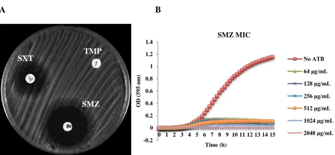
21 -0,2 0 0,2 0,4 0,6 0,8 1 1,2 0 1 2 3 4 5 6 7 8 9 10 11 12 13 14 15 O D (5 9 5 n m ) Time (h) SMZ MIC No ATB 64 ug/mL 128 ug/mL 256 ug/mL 512 ug/mL 1024 ug/mL 2048 ug/mL
CHAPTER III – RESULTS
1. Antibiotic resistance and screening of TMP resistance genes
The 140 S. aureus, initially selected for being previously reported as resistant to SXT, were analysed.
1.1. Antimicrobial susceptibility testing and MIC
All 122 isolates recovered from the African continent were confirmed to be full resistant to TMP, presenting a MIC ≥ 1024 μg/mL, with the exception of one isolate from São Tomé and Príncipe, that presented a MIC of 256 μg/mL – Annex 1/Table 1.
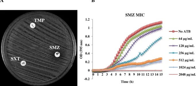
However, full resistance to the combination of TMP and SMZ (resistance to SXT) was only found in 20 isolates (16.4%). The MIC for SXT ranged from 6 to >32 μg/mL, and from 1024 to 2048 μg/mL for SMZ (Figure 4). Interestingly, these isolates (16 MRSA and 4 MSSA) belonged to the same clonal type (PFGE C-ST8-SCCmec IVc/IVd/IVg/V) and were recovered
from two different countries (Angola and São Tomé and Príncipe) – Annex 1/Table 1.
A B
Figure 4 – Antimicrobial susceptibility testing of an isolate fully resistant to TMP, sulfonamides and SXT by A) disk diffusion test; B) MIC determination for SMZ determined with the Tecan spectrophotometer. SMZ TMP SXT 64 μg/mL 128 μg/mL 256 μg/mL 512 μg/mL 1024 μg/mL 2048 μg/mL -0.2 0.2 0.4 0.6 0.8 1.2
22 Interestingly, the remaining 102 African isolates (83.6%) presented a double halo for SXT showing some growth inside (Figure 5A). Of these, 58 isolates were considered hetero-resistant for sulfonamides presenting also double halo phenotype (Figure 5A) and showing a MIC for SMZ ranging between 1024 to 2048 μg/mL (Annex 1/Table 1). The SMZ MIC determination with the Tecan equipment showed that only a small proportion of the original culture could grow even in the lower concentration of antibiotic (Figure 5B). Accordingly, the hetero-resistance to sulfonamides in the presence of full TMP resistance was not sufficient to render S. aureus isolates fully resistant to SXT - Figure 5 and Table 1/Annex 1.
A B
Figure 5 – Antimicrobial susceptibility testing of an isolate fully resistant to TMP and hetero-resistant to sulfonamides and SXT by A) disk diffusion test; B) MIC determination for SMZ determined with the Tecan spectrophotometer.
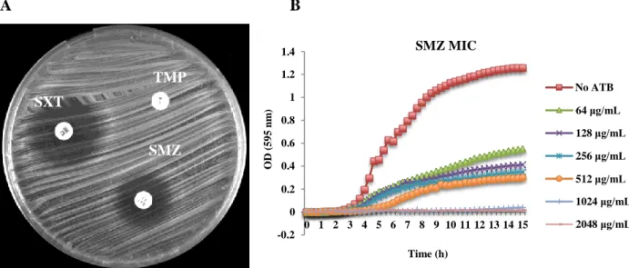
The remaining 44 isolates hetero-resistant for SXT were fully susceptible to sulfonamides, presenting a clear halo on the disk diffusion test and the MIC for SMZ ranged from ≤64 to 256 μg/mL – Figure 6 and Annex 1/Table 1.
-0,2 0 0,2 0,4 0,6 0,8 1 1,2 1,4 0 1 2 3 4 5 6 7 8 9 10 11 12 13 14 15 OD ( 5 9 5 n m ) Time (h) SMZ MIC No ATB 64 ug/mL 128 ug/mL 256 ug/mL 512 ug/mL 1024 ug/mL 2048 ug/mL SXT TMP SMZ 64 μg/mL 128 μg/mL 256 μg/mL 512 μg/mL 1024 μg/mL 2048 μg/mL -0.2 0.2 0.4 0.6 0.8 1.2 1.4
23 -0,2 0 0,2 0,4 0,6 0,8 1 1,2 1,4 0 1 2 3 4 5 6 7 8 9 10 11 12 13 14 15 OD ( 5 9 5 n m ) Time (h) SMZ MIC No ATB 64 ug/mL 128 ug/mL 256 ug/mL 512 ug/mL 1024 ug/mL 2048 ug/mL A B
Figure 6 – Antimicrobial susceptibility testing of an isolate fully resistant to TMP, hetero-resistant to SXT and susceptible to sulfonamides by A) disk diffusion test; B) MIC determination for SMZ determined with the Tecan spectrophotometer.
All 18 MRSA isolates recovered from South America, Portugal and Taiwan were full resistant to TMP, SMZ and SXT presenting MICs ranging from 256 to >1024 μg/mL for TMP, from 1024 to 2048 μg/mL for SMZ and from 24 to >32 μg/mL for SXT – Table 1/Annex 1.
1.2. Detection of TMP resistance genes
The 122 African isolates were tested for the presence of TMP resistance genes dfrG and dfrA. The dfrG gene was the most prevalent gene (81.1%) and was detected in 99 isolates (62 MRSA and 37 MSSA). It was not clustered to any particular S. aureus clone, being present in almost all clonal types. The dfrA gene was detected in 27 MRSA isolates (22.1%). It was mainly associated with clones PFGE B-ST88-SCCmec IVa (n=20, 100%) and PFGE ABB-ST5-SCCmec VI (n=2, 100%) but was also found among three other clonal types: PFGE AQ-ST8-SCCmec VII (n=2, 66.7%) and PFGE C-AQ-ST8-SCCmec IVd (n=3, 15%). Although the overwhelming majority of the isolates presenting dfrG gene lacked dfrA gene, four MRSA isolates belonging to clonal types B (n=2) and AQ (n=2) contained both genes. Since all
SMZ TMP SXT 2048 μg/mL 1024 μg/mL 512 μg/mL 256 μg/mL 128 μg/mL 64 μg/mL -0.2 0.2 0.4 0.6 0.8 1.2 1.4
24 African isolates harboured dfrG or dfrA genes, the screening for the presence of dfrK and dfrB was not performed in these isolates – Table 1/Annex 1.
Regarding the 18 MRSA isolates from other continents, only the two ST241 isolates from Taiwan presented dfrG gene; dfrA gene was only found in one isolate from Portugal. The presence of dfrK gene was screened in the remaining 15 isolates but none was found positive. However, 10/15 isolates harboured dfrB, which was further sequenced, presenting three synonymous and two non-synonymous mutations (F99Y and R150H) in all isolates. For each non-synonymous mutation, the aminoacid was replaced by another aminoacid of the same family: phenylalanine (F) and tyrosine (Y) belonged to the aromatic R group, and Arginine (R) and Histidine (H) belonged to the positively charged R group. A total of five isolates, recovered from Brazil (n=2), Argentina (n=1), Taiwan (n=1) and Portugal (n=1) did not amplify any of the TMP resistance determinants described so far.
2. Comparison of major SXT-resistant MRSA clonal types recovered from São Tomé and Príncipe against major SXT-resistant MRSA ST239/241 international clones
2.1 Fitness experiments
Independent growth experiments showed that the three African isolates (STP33, STP46A and STP151) had shorter lag phases and higher growth rates than TAW10 and HSJ216 - Figure 7.
There was no significant difference between the three isolates recovered from São Tomé and Príncipe. Therefore, the full resistance to SXT and sulfonamides of STP151 did not seem to cause any advantage or any fitness cost to the strain, when compared to the hetero-resistant isolates STP33 and STP46A.
25 Figure 7 – Independent growth curves of selected MRSA strains resistant and hetero-resistant to SXT: STP33, STP46A, STP151, TAW10 and HSJ216.
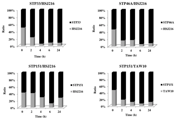
In the co-culture experiments, the higher growth rates of STP33, STP46A and STP151 led to outcompete HSJ216 and TAW10. In fact, while all strains were initially inoculated in almost equal concentrations (Initial ratios: ST33/HSJ216 = 0.97; STP46A/HSJ216 = 1.15; ST151/HSJ216 = 1.33; STP151/TAW10 = 1.10), the African isolates represented 72 to 91% of the population after a 24 h incubation period (Final ratios: ST33/HSJ216 = 10.59; STP46A/HSJ216 = 10.51; ST151/HSJ216 = 2.58; STP151/TAW10 = 7.30). Interestingly, the final ratio was higher for the hetero-resistant isolates (STP33 and STP46A) compared with the full SXT-resistant STP151 isolate when co-cultured against HSJ216 – Figure 8 and Table 5.
Furthermore, the competition assay showed that the African strains exhibited a fitness advantage over HSJ216 and TAW10 isolates, with a relative competitive fitness varying from 1.07 to 1.30 – Table 5. 0 0,2 0,4 0,6 0,8 1 1,2 1,4 1,6 1,8 0 1 2 3 4 5 6 7 8 9 10 11 12 13 14 15 16 17 18 O D (5 9 5 nm ) Time (h) STP33 STP46A STP151 TAW10 HSJ216 0.2 0.4 0.6 0.8 1.2 1.4 1.6 1.8
26 0% 20% 40% 60% 80% 100% 0 2 4 6 24 R a ti o Time (h) STP46A/HSJ216 STP46A HSJ216 0% 20% 40% 60% 80% 100% 0 2 4 6 24 R a ti o Time (h) STP33/HSJ216 STP33 HSJ216 0% 20% 40% 60% 80% 100% 0 2 4 6 24 R a ti o Time (h) STP151/HSJ216 STP151 HSJ216 0% 20% 40% 60% 80% 100% 0 2 4 6 24 R a ti o Time (h) STP151/TAW10 STP151 TAW10
Figure 8 – Co-culture growth of the strain pairs STP33/HSJ216, STP46A/HSJ216, STP151/HSJ216 and STP151/TAW10.
Table 5 – Viable cell ratio and the relative fitness of each strain pairs.
Strain pairs Viable cell ratio Relative fitness, F
Initial ratio (0 h) Final ratio (24 h)
STP33/HSJ216 0.97 10.59 1.30
STP46A/HSJ216 1.15 10.51 1.27
STP151/HSJ216 1.33 2.58 1.07
STP151/TAW10 1.10 7.30 1.21
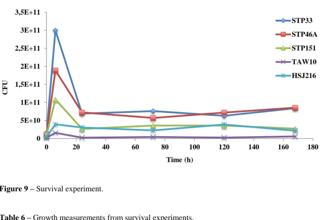
Concerning the survival assay, a significantly increase in cfu count was observed for the African isolates when compared with TAW10 and HSJ216, after 6 h. Furthermore, after seven days, the two SXT hetero-resistant isolates (STP33 and STP46A) survived desiccation better, probably providing them an advantage in a model mimicking survival on hospital surface whereas the three full SXT-resistant isolates (STP151, TAW10 and HSJ216) presented
27 a lower survival rate, which might constitute a disadvantage regarding survival on dry surfaces. In addition, the daily death rate after day 1 seemed to be higher for STP151 and TAW10 – Figure 9 and Table 6.
Figure 9 – Survival experiment.
Table 6 – Growth measurements from survival experiments.
Strain Percentage of inoculum surviving to 6 h Percentage of inoculum surviving to 7 days (168 h)
Daily death rate after day 1 STP33 1801.8 504.5 -1.42 STP46A 1766.4 799.1 -1.91 STP151 1112.8 280.5 -1.00 TAW10 648.8 237.6 -0.05 HSJ216 798.0 433.3 -1.81
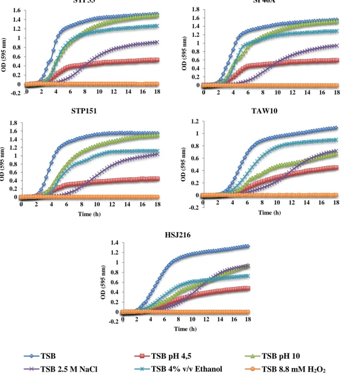
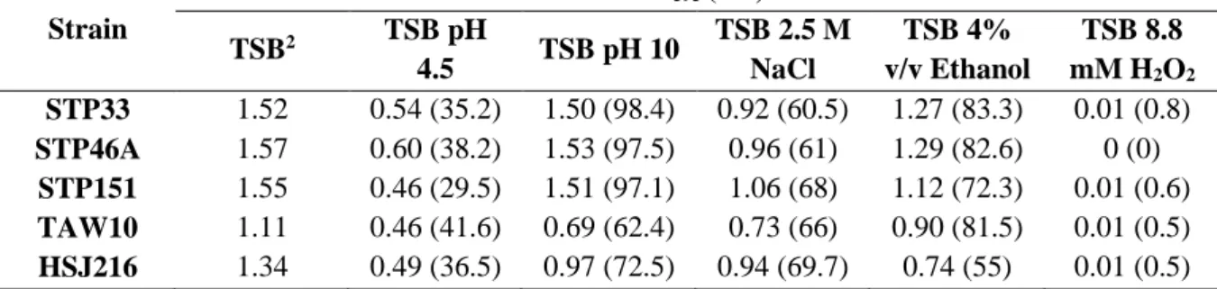
2.2 Resistance to chemical stresses
The selected five isolates were tested under different stress conditions compared with their independent growth on TSB. A moderate to very high growth reduction was observed for all isolates in the presence of 4% (v/v) ethanol, in high saline (2.5 M NaCl) and in acidic (pH
0 5E+10 1E+11 1,5E+11 2E+11 2,5E+11 3E+11 3,5E+11 0 20 40 60 80 100 120 140 160 180 CF U Time (h) STP33 STP46A STP151 TAW10 HSJ216
28 -0,2 0 0,2 0,4 0,6 0,8 1 1,2 1,4 1,6 0 2 4 6 8 10 12 14 16 18 OD ( 5 9 5 n m ) Time (h) STP33 -0,2 0 0,2 0,4 0,6 0,8 1 1,2 1,4 1,6 1,8 0 2 4 6 8 10 12 14 16 18 OD ( 5 9 5 n m ) Time (h) SP46A -0,2 0 0,2 0,4 0,6 0,8 1 1,2 1,4 1,6 1,8 0 2 4 6 8 10 12 14 16 18 OD ( 5 9 5 n m ) Time (h) STP151 -0,2 0 0,2 0,4 0,6 0,8 1 1,2 0 2 4 6 8 10 12 14 16 18 O D (5 9 5 n m ) Time (h) TAW10 -0,2 0 0,2 0,4 0,6 0,8 1 1,2 1,4 0 2 4 6 8 10 12 14 16 18 OD ( 5 9 5 n m ) Time (h) HSJ216 TSB TSB pH 4,5 TSB pH 10
TSB pH 2,5M NaCl TSB 4% v/v Ethanol TSB 8,8mM H2O2
4.5) conditions, respectively. In addition, the presence of 8.8 mM H2O2 in the medium
completely inhibited the growth of all isolates – Figure 10 and Table 7.
Although no apparent difference in the growth was observed for the African isolates in alkaline conditions (pH 10), the percentage of growth of HSJ216 and TAW10 was significantly affected, being reduced to 62 and 72% respectively – Figure 10 and Table 7.
Figure 10 – Growth in the presence of several chemical stresses.
-0.2 0.2 0.4 0.6 0.8 1.2 1.4 0.2 0.4 0.6 0.8 1.2 1.4 1.6 1.8 -0.2 0.2 0.4 0.6 0.8 1.2 1.4 1.6 1.8 1.6 1.4 1.2 0.8 0.6 0.4 0.2 1.2 0.8 0.6 0.4 0.2 -0.2 TSB 2.5 M NaCl TSB 8.8 mM H2O2