UNIVERSIDADE DE LISBOA
FACULDADE DE MEDICINA
Molecular mechanisms by which low doses of ionizing radiation
promote neovascularization in ischemic tissues
Paula Alexandra Gomes de Oliveira
Orientadora: Professora Doutora Susana Constantino Rosa Santos
Tese especialmente elaborada para a obtenção do grau de Doutor em Ciências Biomédicas - Especialidade em Biologia Celular e Molecular
UNIVERSIDADE DE LISBOA
FACULDADE DE MEDICINA
Molecular mechanisms by which low doses of ionizing radiation
promote neovascularization in ischemic tissues
Paula Alexandra Gomes de Oliveira
Orientadora: Professora Doutora Susana Constantino Rosa Santos Tese especialmente elaborada para a obtenção do grau de Doutor em Ciências
Biomédicas - Especialidade em Biologia Celular e Molecular Júri:
Presidente: Doutor José Luís Bliebernicht Ducla Soares, Professor Catedrático em regime de tenure e Vice-Presidente do Conselho Científico da Faculdade de Medicina da Universidade de Lisboa, Presidente do Júri
Vogais: Doutora Raquel Ângela Silva Soares Lino, Professora Catedrática da Faculdade de Medicina da Universidade do Porto;
Doutora Maria José Cardoso Oliveira, Professora Auxiliar Convidada da Faculdade de Ciências da Universidade do Porto;
Doutor Cláudio Areias Franco, Investigador do Instituto de Medicina Molecular, unidade de investigação associada à Faculdade de Medicina da Universidade de Lisboa;
Doutora Maria Isabel de Freitas Ferreira Queimado Monteiro Grillo, Professora Associada Convidada Aposentada da Faculdade de Medicina da Universidade de Lisboa;
Doutor Sérgio Jerónimo Rodrigues Dias, Professor Associado Convidado da Faculdade de Medicina da Universidade de Lisboa;
Doutora Susana Constantino Rosa Santos, Professora Auxiliar da Faculdade de Medicina da Universidade de Lisboa; (Orientadora)
Bolsa de Doutoramento SFRH/BD/80483/2011
financiada pela
Fundação para a Ciência e TecnologiaPreface
The work presented in this Thesis is derived from my Ph.D. research, made possible primarily by the Ph.D. fellowship ref. SFRH/BD/80483/2011, from Fundação para a Ciência e a Tecnologia (FCT), Ministério da Ciência, Tecnologia e Ensino Superior (MCTES), Portugal. This study, carried out between 2012 and 2016, was integrated in the Ph.D. program of Centro Académico de Medicina de Lisboa (CAML), which is shared between Instituto de Medicina Molecular (IMM, Lisbon, Portugal), Faculdade de Medicina da Universidade de Lisboa (FMUL, Lisbon, Portugal) and Hospital Santa Maria, Centro Hospitalar Lisboa Norte (HSM-CHLN, Lisbon, Portugal). The work took place at the Angiogenesis Lab, at Centro Cardiovascular da Universidade de Lisboa (CCUL), under the supervision of Prof. Susana Constantino (CCUL/FMUL).
This thesis is constituted by five chapters, which are preceded by a summary, both in Portuguese and in English. Chapter I consists of a general introduction to blood vessels, with particular emphasis on the angiogenic process, approached from the early embryonic development to adulthood, in physiology and pathology, arteriogenesis and postnatal neovascularization. A brief overview about the cellular and molecular effects of ionizing radiation is also presented. Chapter II specifically indicates the main objectives of the research proposal that led to the work presented in this thesis. Chapter III and IV include the experimental work developed through the research project. Chapter III, Low-dose ionizing radiation induces therapeutic neovascularization in a pre-clinical model of hindlimb ischemia, includes already published work (presented in word format) in Cardiovascular Research journal. Chapter IV, Low-dose ionizing radiation promotes
neovascularization in experimentally induced diabetic mice subjected to hindlimb ischemia, includes results from ongoing work that is currently being developed in our lab
and has not yet been published. Both results chapters include an abstract, a specific introduction, the results obtained in the work developed, and a focused discussion, as well as the methods and the references. In Chapter V, conclusions are integrated and discussed, generating new testable hypotheses which comprise the foreseen future directions of our research in this field.
As opiniões expressas nesta publicação são da exclusiva responsabilidade do
seu autor.
A impressão desta Tese foi aprovada pelo Conselho Científico da Faculdade
de Medicina de Lisboa em reunião de 18 de Julho de 2017.
Acknowledgements
Ao longo deste doutoramento muitas foram as pessoas com quem me cruzei e que contribuíram para o sucesso deste trabalho. Nesta secção da minha tese quero deixar um agradecimento especial a todos os que considerei fundamentais para a realização deste trabalho. Foram anos de muita aprendizagem e crescimento científico, mas principalmente humano. Agora sei com mais certeza que não há caminho, o caminho faz-se a andar e sem dúvida que a vida se resolve sozinha!
Começo por agradecer à minha orientadora, a Professora Susana Constantino, por me ter recebido na Unidade de Angiogénese e por ter acreditado em mim dando-me a oportunidade de crescer a nível científico. Obrigada pelo apoio, profissional, mas também pessoal, que sempre demonstrou ao longo destes anos permitindo-me desenvolver este trabalho com espírito crítico e perfecionismo.
À Fundação para a Ciência e Tecnologia, pela minha bolsa de doutoramento SFRH/BD/80483/2011, financiada por fundos nacionais do Ministério da Ciência, Tecnologia e Ensino Superior.
Ao serviço de Radioterapia, do Hospital de Santa Maria, sem o qual não teria sido possível realizar este trabalho. Gostaria de agradecer particularmente o apoio da Doutora Isabel Monteiro Grilo, na qualidade de ex-diretora do serviço de Radioterapia, que abraçou este projeto e possibilitou o seu início, da Doutora Marília Jorge, também na qualidade de ex-diretora do serviço, e que proporcionou a continuidade do trabalho apoiando-o incondicionalmente, e da Doutora Filomena Pina, na qualidade de atual diretora do serviço de Radioterapia, que sempre demonstrou interesse no trabalho possibilitando também a sua continuidade. Obrigada à Doutora Esmeralda Poli, na qualidade de responsável da Unidade de Física Médica, e à sua equipa por todo o apoio. Obrigada à técnica Ana Duarte pela ajuda no planeamento e dosimetria. Obrigada à técnica coordenadora Isabel Diegues e a toda a sua equipa pela ajuda nas sessões de irradiação ao longo destes anos.
Ao biotério de Roedores do Instituto de Medicina Molecular, com especial agradecimento à Joana, Dolores, Iolanda e Carlos, por se mostrarem sempre disponíveis e por todo o apoio ao longo destes anos.
Ao Laboratório de Histologia Patologia Comparada do Instituto de Medicina Molecular, Tânia, Andreia, Ana e Bruna, obrigada pelo apoio e pela ajuda nas muitas urgências que surgiram.
Um agradecimento especial para ti Andreia! Conhecemo-nos através de um crióstato e um músculo congelado mas hoje somos muito mais. Obrigada por sempre arranjares um tempinho para os meus cortes, pelas dicas de trabalho, mas principalmente pela amizade, pelas conversas tanto sérias como triviais. É tão bom partilhar histórias de crianças e melhor ainda vê-los juntos!
Ao serviço de Citometria de Fluxo do Instituto de Medicina Molecular, Maria Soares e Ana Vieira, obrigada pelo apoio e ensinamentos sobre FACS.
Ao serviço de Bioimagem do Instituto de Medicina Molecular, Rino, António e Ana, obrigada por todo o apoio em todos os microscópios que usei. Foram incansáveis na procura da melhor imagem.
Ao Professor João O’Neill, da Faculdade de Ciências Médicas da Universidade Nova de Lisboa, obrigada por todo o apoio e dedicação na implementação da técnica de diafanização em pequenos animais possibilitando a obtenção de excelentes imagens. Foi um prazer conhecer o seu laboratório e o trabalho que tem vindo a realizar.
A todos os membros da Unidade de Agiogénese que conheci ao longo destes anos e com quem partilhei o meu dia: André, Adriana, Andreia, Carolina, Joana, Liliana, Teresa Freitas (tem sido um apoio para mim desde o primeiro dia, obrigada por partilhar o dia comigo), Carolina Cardina (tens um lugar especial no meu coração), Rita (gostei muito de te conhecer).
Um especial agradecimento à Raquel, foi contigo que tudo começou, deste-me as primeiras introduções sobre o mundo da angiogénese e foi tão bom partilhar o laboratório e o dia contigo. A tua boa disposição era contagiante, tive saudades tuas ao longo deste tempo. Augusto, também para ti um especial obrigada, pela disponibilidade imediata sempre que programávamos isquémias, claro que tínhamos de começar o dia sempre de madrugada mas a boa disposição compensava e o tempo passava sem darmos conta.
Filipa, foste a minha companheira de laboratório durante estes anos, partilhávamos o gosto em ter tudo arrumado. Um grande obrigada, pela disponibilidade que sempre tiveste para me ajudar e lembro-me particularmente duma noite complicada em que fizeste de babysistter e saíste-te muito bem, o miúdo gosta de ti! Obrigada também pelos cafés, pelas conversas e pelos olhares!
Ana, a tua chegada ao grupo é recente, mas sinto que foste fundamental. Muito obrigada pela disponibilidade e apoio, pelas conversas otimistas e pelas correções na escrita desta tese.
Ao meu colega e amigo Daniel pela amizade e companheirismo ao longo dos muitos anos que nos conhecemos. Obrigada pela ajuda, principalmente na formatação desta tese.
À minha amiga Sónia, para mim um modelo de perfecionismo e de rigor. Muito me ensinaste ao longo destes anos, ensinamentos científicos, mas também de vida. E quando a vida complicou tu descobriste a farmácia onde estava aquele leite tão difícil de encontrar… Obrigada por tudo, mantem-te aí!
À minha amiga Filomena, conhecemo-nos há muitos anos e tem sido muito bom saber que te tenho no laboratório/gabinete ao lado. Foram muitas as nossas partilhas ao longo dos últimos anos, científicas, mas também pessoais. Obrigada por me inspirares e por tantas vezes me dizeres para continuar!
Aos meus pais, Hortense e Ivo, pelo exemplo de vida e pelos valores com que me educaram, pelo incentivo e apoio em todos os meus desafios. Pelo imenso amor, por cuidarem sempre tão bem, por estarem sempre presentes, pela ajuda constante, pelas conversas que me levam ou trazem. Um obrigada nunca será suficiente, tentarei sempre fazer o meu melhor!
Ao meu marido Ricardo, mais uma etapa que concluímos juntos. Sabemos que somos uma boa equipa, rearranjamos os dias e modificamos as noites, mas conseguimos. Obrigada pelo amor, carinho e dedicação à nossa família, por tornares tudo mais fácil, por estares disponível sempre que precisei/precisamos. Love you!
Ao meu querido filho Martim, que enche os meus dias de luz e de um amor sem fim. Nesta nossa dança muito me tens ensinado, nasceste no início deste doutoramento e tens sido a melhor companhia, meu menino doce de abraço apertado e gargalhada fácil. Quando nos despedimos de manhã dizes “mãe, faz coisas bonitas no teu trabalho”, a mãe tentará sempre fazer coisas bonitas!
A ti, meu querido Martim, eu dedico esta tese e de agora em diante “vou pedir ao tempo
Resumo
O sistema circulatório ou cardiovascular é formado pelo coração e vasos sanguíneos e a sua função é levar oxigénio e nutrientes aos órgãos e tecidos do corpo. A doença cardiovascular, de que é exemplo a doença arterial periférica (DAP) é uma das principais causas de mortalidade e morbilidade na população mundial. Estima-se que mais de 25 milhões de pessoas na Europa e nos Estados Unidos apresentem DAP, e a sua incidência tende a aumentar. Importantes fatores de risco para a DAP são a diabetes, tabagismo, hipertensão e dislipidemia. A DAP caracteriza-se por uma obstrução do lúmen arterial, resultando num défice de fluxo sanguíneo aos tecidos cuja principal consequência é a presença de sintomas característicos de isquemia. A manifestação mais frequente de DAP é a claudicação intermitente, que é caracterizada por desconforto muscular no membro inferior, produzido pelo exercício, e que alivia com o repouso. A claudicação tem um impacto negativo na qualidade de vida dos doentes, quer a nível profissional, quer interferindo com as suas atividades sociais. A isquemia crítica dos membros inferiores é a manifestação clínica mais grave da DAP, que descreve doentes com dor em repouso ou com lesões tróficas cutâneas, sejam elas úlceras ou gangrena. Aproximadamente 1% dos doentes com DAP apresentam o estado avançado da doença. As limitações dos procedimentos de revascularização, resultantes da extensão e distribuição anatómica da doença arterial, e do tratamento farmacológico, são bem conhecidas. Assim, a amputação surge como última alternativa terapêutica, apesar das taxas de morbilidade e mortalidade associadas. Na última década vários estudos têm vindo a ser desenvolvidos com o objetivo de encontrar tratamentos alternativos, incluindo a angiogénese terapêutica.
A angiogénese terapêutica, a qual pode ser alcançada através da administração local de fatores de crescimento pró-angiogénicos na forma de proteína recombinante ou de células progenitoras endoteliais (CEPs) ou mesmo recorrendo à terapia génica, oferece uma possibilidade de recuperação para estes pacientes. Apesar dos estudos pré-clínicos e de alguns ensaios clínicos em fase I/II mostrarem-se bastante promissores, a progressão desses ensaios clínicos para terapias angiogénicas não têm revelado um efeito benéfico em doentes com isquemia crítica dos membros inferiores. Também o pouco conhecimento sobre os agentes angiogénicos envolvidos, nomeadamente a dose, a frequência e o método de administração têm contribuído para o fracasso dos ensaios clínicos.
No passado, o nosso grupo de investigação demonstrou que baixas doses de radiação ionizante (BDRI) promovem angiogénese durante o desenvolvimento embrionário e no processo de regeneração da barbatana caudal do peixe zebra. Assim, de acordo com os nossos resultados as BDRI induzem angiogénese in vivo, mas não existe prova de que produzam angiogénese terapêutica em doentes com doença isquémica, sendo este o propósito deste trabalho de doutoramento. Assim, o presente trabalho tem como objetivo: 1) identificar os mecanismos celulares e moleculares pelos quais as BDIR poderão
promover angiogénese terapêutica;
2) avaliar se as BDRI promovem a vascularização pós-natal pelo aumento de CEPs em circulação e sua incorporação em locais de neovascularização nos tecidos isquémicos;
3) demonstrar que as BDRI promovem neovascularização num modelo experimental de diabetes.
Um modelo animal de isquémia foi desenvolvido usando fêmeas de ratinhos C57BL/6 e recorrendo a várias técnicas de biologia celular e molecular, nomeadamente RT-PCR, imunohistoquímica, imunofluorescência, citometria de fluxo, ELISA, microscopia de microdissecção a laser e diafanização.
Neste estudo, após a indução de isquémia unilateral, os ratinhos C57BL/6 foram irradiados ou não com 0.3 Gy, durante quatro dias consecutivos.
Os resultados demonstraram que as BDRI, aumentam significativamente a perfusão sanguínea no membro inferior do ratinho sujeito a isquémia, assim como a densidade capilar e a formação de colaterais. Foi também avaliada a vasculatura no membro inferior contralateral que apesar de irradiado não foi sujeito a isquémia. Neste caso particular, nem a densidade capilar, nem a formação de colaterais registaram alterações quando comparadas com animais não irradiados. Estas observações sugerem que em condições não patológicas as BDRI não têm efeito na vascularização.
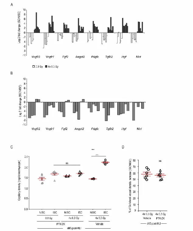
De forma a validar os mecanismos moleculares no nosso modelo isquémico, foram selecionados os melhores candidatos associados a uma resposta pró-angiogénica a partir de uma análise de expressão gênica por microarrays. Os nossos resultados mostraram que a expressão de vários genes pró-angiogénicos nomeadamente, Vegfr2, Vegfr1, Fgf2,
de ratinhos sujeitos a isquémia e comparadas com a expressão em células endoteliais isoladas dos músculos não isquémicos contralaterais. Desta forma, as BDRI sugerem conferir uma resposta pró-angiogénica após indução de isquémia.
Neste trabalho foi também demonstrado que as BDRI aumentam significativamente tanto a concentração de VEGF, PIGF e G-CSF como de CEPs em circulação e medeiam a incorporação destas células nos músculos isquémicos.
Este trabalho revela ainda que não existe diferença de morbilidade nem de mortalidade em ratinhos irradiados quando comparados com ratinhos não irradiados, sendo eles acompanhados durante 52 semanas após exposição a BDRI.
Para complementar este trabalho e sendo a diabetes um importante fator de risco no contexto da DAP, foi observado que em resposta à indução de isquémia as BDRI, administradas durante quatro dias consecutivos, aumentam significativamente a perfusão sanguínea, a densidade capilar e a formação de colaterais num modelo experimental diabético. No entanto estes dados resultam de um estudo preliminar, sendo crucial o aumento do número de animais.
Em conclusão, o presente trabalho demonstra que BDIR induzem angiogénese terapêutica num modelo experimental de isquémia do membro inferior. Este trabalho é relevante pois propõe o uso de BDIR como uma estratégia inovadora e não invasiva no tratamento da isquémia crítica do membro inferior.
Palavras-chave
Neovascularização; Radiação ionizante; Isquémia do membro inferior; Células endoteliais progenitoras; Diabetes
Abstract
Peripheral arterial disease (PAD) is mainly caused by an obstructive artherosclerosis, which results in a mismatch between oxygen supply and demand. Diabetes is an important risk factor for PAD and it is present in almost 50% of the patients with limb ischemia. Critical limb ischemia (CLI) is the end stage of PAD, being characterized by severe obstruction of blood flow to the affected extremity, which results in ischemic rest pain, ulcers or gangrene. Despite substantial evidence of their efficacy in preclinical studies, as well as some promising phase I/II clinical trials, larger randomized clinical trials on angiogenic therapies for CLI have been unsatisfactory.
Here, we investigated the ability of low-dose ionizing radiation (LDIR) to stimulate therapeutic neovascularization, in murine models, in a context of hindlimb ischemia (HLI), conjugated or not with diabetes.
We demonstrate that 0.3 Gy, administered for four consecutive days, significantly improves blood perfusion in the murine ischemic limb by stimulating angiogenesis and arteriogenesis, as assessed by laser Doppler flow, capillary density and collateral vessel formation. LDIR significantly increased the circulating levels of VEGF, PlGF and G-CSF, as well as the number of circulating endothelial progenitor cells (EPCs), mediating their incorporation into ischemic muscles. These effects were dependent upon LDIR exposition on the ischemic niche (thigh and shank regions). In irradiated ischemic muscles, these effects were independent of the recruitment of monocytes and macrophages. Also, the vasculature in an irradiated non-ischemic bed was not affected and after 52-week LDIR exposure no differences in the incidence of morbidity and mortality were seen. Additionally, in diabetic mice, our data suggest that 0.3 Gy applied during four consecutive days significantly promote blood perfusion, capillary and collateral vessel densities in response to HLI induction. These findings disclose an innovative and non-invasive strategy to induce therapeutic angiogenesis in a murine model of severe HLI, emerging as a novel approach in the treatment of CLI.
Keywords
Neovascularization; Hindlimb ischemia; Ionizing radiation; Endothelial progenitor cell; Diabetes
Abbreviations
AKT protein kinase B
ALK activin receptor‐like kinase
ANGPT angiopoietin
BM-MNCs bone marrow-derived mononuclear cells BM-MSCs bone marrow-derived mesenchymal stem cells
CLI critical limb ischemia
CoCl2 cobalt chloride
COUP-TFII chicken ovalbumin upstream promoter transcription factor II
CVD collateral vessel density
CXCR4 cell-derived factor-1 receptor
DLL4 delta‐like‐4
DNA deoxyribonucleic acid
ECs endothelial cells
ECM extracellular matrix
ELISA enzyme linked immunosorbent assay EPCs endothelial progenitor cells
ERK extracellular signal-regulated kinase FACS fluorescence activated cell sorting
FAK focal adhesion kinase
FGF fibroblast growth factor
FGFR fibroblast growth factor receptor
Fox c forkhead C
FSS fluid shear stress
G-CSF granulocyte-colony stimulating factor
Gy Gray
HGF hepatocyte growth factor
HLI hindlimb ischemia
HMVEC-L lung microvascular endothelial cells
i.p. intraperitoneal
IL interleukin
iMM Instituto de Medicina Molecular
LCM laser capture microdissection microscope LDIR low-doses ionizing radiation
LFA lymphocyte function-associated antigen
Mac1 macrophage 1 antigen
MAPK mitogen-activated protein kinase MCP1 monocyte chemoattractant protein 1 MET hepatocyte growth factor receptor
MMPs matrix metalloproteinases
MSCs mesenchymal stem cells
NO nitric oxide
NOS nitric oxide synthase
NRPs neuropilins
PAD peripheral arterial disease
PB-MNCs peripheral blood-derived mononuclear cells
PCA principal component analysis
PDGF platelet-derived growth factor
PDGFR platelet-derived growth factor receptor PECAM-1 platelet endothelial cell adhesion molecule 1 PI3K phosphatidylinositl-3-kinase
PIGF placental growth factor
PSGL-1 P-selectin glycoprotein ligand 1
PTK/ZK PTK787/ZK222584
ROI region of interest
RT‐PCR real-time polymerase chain reaction S1PR sphingosine-1-phosphatase receptor SDF-1 cell-derived factor-1
SMCs smooth muscle cells
STZ streptozotocin
TGFβR transforming Growth Factor Receptor β TIE2 tyrosine-protein kinase receptor for ANG
TNFα tumor necrosis factor α
TPS treatment planning system
TSP1 thrombospondin 1
uPA urokinase-type plasminogen activator VCAM vascular cell adhesion molecule VE-cadherin vascular endothelial cadherin VEGF vascular endothelial growth factor
Table of contents
Perface ... i Acknowledgements ... iii Resumo ... vii Abstract ... xi Abbreviations ... xiiiTable of contents ... xvii
CHAPTER I ... 1
1. Blood vessels: Structure and Function ... 1
1.1. Endothelial Cell Specification ... 3
2. Vasculogenesis ... 5
2.1. Vasculogenesis during embryo development ... 5
2.2. Postnatal Vasculogenesis ... 5
2.2.1. EPCs phenotypic identification and characterization... 6
2.2.2. Mobilization and Recruitment of EPCs ... 7
3. Arteriogenesis ... 9
4. Angiogenesis ... 11
4.1 Physiological Angiogenesis ... 15
4.1.1 Angiogenic Regulators ... 15
4.2. Pathological Angiogenesis ... 25
4.2.1. Critical Limb Ischemia ... 25
5. Pro‐angiogenic therapy ... 27
5.1. Local administration of pro-angiogenic growth factors ... 27
5.2 Cell-based therapeutic strategy ... 28
6. References ... 32
CHAPTER II ... 47
Aims ... 47
CHAPTER III ... 51
Low-dose ionizing radiation induces therapeutic neovascularization in a pre-clinical model of hindlimb ischemia. ... 51
Abstract ... 53 Introduction ... 54 Methods ... 55 Results ... 56 Discussion ... 71 References ... 77 Supplementary Material ... 80 CHAPTER IV ... 103
Low-dose ionizing radiation promotes neovascularization in experimentally induced diabetic mice subjected to hindlimb ischemia ... 103
Abstract ... 105 Introduction ... 106 Methods ... 108 Results ... 113 Discussion ... 116 References ... 118 CHAPTER V ... 121
Concluding Remarks and Future Perspectives ... 121
Chapter I
1. Blood vessels: Structure and Function
Contrarily to primitive and smaller organisms, the complex body architecture of vertebrates requires an efficient circulatory system, able to distribute oxygen and nutrients to tissues and remove carbon dioxide and other metabolic waste products throughout the body.
The circulatory system carries out two main networks: the blood-vascular and the lymphatic systems, both formed by endothelial cells (EC). The cardiovascular system allows blood to circulate and transport nutrients, oxygen, carbon dioxide and other molecules throughout all cells in the body1, while the lymphatic system drains extravassed fluid, the lymph, from the extracellular space and returns it into the venous circulation. The lymphatic vasculature is also essential for the immune defense, since any foreign material present in the lymph is filtered through the chain of lymph nodes2. Both networks are essential for homeostasis of a healthy organism, and their malformation or dysfunction contributes to many diseases1.
Blood vessels are divided into three main groups: arteries, veins, and capillaries3. Briefly, blood full of oxygen and nutrients is pumped from the heart to the tissues, through the arteries that ramify into smaller arterioles and into capillary beds, while blood enriched in carbon dioxide and waste returns to the heart through venules and veins1.
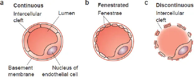
Arteries and veins are further divided according to their caliber, into large, medium and small blood vessels. The microvasculature composed of the smallest vessels (arterioles, venules and capillaries) is a very dynamic and complex system, capable of constant change, while the larger blood vessels are more permanent structures with reduced plasticity3. Capillaries, the most abundant vessels in our body, are also one of the most important vessels of the cardiovascular system, since their thin walls allow the exchange of oxygen and nutrients between blood and tissues3. Capillaries are composed of endothelial cells surrounded by basement membrane and a sparse layer of pericytes embedded within the EC basement membrane. Pericytes, due to their contractile fibers, possess a cell body with prominent nucleus and a small content of cytoplasm. They are functionally significant, since when vessels lose pericytes, they become hemorrhagic and hyper dilated, leading to conditions such as edema, diabetic retinopathy, and even embryonic lethality4. Capillaries in different tissues exhibit different cellular morphology, associated with distinct levels of
permeability (Figure 1): (a) continuous capillaries, characterized by an uninterrupted endothelium and a continuous basement membrane, that exist, for instance, in muscle, lung or the nervous system; (b) fenestrated capillaries, characterized by a continuous basement membrane with fenestrae or pores in the endothelium that allow the rapid passage of macromolecules and exist, for example, in the kidney, intestines or endocrine glands and (c) discontinuous capillaries, characterized by large openings in the endothelium and a discontinuous or absent basement membrane that exist, for instance, in the liver and spleen5.
Figure 1 - Capillary wall morphology. (a) Continuous capillaries have no openings in their wall and are lined continuously with the EC body. (b) Fenestrated capillaries have small openings, called fenestrae that are covered by a small, non-membranous, permeable diaphragm and allow the rapid passage of macromolecules. The basement membrane of ECs is continuous over the fenestrae. (c) Discontinuous capillaries have a large lumen, many fenestrations with no diaphragm and a discontinuous or absent basal lamina. Adapted from reference5.
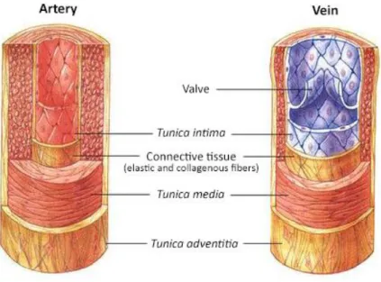
In contrast, the walls of larger vessels (arteries and veins) have three specialized layers (Figure 2). The tunica intima consists of an endothelium, a basement membrane and an internal elastic layer. The tunica media is composed of a thick layer of smooth muscle cells (SMCs) with reticular fibers, elastin and proteoglycans and external elastic lamina, while the tunica adventitia consists of connective tissue with both elastic and collagenous fibers and external elastic lamina5,6. Arterioles and venules have an increased coverage of mural cells when compared to capillaries.
The advential layer has its own blood supply, known as vasa vasorum. SMCs and elastic laminae contribute to the vessel tone and mediate the control of vessel diameter and blood flow. Although the walls of arteries and veins are composed of these same layers, they present some differences as a result of the pressure and direction of the blood flow. Arteries are more robust, with a strong elastic vessel wall to cope with the high arterial blood pressure downstream of the heart. Since veins conduct blood back to the heart, blood flows with a lower pressure in veins and consequently their wall is thinner than the one of arteries. Veins have additional semi-lunar valves, which prevent the blood from flowing backwards5.
Figure 2 - Morphology and wall composition of large blood vessels. Adapted from reference7.
1.1. Endothelial Cell Specification
Although arteries and veins are both formed from primitive blood vessels, different hemodynamic changes and physiology lead to their distinct morphology and physiology factors, in a process known as arteriovenous differentiation1,2,8. Hemodynamic forces such as blood flow rate, direction, and pressure are key factors driving the differentiation of vessels into arteries and veins. However, studies have demonstrated that arteries and veins possess distinct molecular identities from a very early stage, suggesting that genetics have a critical role in dictating arterial/venous fate9. Although the molecular processes
underlying arteriovenous differentiation are not fully understood, increasing evidence suggests that this process is regulated by the concerted action of different molecules. The first discovered specific markers that distinguish arteries from veins were two members of the Eph-Ephrin subclass of receptor tyrosine kinases, ephrinB2 and EphB49. EphrinB2 is a transmembrane ligand and is specifically expressed in endothelial precursors that produce arteries, whereas Ephb4, the receptor for Ephrin B2, is found preferentially in veins10. Mice deficient in Ephrin B2 and Ephb4 have similar defects in the remodeling of primary capillary vessels into a mature vascular network8.
It is known that Notch pathway is directly involved in the differentiation of the arterial branch1. In mice, the receptors Notch1 and Notch2 and the ligands Jagged1, Jagged2 and delta‐like‐4 (DLL4) are all expressed in arterial but not in venous ECs. Notch4-/- and Notch1 -/- mice exhibit abnormal vascular development, whereas Dll4-/- mice are not viable, presenting severe vascular defects and showing reduced EphrinB2 expression and increased EphB4 expression, consistent with a failure in arterial differentiation. These data suggest that Notch activity is important for the promotion of arterial cell fate9,11. Upstream from Notch pathway are Foxc1 and Foxc2 (forkhead C1 and C2) that control arterial specification by regulating the expression of the Notch ligand Dll4. Mice with inactivated Foxc1 and Foxc2 develop arterial-venous malformations10.
Additionally, COUP-TFII (chicken ovalbumin upstream promoter transcription factor II) is specifically expressed in venous endothelium. COUP-TFII mutant mice show ectopic expression of arterial markers such as neuropilin 1 (NRP1), Notch1 and EphrinB2 in veins. These studies suggest that COUP-TFII is a crucial regulator of venous cell fate by inhibiting the expression of NRP1 and Notch signaling8,12.
The expansion of larger arteries and veins occurs by acquisition of additional layers of mural cells, extracellular matrix (ECM) and elastic laminae to provide the required viscoelastic properties. Homotypic and heterotypic junctions facilitate cell-to-cell communication and regulate vessel permeability. Vascular endothelial cadherin is an important component of EC-EC junctions, whereas neural cadherin facilitates EC-mural cell communication. Gap junctions (made of connexins) also facilitate communication between ECs and between ECs and perivascular cells. Tight junctions (formed by occludins, claudins and zona occludens) contribute to the blood tissue barrier in the brain and retinal capillaries6.
2. Vasculogenesis
Vasculogenesis is a fundamental process by which new blood vessels are formed, characterized by the differentiation of precursor cells into endothelial cells and the de novo formation of a vascular network.
2.1. Vasculogenesis during embryo development
The development of the vascular system is one of the earliest events in organogenesis13. In vertebrates, one of the mechanisms by which the vascular development proceeds is via
vasculogenesis, the process of de novo blood formation, characterized by the formation of
vessels directly from angioblastic precursors14.
In the early phase of vasculogenesis, hemangioblasts undergo their first critical steps of differentiation within the blood islands, being committed to differentiate into angioblasts and primitive haematopoietic cells. The angioblasts aggregate in the periphery and the hematopoietic precursors accumulate at the center of the blood islands in the yolk sac and embryo15,16. The angioblasts migrate to discrete locations, differentiate in situ and assemble into solid endothelial cords, later forming a plexus with endocardial tubes13. Vascular endothelial growth factor (VEGF) and their receptors, vascular endothelial growth factor receptor 1 and 2 (VEGFR1 and VEGFR2), are key players in embryonic vessel formation. Particularly, VEGFA is required for the chemotaxis and differentiation of endothelial precursor cells, EC proliferation and angiogenic remodeling1. Genetic studies show that the deficiency in one of these VEGF/VEGFR molecules is lethal in mice, due to failed development of the vasculature2,16. The lethality resulting from the loss of a single allele is indicative of a dependent regulation of embryonic vessel development by VEGFA2. The existence of a precursor for these cells types (haemangioblast) is suggested by defects in both the haematopoietic and angioblastic lineages of embryos lacking VEGFR2. Moreover, in the absence of the VEGFR1 mice produce angioblasts but their assembly into functional blood vessels is impaired17.
2.2. Postnatal Vasculogenesis
Until the late 1990s, it was generally accepted that after birth, new blood vessels were developed only by angiogenesis. However, new findings indicate that vasculogenesis is not
restricted to early embryogenesis18. Asahara et al., pioneer in the scientific field of adult vasculogenesis, isolated mononuclear cells from adult peripheral blood and found that those cells had the same characteristics as the embryonic angioblasts, contributing to the revascularization of the ischemic tissue19. These cells were termed “endothelial progenitor cells” (EPCs). Thus, a new concept, describing postnatal neovascularization as a combination of vasculogenesis and angiogenesis, in which EPCs play a crucial role, emerged18.
2.2.1. EPCs phenotypic identification and characterization
The identification of EPCs can be based on their cellular origin, isolation methods and surface marker expression20. Although the analysis of cell surface markers is currently the most used methodology21, EPCs identification is controversial, since there is no unique protein marker that defines them22. The analyzed markers are usually shared with cells from hematopoietic lineage and mature ECs23. Therefore, EPCs combine stem cell markers, such as CD133, c-kit and Sca-1 and endothelial markers, such as VEGFR2, CD31, VE-cadherin and von Willebrand factor20,24. CD34 represents a link between precursor and mature cells, since it is expressed in both cell types. On the other hand, CD133 allows the distinction between progenitor and mature cells, since it is not present on mature ECs22.
Beyond the controversy about the phenotypic identification of EPCs, the heterogeneity of cultured cells has also been a concern. Many studies reported EPCs after using different sources of the cells or method for culture22,25-27. For instance, human CD34+ cells isolated from peripheral blood, umbilical cord blood or bone marrow can all differentiate into ECs. Moreover, studies show that when total mononuclear cells, isolated from peripheral blood, are cultured in vitro, two types of cell populations that appear sequentially can be observed26. The first population, “Early EPCs”, comes after 4-7 days of plating on fibronectin-coated surface with the addition of endothelial growth media, as spindle-shaped cells. “Early EPCs” gradually loose CD45 and CD31 expression and gain low-level expression of VEGFR2 and VE-Cadherin. However, they lose this low-level expression of VEGFR2 and VE-cadherin after 3 weeks, dying after that26. These cells play a role in vasculogenesis by secreting large quantities of angiogenic factors, which act through paracrine mechanisms28. On the other hand, when blood-derived mononuclear cells are
higher and longer sustained expression of VEGFR2 and VE-cadherin26, much higher proliferation potential and are able to differentiate into ECs and promote vascular tube formation22. Thus, “late EPCs” enhance neovasculogenesis, by providing a sufficient number of ECs based on their high proliferation potency26. These two type of cells have the ability to absorb acetylated low-density lipoprotein and bind lectin Ulex europaeus agglutinin I29. Despite such differences in genetic and functional in vitro aspects, they equally contribute to neovasculogenesis26.
2.2.2. Mobilization and Recruitment of EPCs
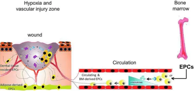
Under physiological conditions, levels of EPCs in the peripheral circulation are low and these cells reside in the bone marrow niche in a quiescent state30. The mobilization of EPCs from the bone marrow into the peripheral circulation is promoted by several growth factors, chemokines and cytokines which are produced in response to a physiological stress (tissue hypoxia and trauma) (Figure 3)31.
One of the most important factors in EPCs mobilization is VEGF. When VEGF interacts with its receptor, VEGFR2, it activates nitric oxide synthase (NOS) in the bone marrow, which produces nitric oxide (NO), a key player for matrix metalloproteinase 9 (MMP) 9 activation. Activated MMP-9 contributes to the transformation of membrane-bound Kit ligand to a soluble form (sKitL) and consequently detaches EPCs from the bone marrow niche to the peripheral circulation28.
Once in circulation, homing of EPCs is activated in response to chemokine gradients that are formed in the tissue undergoing active remodeling. The major chemokine that regulates activation and homing of EPCs is the stromal cell-derived factor1 (SDF1)32,33. In response to an ischemic stimulus, released SDF1 interacts with its receptor (CXCR4), and initiates, not only the mobilization of EPCs from bone marrow, but also their adhesion to ischemic areas. The SDF-1/CXCR4 interaction upregulates the P-selectin glycoprotein ligand 1 (PSGL1) expression on the surface of EPCs, which is the major ligand of P-selectin. The binding contributes to the adhesion of EPCs to the sites of vessel damage and enhances their pro-angiogenic capacity. However, in the absence of injury, the effect of SDF1 is abolished. After homing into the injured vessel wall, EPCs will contribute to new vessel formation and remodeling, but the mechanism behind the functional activity of EPCs is still under investigation. Besides differentiating into ECs, EPCs also produce multiple paracrine
factors, such as VEGF, SDF1, hepatocyte growth factor (HGF), angiopoetin1 (ANGPT1), insulin-like growth factor 1, monocyte chemoattractant protein1 (MCP1) and platelet-derived growth factor (PDGF). These factors can assist different cell types in promoting angiogenesis and tissue regeneration, reflecting the indirect contribution of EPCs to neovascularization34.
EPCs are mobilized from bone marrow into the peripheral circulation and recruited into sites of vessel injury to participate in blood vessel formation, in both physiological and pathological conditions. However, the mechanisms of mobilization and recruitment are still incompletely understood and require further analysis. Nevertheless, several studies suggest that EPCs play a critical role in postnatal neovascularization and vascular homeostasis30.
Figure 3 - EPCs mobilize in response to hypoxia induced by trauma or vascular injury. In normal homeostatic conditions, EPCs reside within a stem cell niche in the bone marrow. Peripheral tissue hypoxia under trauma, wound healing or vascular injury conditions results in increased production of EPC-mobilizing chemokines and growth factors to a concentration greater than that in the bone marrow, causing EPC release and mobilization into the peripheral circulation. Once in circulation, EPCs respond to chemokine signaling in the tissues by undergoing active remodeling and homing to the injury site. Concomitantly, circulating progenitor cells, tissue-resident, and adipose-derived stem cells respond to the chemokine signaling, homing to the active tissue-remodeling site. Adapted from reference20.
3. Arteriogenesis
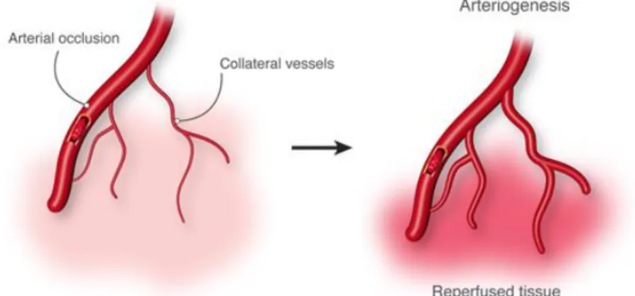
Arteriogenesis, defined as the formation of mature arteries from pre-existing arterioles after an arterial occlusion, needs to be activated to confer proper tissue function35 (Figure 4).
Figure 4 - Arteriogenesis. Arterial occlusion induces a pressure gradient followed by a redistribution of perfusion, an increase in collateral flow and a subsequent outgrowth of the collateral arteriole. Adapted from reference36.
The driving force for arteriogenesis is altered fluid shear stress (FSS), which appears within the collateral arteriole after a blood flow increase. However, the large pressure difference in the pre-existing arterioles connecting upstream with downstream branches, relative to the point of occlusion, also contributes to initiate a complex cascade of molecular and cellular events leading to increased vessel lumen and wall thickness37. The primary physiological response to FSS is the activation of ECs in the collateral wall. After an arterial occlusion, the ECs of collateral vessels appear swollen, as opposed to the completely flat inner surface of normal arteries38. The endothelial activation is indicated by several processes that condition for the attraction of circulating cells: upregulated genes that encode for chemoattractant or activated cytokines, including growth factors or adhesion molecules37. Thus, changes in the expression and conformation of adhesion molecules converts the collateral endothelium from a quiescent vessel layer with very low adhesion tendency into a highly activated one, which is now supporting attraction, adhesion and invasion of leukocytes37. Also, several molecules involved in cell proliferation and migration were found up-regulated, including MMP2, MMP9, urokinase-type plasminogen activator (uPA), focal adhesion kinase (FAK) and integrins (α5β1 and αvβ3)38. Moreover, after an arterial obstruction, the altered FSS induces at least two signaling pathways: (1) the
attraction, adhesion and invasion of monocytes that are required for structural remodeling and (2) the pathway that causes ECs and SMCs to enter the cell cycle, leading to proliferation. Thus, in response to altered FSS, NO and VEGF are induced and, together with calcium-activated ion channels, interfere with osmotic regulation of the endothelium38,39. This occurs along with monocyte adhesion, since the activated endothelium produces MCP1, leading to the recruitment of monocytes to the sites of proliferation40. After the initial monocyte-endothelium interaction, which is mediated by several selectins, the tight monocyte adhesion to the endothelium is triggered by macrophage-1 antigen (Mac1) and
lymphocyte function-associated antigen (LFA). These integrins interact with their corresponding adhesion molecules (intercellular adhesion molecule 1 and 2 (ICAM1), (ICAM2) and vascular cell adhesion molecule 1 (VCAM1), (respectively) on the endothelial cell surface41. After adhesion, migration into deeper parts of the collateral wall and surrounding areas can be observed. During migration, monocytes should overcome barriers, like the internal elastic lamina and ECM. However, as monocytes are producers of proteases, such as MMPs and uPA, they enable the proteolytic degradation of these barriers and could therefore create a gap by which monocytes could invade the vascular wall. At the same time, these events may also generate a proliferation signal for SMC37. Furthermore, lymphocytes (natural killer family, CD4 and CD8 cells) also play a role in arteriogenesis. Studies from Stephen Epstein´s group showed that in mice with a genetic deficiency in the T-cell marker CD4, arteriogenesis was inhibited in the hindlimb ischemia model, but could be rescued by an injection of purified CD4 positive cells. Additionally, the lack of CD4 positive T-cells led to a reduced inflammatory response in the same model including a consistent reduction in the number of monocytes37. As mentioned before, remodeling from a small pre-existing arteriole to a large collateral artery is facilitated by a complex cascade of processes. The signaling cascade uses the mitogen activated protein kinases with activation of the RAS-ERK- and the Rho pathway41. ECs mitosis precedes that of SMCs by a few hours and growth factors are released from monocytes such as MMPs, u-PA and fibroblast growth factor 2 (FGF2) during that time38. Also, degradation of extracellular structures leads to the release of additional matrix-bound growth factors. The increase in cell mitosis in SMCs occurs together with a morphological change: their transformation from a contractile into a proliferative/synthetic phenotype. Elastin is
stimulate SMCs proliferation41. Subsequently, SMCs migrate and rearrange accordingly to the increasing vessel lumen and wall thickness. The controlled destruction of the vascular scaffolding paves the way for the expansion and outward growth of collateral vessels. Moreover, apoptosis of SMCs may facilitate the renewal of the vascular wall38. Finally, remodeling enters in a maturation state when both elastic lamina and ECM components are rearranged. In this process, laminin and collagen IV are crucial since they promote the differentiation and inhibit the proliferation of SMCs37.
4. Angiogenesis
After the formation of a primitive vascular plexus during vasculogenesis, the growth, expansion and remodeling of primitive vessels into a mature vascular network (including arteries, veins, and capillaries) is initiated. This process, named angiogenesis, occurs by intussusception, in which interstitial tissue columns are inserted into the lumen of pre-existing vessels, inducing partition of the vessel lumen, or by sprouting of new vessels from the ends and sides of the pre-existing ones17,42-44.
The sprouting angiogenesis is facilitated by hypoxia, which regulates the expression of several genes involved in vessel formation, patterning and maturation, such as NOS, VEGF and ANGPT26. The existing vessels dilate and become leaky in response to NO and VEGF, respectively. At the same time, redistribution of intercellular adhesion molecules (platelet endothelial cell adhesion molecule 1(PECAM1), vascular endothelial cadherin (VE cadherin)) and alteration in cell membrane structure occur16. Consequently, the extracellular matrix ECM dissolves in response to activation of MMPs (MMP2, MMP3 and MMP9) and suppression of protease inhibitors. Degradation of ECM also results in the release of growth factors, including FGF2, VEGF and insulin-like growth factor, which otherwise remain stored within the matrix. As the physical barriers are dissolved, ECs can migrate to distant sites, through interactions between integrins (αvβ3 and α5β1) and the matrix proteins, and proliferate in response to VEGF and other endothelial mitogens (ANGPTs, FGFs and PDGF)6,16.
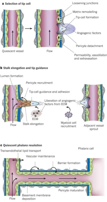
The different steps of angiogenic process are explained in Figure 5. Briefly, the ECs, also known as “tip cells”, become selected to lead the tip in the presence of factors such as: VEGF receptors, NRPs and NOTCH ligands. The neighbors of the “tip cell” assume positions
as “stalk cells”, which divide to support sprout elongation and establish the lumen3,44,45. At this point, the vessel sprouting remodels into a highly organized vascular network of larger vessels ramifying into smaller ones160. Then, the maturation process begins, described as the stepwise transition from an actively growing vascular bed to a quiescent functional network. This process involves the suppression of endothelial proliferation and sprouting, and the stabilization of existing vascular tubes through the recruitment of mural cells. Pericytes establish direct cell-cell contact with ECs in capillaries and immature vessels, whereas SMCs cover large diameter vessels (arteries and veins) and are separated from ECs by a matrix1. During this process, the pericytes and SMCs contribute to the deposition of ECM. This is critical for normal vessel growth and maintenance by providing the solid scaffold through which new vessels may migrate and store growth factors and pro-enzymes involved in vessel development. Thus, the recruitment of these mural cells is crucial for the maturation and stability of the new vasculature44.
The regulation of the angiogenic process involves at least four molecular pathways: (1) PDGF/platelet-derived growth factor receptor (PDGFR); (2) ANGPT1/Tie2; (3) Transforming growth factor β (TGFβ) and (4) Sphingosine 1 phosphatase receptor (S1PR) signaling. The PDGF/PDGFR signaling is an important regulator of this process. PDGFβ is secreted by ECs in response to VEGF, recruiting pericytes and SMC and allowing their proliferation and migration during vascular maturation6. Pdgf mutations leads to poorly covered vessels with excessive endothelial sprouting, microaneurysms, leakage and haemorrhaging1.
Also, critical for vessel formation and stability are ANGPT1 and ANGPT2 and their Tie receptors. The Tie2 receptor is expressed in ECs and is stimulated by ANGPT1 and ANGPT2, expressed by mural cells and ECs, respectively1,46. ANGPT1 stabilizes nascent vessels and makes them leak-resistant through the communication between ECs and mural cells6. In agreement with this, mouse mutants for Tie2 or ANGPT1 have similar behaviors, presenting severe vascular defects, and are unable to recruit pericytes47. The role of ANGPT2 is dependent on the presence or absence of VEGF. When VEGF is present, ANGPT2 promotes blood vessel growth and sprouting; the absence of VEGF leads to endothelial cell death and vessel regression48,49.
Additionally, TGFβ1 is also involved in the stabilization of immature vasculature. It is a multifunctional cytokine that promotes the maturation of vessels through the stimulation
migration. TGFβ1 is expressed in ECs and mural cells and can be either pro or anti-angiogenic, depending on context and concentration 6. Loss of function of TGFβR2 in mice causes vessel fragility due to impaired mural cells development50.
Also, S1PR signaling controls EC/mural cell interaction. Endothelial-derived S1P binds to G protein-coupled S1PRs and triggers cytoskeletal, adhesive and junctional changes, affecting cell migration, proliferation and survival. Disruption on S1PR1 or loss of both S1PR2 and S1PR3 in mice causes defective coverage of vascular SMCs50.
In the end of the maturation process ECs acquire highly specialized characteristics to provide the functional needs within specific tissues and organs16. This process includes arterio-venous determination, formation of homotypic and heterotypic junctions and ECs differentiation to form organ-specific capillary structures6.
Figure 5 - Angiogenesis. (a) After stimulation with angiogenic factors, the quiescent vessel dilates and an EC tip cell is selected to ensure branch formation. Tip cell formation requires degradation of the basement membrane, pericyte detachment and loosening of endothelial cell junctions. Increased permeability permits extravasation of plasma proteins to deposit a provisional matrix layer, and proteases remodel the pre-existing interstitial matrix, all enabling cell migration. (b) Tip cells navigate in response to guidance signals and adhere to the ECM to migrate. Stalk cells behind the tip cell proliferate, elongate and form a lumen, and sprouts fuse to establish a perfused neovessel. Proliferating stalk cells attract pericytes and deposit basement membranes to become stabilized. (c) After fusion of neighboring branches, lumen formation allows perfusion of the neovessel, which resumes quiescence by the promotion of a phalanx phenotype, re-establishment
of junctions, deposition of basement membrane, maturation of pericytes and production of vascular maintenance signals. Adapted from reference45.
4.1 Physiological Angiogenesis
After birth, angiogenesis still contributes to organ growth, but once the new vessels are assembled, the ECs become notably resistant to exogenous factors and most blood vessels remain quiescent16,43. Accordingly, in adults and in physiological conditions, angiogenesis occurs only in specific situations, such as wound healing, regeneration of the endometrium during the menstrual cycle or in the placenta during pregnancy6,51.
Quiescent ECs retain their remarkable ability to divide rapidly in response to a physiological stimulus, such as hypoxia, low pH or shear stress, and therefore influence the formation, maturation and remodeling of small and large vessels6,51. Thus, in physiological situations, it is reasonable to assume that molecules involved in vessel formation and maturation during embryonic development are also involved in the postnatal period, but their precise role is not yet known because most knockout mice die pre- or perinatally6.
4.1.1 Angiogenic Regulators
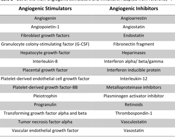
In multicellular organisms, the process of angiogenesis is regulated by a complex and tight balance between pro- and anti-angiogenic molecules43. Physiologically, the body controls angiogenesis through a series of “on” and “off” regulatory switches. The main “on” switches are known as angiogenesis growth factors and the main “off” switches are known as endogenous angiogenesis inhibitors. A selective list of the main stimulators and inhibitors that take part in the angiogenic process is shown in Table 1 and some of these factors will be briefly detailed below 52.
Table 1 - List of the main angiogenic stimulators and inhibitors. Adapted from reference52.
Angiogenic Stimulators
Angiogenic Inhibitors
Angiogenin Angioarrestin
Angiopoietin-1 Angiostatin
Fibroblast growth factors Endostatin
Granulocyte colony-stimulating factor (G-CSF) Fibronectin fragment
Hepatocyte growth factor Heparinases
Interleukin-8 Interferon alpha/ beta/gamma
Placental growth factor Interferon inducible protein Platelet-derived endothelial cell growth factor Interleukin-12
Platelet-derived growth factor-BB Metalloproteinase inhibitors
Pleiotrophin Plasminogen activator inhibitor
Progranulin Retinoids
Transforming growth factor alpha and beta Thrombospondin-1
Tumor necrosis factor-alpha Vasculostatin
Vascular endothelial growth factor Vasostatin
Vascular Endothelial Growth Factor
VEGF family consist of six members: VEGFA, also called VEGF, VEGFB, VEGFC, VEGFD, PIGF and virus VEGF (VEGFE)53. VEGF was the first identified and the most studied of the VEGF family members54. VEGF is expressed in different tissues including brain, kidney, liver, spleen and by many different cell types55. Several in vitro studies have shown that VEGF stimulates microvascular EC proliferation, enhancing migration and inhibiting apoptosis. Additionally, VEGF induces the growth of new capillaries from preexisting vasculature. In vivo studies also showed that VEGF regulates vascular permeability, which is important for the initiation of angiogenesis56-59. VEGF causes vasodilatation by inducing endothelial nitric oxide synthase and thus increasing NO production60, and it also plays a crucial role in several angiogenic processes, namely wound healing, ovulation, maintenance of blood pressure, menstruation and pregnancy61. In humans, VEGF is expressed in almost every solid tumors as well as in some hematological malignances62.
Different growth factors and cytokines such as PDGF, tumor necrosis factor α (TNFα), TGFβ and interleukin 1β (IL1β) induce transcription of VEGF mRNA, thus VEGF may function as a mediator for indirect action of angiogenic factors53. VEGF levels can also be regulated by tissue oxygen tension, since exposure to hypoxia induces VEGF expression. In contrast, normoxia downregulates VEGF production and even causes regression of some newly formed blood vessels63.
The expression of VEGF has been shown to be associated with significant steps in angiogenesis and physiological vasculogenesis. According to this, several in vivo studies demonstrate that, in mice, deletion of the VEGF gene leads to embryonic death, resulting in vascular defects and cardiovascular abnormalities64.There are currently six isoforms of VEGF resulting from alternative splicing of a single gene (VEGF121, VEGF145, VEGF165, VEGF183, VEGF189 and VEGF206). Some of these isoforms remain associated with cells or membranes, while others are extracellularly released. Despite this difference, all of them have identical biological activities65,66.
Two high affinity binding sites for VEGF have been identified on vascular endothelium: VEGFR1 (Flt1) and VEGFR2 (KDR or Flk1). An additional member of this family, VEGFR3 (Flt4) is not a receptor for VEGF but binds VEGFC and VEGFD55. All VEGF receptors have seven immunoglobulin domains in their extracellular part and an intracellular tyrosine kinase domain65. Also, all VEGF receptors are expressed in high levels during embryogenesis, and in adults VEGFR1 and VEGFR2 are mainly expressed in the blood vascular system, whereas VEGFR3 is restricted to the lymphatic endothelium67. Knockout mice for VEGFR1 or VEGFR2 are embryonically lethal, being the blood vessels completely absent, which suggest that both receptors are essential for normal development of the embryonic vasculature68,69. Neuropilin, another receptor for VEGF, was primarily identified as a receptor for members of the collapsing/semaphoring family on neuronal cells, but it is also expressed on ECs. Neuropilin is an isoform specific receptor that binds to VEGF16565.
VEGFR1 strongly interacts with VEGF, but this interaction plays a minor role in angiogenesis. However, interaction of VEGFR2 with VEGF is a major contributor to the mitogenic, chemotactic, angiogenic and increased permeability effects of VEGF70. Once VEGF binds to the extracellular domain of the receptor, after the dimerization and auto-phosphorylation of the intracellular receptor tyrosine kinases, a cascade of downstream proteins are activated54.
Due to the key role of VEGF in angiogenesis, the use of anti-VEGF drugs has been applied in many different fields of medicine, such as prevention of angiogenesis associated with tumors and induction of neovasculature in ischemic diseases3.
Angiopoietins
ANGPT family consists of four ligands: ANGPT1, 2, 3 and 4; and two tyrosine kinase receptors, TIE1 and TIE245.
ANGs are paracrine growth factors that act as ligands for the TIE receptors, specifically on ECs, and are involved in the maintenance, growth and stabilization of the vessels71,72. The most important and well characterized ANGPTs that play a role in angiogenesis are ANGPT1 and ANGPT2. Both ANGPT1 and ANGPT2 bind to TIE2 receptors, but only the binding of ANGPT1 results on signal transduction and regulation of blood vessel maturation55. Thus, the interaction between ANGPT1 and TIE2 receptor induces the remodeling and stabilization of blood vessels. ANGPT1 is expressed by mural and tumor cells, whereas ANGPT2 is released from angiogenic tip cells. In confluent endothelium, ANGPT1 induces the maintenance of quiescent endothelial cells through cell-cell junction. Also, ANGPT1 stimulates mural coverage and basement membrane deposition, thereby promoting vessel tightness45. Studies have shown that knockout mice for ANGPT1 or TIE2 exhibit similar phenotypes, with prominent endocardial and myocardial defects. The primary vasculature of these mice develops normally, but the stabilization and remodeling of the vessels fails, leading to embryonic death65. On the other hand, overexpression of ANGPT1 in transgenic mice causes excessive hypervascularization, namely a greater number of blood vessels with large diameters73. Concerning ANGPT2, its role in angiogenesis is more complex. ANGPT2 can act as a pro- or anti-angiogenic factor, dependent on co-stimulatory molecules, such as VEGF. At first, ANGPT2 was considered as a natural antagonist of ANGPT1, since it blocks ANGPT1-induced TIE2 autophosphorylation in ECs49. However, recent new studies suggest that in the presence of VEGF, ANGPT2 mediates an increase in the capillary diameter, induces migration and proliferation of ECs and stimulates sprouting of new blood vessels, while in its absence, ANGPT2 causes apoptosis of ECs and regression of blood vessels74. Despite the current difficulty to understand ANGPT2 activity, the selective inhibition of ANGPT2 may have clinical value. Several studies have shown ANGPT2 upregulation in some
osteoarthritis and psoriasis75-77. Additionally, various agents that block either TIE2 or ANGPT2 are being evaluated in early-phase clinical trials45. Regarding TIE1 receptor, since no ligand has been identified, it may act as a negative regulator of TIE2, but its role remains undefined.
Fibroblast Growth Factors
FGFs are soluble growth factors that stimulate EC proliferation and migration, as well as production of collagenase and the plasminogen activator54. FGF family is known to contain at least 20 factors, numbered FGF1 to FGF20, and 4 different tyrosine kinase receptors (FGFR), numbered FGFR1 to FGFR4.
The most studied forms are FGF1 (acid FGF) and FGF2 (basic FGF), which were among the first growth factors that were known to stimulate angiogenesis.
They are both secreted by a wide range of cell types and bind to all four FGFR65. In addition, FGFs also bind, with high affinity, to heparin sulfate proteoglycans (HSPGs), which act as co-receptors. HSPGs are located on cell surface and within the ECM, modulating the effects of FGF both in vitro and in vivo78,79. Heparin induces oligomerization of FGF2, which might be important for receptor dimerization and activation. FGFR activation will then trigger an intracellular signal cascade leading to multiple biological responses, including ECs proliferation, migration, differentiation, protease production and angiogenesis55.
FGFs stimulate angiogenesis directly by activating their receptors on ECs or indirectly by inducing the release of angiogenic factors from other cell types. For instance, in the heart, FGF-mediated signaling induces vessel growth by stimulating the release of hedgehog, ANGPT2 and VEGFB.
FGF1 and FGF2 knockout mice develop and grow normally without any evident pathological phenotype, being their organogenesis and life span unaffected. However, mice lacking FGF2 show neuronal defects and exhibit delays in wound healing, suggesting that FGF2 might regulate sprouting of new sites of tissue repair80,81. Additionally, tube formation stimulated by VEGF is totally abolished when neutralizing antibodies against FGF2 are added to the system, showing that in this particular setting, VEGF requires the presence of FGF2 to promote vessel assembly82. FGF signaling also contributes to the proliferation of tumor cells, either by an autocrine or paracrine mechanism82,83.
Moreover, disruption of the genes encoding FGFR1 or FGFR2 leads to embryonic death, which makes impossible to define the role of these receptors in the later stages of development and in angiogenesis84,85.
Transforming Growth Factor β
TGFβs are a family of homodimeric cytokines that include TGFβ1, 2, 3, 4 and 5. TGFβ binds to two different types of serine-threonine kinase receptors, known as type I (TGFβR1) and type II (TGFβR2). TGFβR1 and TGFβR2 are interdependent, meaning that TGFβR1 requires TGFβR2 to bind to TGFβ and TGFβR2 requires TGFβR1 for signaling. ECs also present another specific type III (co)receptor, endoglin, which is upregulated during angiogenesis. TGFβ helps to control many different cellular processes, including angiogenesis3. TGFβs are normally found in the ECM of many different cells types. In the microvasculature both EC and pericytes express TGFβRs and produce TGFβ, suggesting an auto- or paracrine loop for TGFβ86.
An interesting property of TGFβ is that it is secreted as inactive precursors which need to become activated87. In vitro studies have shown their activation by heat, acidification and proteases88. Additionally, in vivo, activation of TGFβ by proteases could be a regulatory mechanism for TGFβ-mediated activity89.
To date, both pro- and anti-angiogenic properties have been attributed to TGFβ, depending on the context. At low doses (<0.5 ng/ml), TGFβ helps to initiate the angiogenic switch by upregulating angiogenic factors and proteinases. However, at high doses, it inhibits EC growth, promotes basement membrane reformation and stimulates SMCs differentiation and recruitment71.
Additionally, TGFβ reduces the degradation of the perivascular ECM by the induction of protease inhibitors and the reduction of proteases. On the other hand, TGFβ stimulates angiogenesis in vivo, leading to the formation of highly vascular granulation tissue two or three days after mice being subcutaneously injected90.
Since TGFβ is not an EC mitogen, angiogenesis is indirectly stimulated through the recruitment of inflammatory cells, which in turn release pro-angiogenic cytokines65. TGFβ is highly chemotactic for monocytes, which under the influence of TGFβ are able to infiltrate a wound site and produce angiogenic factors91. However, a direct action is also