Vol.48, Special : pp. 185-190, October 2005
ISSN 1516-8913 Printed in Brazil
BRAZILIAN ARCHIVES OF
BIOLOGY AND TECHNOLOGY
A N I N T E R N A T I O N A L J O U R N A L
Effects of Low-Dose Ionising Radiation on Pituitary
Adenoma: is there a Role for L-type Calcium Channel?
Marcella Araugio Soares and Raquel Gouvêa dos Santos
*Laboratório de Radiobiologia; Centro de Desenvolvimento da Tecnologia Nuclear-CDTN/CNEN; C. P. 941; 30123-970; santosr@cdtn.br; Belo Horizonte - MG - Brasil
ABSTRACT
Pituitary adenomas constitute about 6–18% of brain tumours in adults. Activation of voltage gated calcium currents can account for growth hormone oversecretion in some GH-secreting pituitary adenomas that produce an acromegaly appearance and increase mortality. Ca2+ ions, as mediators of intracellular signalling, are crucial for the development of apoptosis. However, the role of [Ca2+] in the development of apoptosis is ambiguous. In this study, the effects of low-dose ionising gamma radiation (60Co) on rat pituitary adenoma cells survival and proliferation and the role of calcium channels on the apoptosis radio-induced were evaluated. Doses as low as 3 Gy were found to inhibit GH3 cell proliferation. Even though there was a significant number of live cells,168 hours following irradiation, they were not able to proliferate. The results indicate that the blockade of extracellular calcium influx through these channels does not interfere in the radiation-induced apoptosis in GH3 cells.
Key words: Calcium channels; ionising radiation; pituitary adenoma cells
* Author for correspondence
INTRODUCTION
Pituitary adenomas constitute about 6–18% of brain tumours in adults (Frohman, 1995). Despite the fact that pituitary adenoma is a benign tumour, patients can develop acromegaly due to hypersecretion of growth hormone that, not only alters appearance of these patients (acromegalic appearance), but also increases mortality (Klibanski et al, 1995). Currently, only 20-50% of patients can achieve the control of hormone activity after surgery. Somatostatin analogues are used for acromegaly treatment; however, life-long therapy is both inconvenient and very expensive. For this reason, radiotherapy has been widely used as adjuvant treatment for residual adenome (Melmed, 1990).
The major risk from gamma knife radiosurgery is radiation damage to the optic chiasm. To reduce this unwanted radiation effect, searches for radioprotective agents and alternative therapies have been performed. In a search for radioprotective agents, diltiazem, a calcium antagonist with a benzothiazepine structure, was found to protect mice against a lethal gamma radiation dose (Floersheim, 1992).
The role of [Ca2+](i) in apoptosis induced by
agents that do not immediately increase [Ca2+](i), such as 5-FdUr, TGF beta-1, doxorubicin, or radiation, is far more controversial (Tombal et al, 2002). [Ca2+]
(i) homeostasis is achieved by
intracytoplasmic Ca2+ stores (Endoplasmic
Reticulum and mitochondria) and Ca2+ influx
Ca2+ influx responsible for hormone secretion. In a previous report (Lomeo et al, 2004), the presence of Cav1 (L-type Cav) in GH3 cells, an immortalized cell line derived from a rat pituitary adenoma, was shown. Calcium is required as a cofactor by primer recognition proteins involved in DNA synthesis and by protein kinase C (PKC), which is activated by ionising radiation (Hallahan et al, 1994). However, the role of Ca2+ influx through Cav1 on the radiation effects has not been evaluated. Because these processes may be involved in radiation-mediated regulation of the progression of cells through the phases of the cell cycle, the effects of the Cav1 antagonists on the pituitary adenoma radio-induced cell death were studied.
MATERIALS AND METHODS
Cell Culture
GH3 cells from rat pituitary adenoma (ATCC, Rockville, MD, USA) were grown in a humidified
atmosphere of 5% CO2/95% air, at 37 °C. Cells
were maintained in Dulbecco’s modified Eagle medium (DMEM) supplemented with 10% Fetal Bovine Serum (FBS) and 0.1 % antibiotics.
Gamma Irradiation
GH3 cells were irradiated at a rate of 0.3 Gy/minute in a GammaCell irradiator (60Co; LIG, CDTN/CNEN, Brazil) at room temperature in the presence of molecular oxygen. Following irradiation, cells were incubatedat 37 °C.
Calcium channel antagonist
Nifedipine, a dihydropyridine calcium antagonist, was used as calcium channel blocker (5µM). The cells were incubated with Nifedipine one hour before irradiation (3Gy). Nifedipine was replaced every 24 hours.
Growth-inhibition Assay
Exponentially growing GH3 cells were plated in
40 mm Petri dishes at 105 cells/dish. After
attachment, cells were gamma irradiated (IR, 3-12 Gy) in the presence or absence of Nifedipine.
✁ ✂✄☎✆
After exposure to IR, the cells were returned to the CO2 incubator. Cell cytotoxicity was determined by evaluation of the membrane integrity after different times using Trypan blue exclusion.
Metabolic viability assay
GH3 cells (0.5x10⁴ cells/well) were plated in 24 well dishes and treated with gamma radiation at 3-12 Gy. Cellular metabolic viability was measured photometrically after 24, 48, 144 and 216 hours by MTT assay.
Clonogenic assay
GH3 cells were initially plated in 60 mm-Petri
dishes at a density of 104 cells per dish and
allowed to attach overnight. Cells were gamma irradiated at 3-12 Gy and incubated for colony formation. After formation of colonies, they were stained with Giemsa. Colonies with more than 50 cells were counted. The loss of clonogenic survival was determined from the ratio of colonies in the treated cell group/colonies in the control group. All of the experiments were done in triplicate and performed at least three times.
Statistical analysis
Data were expressed as means + S.D. Statistical significance of differences between means was determined by Student´s t test. Differences were considered significant at the level p < 0.05.
RESULTS AND DISCUSSION
Ionising radiation has been used to treat acromegaly for many years. In general, pituitary tumours respond to ionising radiation therapy and such therapy can lead to disease-free remission. Following radiotherapy in acromegalic patients, hormone concentrations decrease to normal levels in ~80% of the patients, with a decrease rate of ~20% per year (Becker et al, 2002). With the use of the gamma knife, the median latency is 12-15 months shorter (Vladyka et al, 2000). The mechanisms involved in this difference between classic and focal irradiation are not known. In addition, the knowledge about the mechanism of the effect of ionising gamma radiation on endocrine pituitary cells is not completely established. In this study the effect of low dose ionising radiation on GH3 cells and the involvement of Cav1 was examined.
cycle checkpoint mechanisms, which have been studied in many different cell lines.
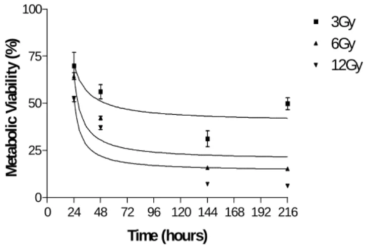
The effect of gamma irradiation by doses of 3 to 12 Gy on the cellular viability of GH3 cells is shown in Fig.1. After 24 hours, plates exposed to three Gy showed only 52.84% of viable cells, which were not altered significantly after this time. At six Gy, only 27.97% of viable cells were found
after 24 hours. This effect progressed to 11.31% after 168 hours. Those cells exposed to twelve Gy showed 34.19% of viability after 24 hours, and progressed quickly to 2.28% after 168 hours. The treatment with gamma radiation also altered significantly the metabolic viability of GH3 cells. This effect was time- and dose-dependent.
0 25 50 75 100 125 150 175 200 0
5 10 15 20 25 30
Control 3Gy 6Gy 12Gy
Time after irradiation ( hours)
N
u
m
b
er
of
ce
ll
s
x
1
0
4
Figure 1 - Kinetics of the cytotoxic effect of gamma radiation on GH3 cell line. GH3
cells were exposed to 3-12 Gy of ionising gamma radiation. Numbers of viable cells were determined by trypan blue staining. Cytotoxic effect was dose-dependent. Only a few cells remained alive after 12 Gy. (n=3)
0 24 48 72 96 120 144 168 192 216 0
25 50 75 100
3Gy 6Gy 12Gy
Time (hours)
M
et
a
bo
li
c
V
ia
b
ili
ty
(%
)
Figure 2 - Kinetics of the effect of gamma radiation on the metabolic viability of GH3 cell line. GH3 cells were exposed to 3-12Gy of ionising gamma radiation. The
0 20 40 60 80 100 120
%
o
f
c
e
ll
s
fo
rm
ing
c
o
lo
n
ie
s
Control 3Gy 6Gy 12Gy
*p<0,01
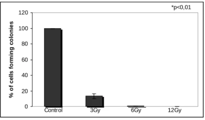
Figure 3 - Proliferation of GH3 cells exposed to gamma rays. GH3 cellswere irradiated
and the number of colonies was counted after 10 days of incubation. Gamma rays reduced proliferation in a dose-dependent way (n=3), p< 0.01.
Figure 4 - Clonogenic assay. (A) control, (B) cells exposed to 3 Gy (C), cells exposed to 6
Gy, (D) cells exposed to 12 Gy, (E) cells exposed to 103 ng/mL of Crotalus venom and (F) cells exposed to 104 ng /mL. Cells exposed to 6 and 12 Gy of gamma radiation presented an increased cytoplasmatic volume (Optic microscope: 40x).
As can be seen in Figs. 3 and 4, gamma radiation inhibited proliferation rate of GH3 cells. The cells exposed to 12 Gy did not form colonies and those ones exposed to 6 Gy showed only 0.46% of survival rate. Furthermore, the cells exposed to both doses presented an increased cytoplasmatic body (Fig.4). The cells exposed to 3 Gy showed 13.22% of survival rate. It has been proposed that calcium activates endonucleases that cause DNA fragmentation and death in apoptotic cells. γ irradiation is an inducer of apoptosis that leads to increases in [Ca 2+](i) (Story et al, 1992).
Ca2+ ions as mediators of intracellular signaling are crucial for the development of apoptosis. An
increase in [Ca2+](i) of apoptosing cells
(McConkey et al, 1996) due to emptying of Ca2+ stores and to Ca2+ influx from extracellular medium (Bian et al, 1997) is a general phenomenon, independent of the apoptotic
stimulus. However, the role of Ca2+ in the
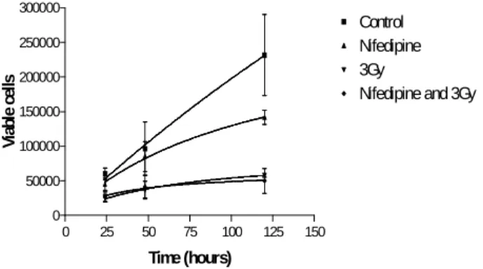
to proliferate. The metabolic activity was also significantly decreased by gamma radiation. Nifedipine (calcium channel blocker) did not interfere significantly with the proliferative rate or with the radiation-induced cell death of GH3 cells (Fig. 5). Thus, calcium influx through Cav 1 does not appear to be involved in this effect. These results are in agreement with those of
Marekova et al. (2003) showing that GH3 cells are radiosensitive. On the other hand, these findings differ from those of Floersheim (1992) in terms of the fundamental observation that the calcium channel antagonist does not protect γ
irradiation-triggered cell death.
0 25 50 75 100 125 150 0
50000 100000 150000 200000 250000 300000
Control Nifedipine 3Gy
Nifedipine and 3Gy
Time (hours)
V
ia
b
le ce
lls
Figure 5 - Interference of Nifedipine on the radiation induced apoptosis of GH3 cells.
GH3 cells were exposed one hour to Nifedipine (5µM) and then exposed to 3 Gy of ionising gamma radiation. Numbers of viable cells were determined by trypan blue staining. Calcium channel blocker did not interfere in the proliferative rate or in the radiation induced apoptosis of GH3 cells (n=3).
Apoptosis of cancer cells caused by irradiation requires calcium influx by an unknown pathway (Prevarskaya et al., 2004). It has been suggested that some threshold level of cellular calcium must be reached to induce cell death (He et al., 1997). One of the numerous calcium-involving processes in mammalian cells is store operated calcium entry (SOCE) — the process in which depletion of calcium stores in the endoplasmic reticulum (ER) induces calcium influx from the extracellular space.
SOCE is part of the calcium influx stimulated by thyrotropin-releasing hormone (TRH) in GH3 pituitary cells and regulates hypophyseal secretion of prolactin (Villalobos & Garcia-Sancho, 1995). GH3 presents other calcium channels than Cav1 (Lomeo et al, 2004), such as Cav2 (Safa et al,
2001) and store-operated Ca2+ channels
(Villalobos & Garcia-Sancho, 1995). The results obtained in this study argue against the involvement of Cav1 in the low dose ionising radiation effect on GH3 cells. If calcium influx from the extracellular space has a role in this effect, SOC channels can not be ruled out.
RESUMO
Adenomas de pituitária constituem cerca de 6-18% dos tumores cerebrais em adultos. A ativação de correntes de cálcio dependentes de voltagem podem levar à super-excreção de hormônio do crescimento produzindo acromegalia e
aumentando a mortalidade. Íons Ca2+ como
mediadores de sinalização intracelular são cruciais no desenvolvimento da apoptose. No entanto, o papel da [Ca 2+] no desenvolvimento da apoptose é ambíguo. Neste estudo nós avaliamos os efeitos de
baixas doses de radiação gama (60Co) na
cálcio extracelular não interfere na apoptose induzida pela radiação.
REFERENCES
Becker, G.; Kocher, M.; Kortmann, R. D.; Paulsen, F.; Jeremie, B.; Muller, R. P. and Bamberg, M. (2002), Radiation therapy in the multimodal treatment approach of pituitary adenoma. Strahlenther Onkol.,
178, 173-186.
Floersheim, G. L. (1992), Calcium antagonists protect mice against lethal doses of ionising radiation. Brit. J. Radiol., 65, 1025-1029.
Frohman, L. A. (1995), Diseases of the anterior pituitary. Endocrinology and Metabolism. New York.
pp. 289-384.
Hallahan, D. E.; Bleakman, D.; Virudachalam, S.; Lee, D.; Grdina, D.; Kufe, D. W. and Weichselbaum, R. R. (1994), The role of intracellular calcium in the cellular response to ionising radiation. Radiat Res.,
138, 392-400.
He, H.; Lam, M.; McCormick, T. S. and Distelhorst, C. W. (1997), Maintenance of calcium homeostasis in the endoplasmic reticulum by Bcl-2. J. Cell. Biol.,138, 1219-1228.
Klibanski, A.; Reichlin S. and Thorner M. J. (1995), Clinical review. J. Clin. Endocrinol. Metab., 80,
3395-3402.
Lomeo, R.; Santos, R. G.; Cruz, J. S.; Mafra, R.; Soares, M. A.; Pimenta, A. M.; Nascimento, M. C. and Lima, M. E. (2004), Tx1, a toxin from the venom of the spider Phoneutria nigriventer, blocks L-type calcium chanells in GH3 cells In: Congresso da Sociedade Brasileira de Toxinologia, 8., Angra dos Reis. J. Venom. Anim. Toxins, 10, 500.
Melmed, S. (1990), Acromegaly. N. Engl. J. Med., 322, 966-977.
Prevarskaya, N.; Skryma, R. and Shuba, Y. (2004), Ca2+ homeostasis in apoptotic resistance of prostate cancer cells. Biochem. Biophys. Res. Commun.,322,
1326-1335.
Safa, P.; Boulter, J. and Hales, T. G. (2001), Functional properties of CaV1.3 (_1D) L-type Ca2_ channel splice variants expressed by rat brain and neuroendocrine GH3 cells. 276 : (42), 38727-38737.
Story, M. D.; Stephens, L. C.; Tomasovic, S. P. and Meyn, R. E. (1992), A role for calcium in regulating apoptosis in rat thymocites irradiated in vitro. Int. J. Radiat. Biol., 61, 243-251.
Tombal, B.; Denmeade, S. R.; Gillis, J. M. and Isaacs, J. T. (2002), A supramicromolar elevation of intracellular free calcium ([Ca(2+)](i)) is consistently required to induce the execution phase of apoptosis.
Cell. Death Differ., 9, 561-73.
Villalobos, C. and Garcia-Sancho, J., (1995), Capacitative Ca2+ entry contributes to the Ca2+ influx induced by thyrotropin-releasing hormone (TRH) in GH3 pituitary cells. Pflugers Arch.,430, 923-935.
Vladyka, V.; Liscar, R.; Simonova, G.; Urgosik, D.; Chytka, T.; Vymazal, J.; Novotny, J.; Marek, J. and Hana, V. (2000), Gamma knife radiosurgery for pituitary adenomas: evaluation of a series of 163 patients. J. Radiosurgery, 3, 113-131.