Faculdade de Engenharia da Universidade do Porto
Diabetic foot thermophisiology characterization
Ana Rita Soares Marques
Dissertation
Mestrado Integrado em Bioengenharia
Ramo Engenharia Biomédica
Supervisor: Prof. Dr. Ricardo Ângelo Rosa Vardasca, PhD AMBCS
ii
iii
“Para mantermos um diabético a caminhar sobre os seus pés há que lhes prestar, nós, ele e os seus familiares mais chegados, todas as atenções. Quase como se fossem de vidro…”
v
Abstract
Diabetes affects around 13% of the Portuguese population,one in seven patients are at high risk of developing diabetic foot. This condition involves several anatomopathological and peripheral neurological disorders, which affect the peripheral microcirculation. This might have serious consequences such as: ulcers, ischaemia and amputations. The monitoring and assessment of the treatments are important to evaluate its progression, and to promptly act in case of regression.
Medical thermography is a noninvasive, nonionising, safe and accurate medical imaging method that enables to monitor the temperature distribution on the surface of the skin. This information is directly related to the physiology, allowing for real-time assessment, in particular microvascular peripheral and autonomic nervous systems. The diabetic foot has manifestations in these two systems and is therefore capable of being monitored by the proposed technique.
The present project aims to investigate the use of medical thermography in the physiological characterization of patients who already were diagnosed with diabetic foot. Moreover, this research should result in the definition of a protocol for the collection and analysis of thermographic images of diabetic foot. The results of this project will allow the study of diabetic foot disease more extensively, enabling a better knowledge of its physiology. The potential of thermography as a tool for folow-up of treatments was evaluated. The data collections were perfomed at the Diabetic Foot Clinic at the Hospitalar Center of Porto.
The obtained results can be used as reference for future research studies in the area, the proposed methodology can be improved to become used in daily practice, helping clinical professionals in the diagnosis and treatment assessment, providing better care and reducing the associated costs.
vii
Resumo
A diabetes afeta cerca de 13% da população portuguesa, cerca de um em cada sete doentes com diabetes, está em sério risco de desenvolver pé diabético. Trata-se de uma série de alterações anatomopatológicas e neurológicas periféricas que afetam a microcirculação periférica e podem ter como consequências graves: úlceras, isquemias e amputações. Pelo que a caracterização e monitorização destes sistemas é importante de forma a evitar a sua progressão.
A termografia médica é um método de imagem clínica não invasivo, não ionizante e preciso que permite monitorizar a distribuição da temperatura à superfície da pele. Esta informação fisiológica pode ser avaliada em tempo real, monitorizando os sistemas microvascular periférico e nervoso autónomo. O fenómeno do pé diabético tem manifestações nesses dois sistemas e é portanto passível de ser monitorizado por essa técnica.
Este trabalho de investigação tem como finalidade caracterizar a distribuição da temperature à superfície da vista plantar do pé em doentes, bem como o desenvolvimento de uma metodologia para a sua monitorização. O trabalho de campo foi realizado na Clínica do pé diabético do Centro Hospitalar do Porto.
Os resultados deste projeto permitirão efetuar estudos de maior dimensão na área da patologia do pé diabético, possibilitando conhecer melhor a sua fisiologia, auxiliar no diagnóstico e conduzir para um tratamento mais adequado, permitindo a avaliação contínua destes tratamentos. Dessa forma contribui para um aumento da qualidade de vida dos doentes de pé diabético e permite reduzir de forma eficaz os custos associados à patologia.
ix
Agradecimentos
Gostaria de agradecer ao meu orientador Prof. Ricardo Vardasca pela oportunidade de elaborar este estudo, e pelo seu acompanhamento, críticas e sugestões ao longo do mesmo, de forma a que fossem cumpridos todos os objetivos. E também ao Prof. Joaquim Gabriel pelo incentivo e simpatia.
Um enorme agradecimento a todos os médicos, enfermeiros e auxiliares do serviço da Clínica do pé diabético do Centro Hospitalar do Porto, em especial ao Dr.Rui Morais Carvalho e a Dra.Helena Neto, pelos seus conselhos e ensinamentos.
Ao longo das semanas que estive presente no CHP pude observar e comprovar os seus esforços inexcedíveis para com os doentes que passam por este serviço. É sem dúvida muito gratificante puder acompanhar o seu trabalho, o modo como tratam os seus pacientes, e a forma como tentam incutir-lhes um maior auto-cuidado e prática de vida mais saudável.
A todas as pessoas que participaram neste projeto, quer voluntários da Feup ou do CHP, um muito obrigado, pois sem a sua cooperação nada seria possível.
Às minhas melhores amigas Maria João e Alexandra, por todo o apoio e por estarem sempre presentes na minha vida, mesmo apesar da distância.
À Liliana, por me acompanhar ao longo desta jornada e pela ajuda neste projeto, obrigada.
A todos os restantes bons amigos que fiz durante a faculdade, apesar de já não estarmos todos em Vila Real, é bom perceber como mantivemos sempre a amizade e como foram e serão sempre parte integrante da minha vida.
E por último, mas as pessoas mais importantes da minha vida, quero agradecer aos meus pais Orlando e Dalila e o meu irmão Tiago, por serem a minha força motriz, pelo carinho, compreensão e apoio imensurável ao logo de todo a minha vida mas principalmente durante o meu percurso académico. É dedicado a vós este trabalho, pois uma vida não é suficiente para vos agradecer tudo o que fazem por mim.
xi
Contents
Abstract ... v
Resumo ... vii
Agradecimentos ... ix
Contents ... xi
List of figures ... xiii
List of tables ... xv
Abbreviations ... xvii
Chapter 1 ... 1
Introduction ... 1 1.1 - Motivation ... 2 1.2 - Aim ... 31.3 - Structure of the Dissertation ... 4
Chapter 2 ... 7
Literature Review ... 7
2.1. Diabetes and Diabetic Foot ... 7
2.1.1. Definition and classification of diabetes ... 7
2.1.2. Definition of diabetic foot ... 9
2.1.3. Epidemiology ... 10
2.1.4. Pathophysiology ... 10
2.1.5. Innervation and vascularization of foot ... 13
2.1.6. Diagnosis ... 14
2.1.7. Prevention ... 16
2.2. Thermography ... 16
2.2.1.Thermology: principles and procedures ... 16
2.2.2. Thermography and its application ... 17
2.2.3. Thermoregulation and heat transfer mechanisms ... 18
2.2.4.Infrared radiation and thermography ... 20
2.2.5. Previous Studies ... 21
xii
Methodology ... 27
3.1. Sample and capture images protocol ... 27
3.2. Evaluation instruments ... 31
3.2.1. FLIR Thermacam Researcher Pro ... 31
3.3. Materials ... 31
3.4. Procedures ... 32
3.4.1. Data collection procedure ... 32
3.4.2. Data analysis procedure ... 34
3.4.2.1.Selection of Regions of interest (ROIs) ... 34
Chapter 4 ... 37
Results ... 37
4.1.1. Sample temperature for 1st stage ... 38
4.1.2. Profile temperature for 2nd stage ... 42
4.1.3. Profile temperature for 3rd stage ... 44
4.2.1. Ulcer neuropathic vs ulcer ischaemic ... 49
4.2.2. Ulcer neuropathic vs ulcer neuroischaemic ... 50
4.2.3. Ulcer ischaemic vs ulcer neuroischaemic ... 51
4.3.1. Different type of diabetes ... 52
4.3.3. Different BMI classification ... 55
4.3.4. Different age group ... 57
Chapter 5 ... 63
Discussion ... 63Chapter 6 ... 67
Conclusion... 67References ... 69
APPENDIX A ... 73
APPENDIX B ... 76
APPENDIX C ... 77
APPENDIX D ... 81
APPENDIX E ... 84
APPENDIX F ... 86
APPENDIX G ... 88
APPENDIX H ... 90
xiii
List of figures
Figure 1 - Distribution of diabetes in many countries worldwide in 2012. ... 1
Figure 2 - Number of sales of Insulins and Oral Antidiabetic in Ambulatory within the SNS in Portugal Continental ... 2
Figure 3- Navigation diagram of dissertation. ... 5
Figure 4- Mechanism of appearance of the ulcer ... 11
Figure 5 - Location of plantar nerves (A) ; Location of plantar arteries (B). ... 14
Figure 6 - Evaluation of the diabetic foot using the Semmes Weinstein ... 15
Figure 7- Illustration of vasodilation and vasoconstriction processes, respectively... 19
Figure 8- Wavelength of infrared radiation, divided into short, medium and longe waves . .. 21
Figure 9- Participants distribution, divided in three groups. ... 28
Figure 10- Distribution of diabetic participants by diabetes type... 30
Figure 11- Frequency of diabetic participants by ulcer type... 30
Figure 12- Illustration of ThermaCAM Researcher Pro software for an analysis of foot thermal image. ... 31
Figure 13- Illustration of equipment for capturing thermographic images. The temperature and humidity of the room should be maintained within comfortable limits, and the camera must be positioned perpendicular to the observation surface at a distance of 80-150 cm from the feet of the volunteer to minimize geometric errors in temperature measurement. ... 32
Figure 14-Illustration of thermal imaging over the three stages. ... 33
Figure 15 - ROIs of autonomic nervous system (pallet rain). ... 34
Figure 16– Regions of interest of vascular system (pallet rain). ... 35
Figure 17– Thermal images of the feet of a healthy individual over the three stages. ... 46
xiv
Figure 19- Variation of the mean temperature of ROIs of autonomous nervous system
throughout the three stages of healthy and diabetic feet. ... 46
Figure 20- Variation of the mean temperature of ROIs of vascular system throughout the
three stages of a healthy and diabetic feet. ... 47
Figure 21– Thermal and visual images of a neuropathic, ischaemic and neuroischaemic
ulcers. ... 51
Figure 22- Thermal symmetry for ANS. ... 60 Figure 23- Thermal symmetry for VS. ... 60 Figure 24– Thermal images of healthy individuals (A and B). Thermal images of individuals
with diabetic foot (C and D). ... 61
xv
List of tables
Table 1– Clinical features that distinguish neuropathic and vascular foot ulcers... 15
Table 2- Distribution of participants by gender. ... 28
Table 3- Distribution of participants by interval of age. ... 29
Table 4- Distribution of participants of interval of glicemia. ... 29
Table 5- Distribution of participants by BMI interval. ... 30
Table 6 - Mean and Standard Deviation of humidity and temperature by Group (Diabetics Vs Healthy). ... 34
Table 7 – Temperature distribution in ROIs of the ANS by participant type. ... 38
Table 8 – Temperature distribution in ROIs of the VS by participant type. ... 39
Table 9- Mann Whitney U, Wilcoxon W Z and significancy of Mann Whitney test for ANS ROIs temperatures in all partipants in 1st stage. ... 40
Table 10- Mann Whitney U, Wilcoxon W Z and significancy of Mann Whitney test for VS ROIs temperatures in all partipants in 1st stage. ... 41
Table 11- Temperature distribution in ROIs of the ANS by participants type. ... 42
Table 12- Temperature distribution in ROIs of the VS by participants type. ... 43
Table 13- Temperature distribution in ROIs of the ANS by participants type. ... 44
Table 14- Temperature distribution in ROIs of the VS by participants type. ... 45
Table 15- Difference of mean temperatures of ANS ROIs for healthy and diabetic particpants. ... 48
Table 16- Difference of mean temperatures of VS ROIs for healthy and diabetic particpants. ... 48
Table 17- Mann Whitney U, Wilcoxon W Z and significancy of Mann Whitney test for ANS ROIs temperatures of partipants with neuropathic and ischaemic ulcers... 49
xvi
Table 18- Mann Whitney U, Wilcoxon W Z and significancy of Mann Whitney test for VS
ROIs temperatures of partipants with neuropathic and ischaemic ulcers. ... 50
Table 19 - Mann Whitney test for different type of diabetes in the ANS. ... 52
Table 20- Mann Whitney test for different type of diabetes in the VS. ... 53
Table 21- Different glicemia interval and ANS. ... 54
Table 22- Different glicemia interval and VS. ... 55
Table 23- Different BMI classification and ANS. ... 56
Table 24- Different BMI classification and VS. ... 57
Table 25- Different age groups and ANS ... 58
Table 26- Different age groups and VS. ... 59
xvii
Abbreviations
ABI Ankle-brachial index ANS Autonomic nervous system CHP Centro Hospitalar do Porto DM Diabetes Mellitus
EU European Union
IDF International Diabetes Federation IR Infrared radiation
OECD Organisation for Economic Co-operation and Development ROI Region of interest
SNS Serviço Nacional de Saúde
1
Chapter 1
Introduction
Diabetes mellitus (DM) is a disease characterized by chronic hyperglycemia with
disturbances of the metabolism of carbohydrates, fats and proteins resulting from defects in insulin secretion, insulin action, or both. There is a worldwide prevalence of disease due to aging populations, poor diet, concomitant epidemic of obesity, physical inactivity and unhygienic environment [1].
According to the International Diabetes Federation (IDF), diabetes affects over 371 million people worldwide, accounting for 8.3% of the world population and continues to increase in all countries. In over 50% of people with diabetes it has not been diagnosed, pursuing their silent evolution [2].
Diabetes in 2012 caused the deaths of 4.8 million people, half of whom were under 60 years, and it is estimated that in 2030 the number of people with diabetes worldwide will reach 552 million, representing an increase of 49% of the population affected by the disease [3].
A report published in November 2012 by OECD, shows that Portugal is the European Union (EU) country with the highest prevalence of diabetes (9.7%), a percentage that exceeds by more than three percent the mean of the EU, as shown in Figure 1 [4].
2
1.1 - Motivation
Diabetes affects 12.7% of the portuguese population aged between 20 and 79 years, which corresponds to an estimated 1.003 million individuals [3].
Pathologic changes associated with diabetes are numerous, and the diabetic foot is one of the most complicated health problems. Diabetic neuropathy affects changes in sensitivity of the feet and is mainly responsible for the appearance of these lesions, which are difficult to treat and to prognosis [5].
Since diabetes is a condition that affects a large number of population, the costs associated with this have great economic impact on the Serviço Nacional de Saúde (SNS) and also in the patients self costs. With diabetic population increasing, these costs are also increasing proportionally (Figure 2). It is essential to investigate ways to prevent and deal with diabetes on a cost-effective manner for the general population and the SNS.
Figure 2 - Number of sales of Insulins and Oral Antidiabetic in Ambulatory within the SNS in Portugal
Continental - In Value. (Source: Estatísticas do Medicamento – INFARMED) [3].
However, there are other costs associated with diabetes, such as loss of income, productivity decrease of affected patients and the psychosocial aspect of patients and their families, who despite of being intangible are nonetheless important.
The temperature is a useful indicator of various diseases, the medical use of Thermology techniques, such as infrared-based imaging, has potential of use in medicine [6].
Infrared thermography is a technique that allows mapping the surface temperature of the human body or a region, with the intent of distinguishing areas with different values. Since 1960, there is an increased understanding of the physiology and thermal relation between the skin temperature and blood perfusion. Moreover, the recent advances in the standardization of the technique and the advantages of computer aided medical imaging systems have greatly improved the reliability of this technology in medicine. In diabetology studies it was
3
demonstrated its value and relevance to the clinical evaluation of peripheral perfusion and tissue viability [7].
Medical thermography provides functional information that is not provided by any other medical imaging methods, and has the great advantage of being non-invasive, non-ionizing and accurate, passively capturing the radiant heat emitted from the human body surface [8,9].
In diabetes, vascular activity in the extremities, especially in the feet, can be indicative of autonomous nervous system activity, because the vasomotor function is regulated by autonomic fibers of the sympathetic system, and its dysfunction could be associated with different patterns of temperature in diabetics [10].
Complications affecting the lower limbs are the most common manifestations of diabetes and infrared imaging can be a useful tool for early identification of those conditions and aid to prevent more serious complications, which can lead to amputations, in the most extreme situation.
Due to the severity of problems that may be related to diabetes, and the fact of this condition is increasingly affecting more people worldwide, it is necessary to develop effective methods to timely detect these situations. Medical thermography presents itself as a very credible solution, since it is a painless, non-invasive, non-ionizing and accurate method, since it does not cause any harm or discomfort to the patient, and is economically affordable [11].
1.2 - Aim
The aim of this research is to develop a methodology for characterizing the skin temperature of the plantar feet in diabetic patients.
In addiction to this main goal, the objectives were:
- Design, implement and assess a methodology for studing surface skin temperature of the plantar feet.
- Characterize the thermal profile of plantar feet in a healthy population. - Characterize the temperature profile of plantar feet in diabetic feet patients. - Evaluate quantitative benchmarks to be used as references for future studies. - Evaluate the proposed methodology for frequent follow-up of diabetic foot patients, through a study case.
4
1.3 - Structure of the Dissertation
This document is further organized in five sections:
The literature review is divided in two main topics: firstly diabetes is classified and then diabetic foot and its main complications are presented. The other topic is thermography, where the technique is described, the role of thermoregulation and the relevant studies of thermography in the diabetic foot.
Methodology, the images capture protocol, materials and methods used in this research and the analysis methods.
Results, present the outcomes of all the tests and experiments performed, and also its interpretation and more relevant information.
Discussion, analyzes the significance of findings and the justification of the deviations. Conclusion, states the accomplishment of the aim and the proposed future work. Figure 3 shows the navigation diagram of this dissertation.
5
Figure 3- Navigation diagram of dissertation.
1. Introduction 3. Methodology 4. Results 5. Discussion 6. Conclusion 2.1. Diabetes and Diabetic Foot 2.2. Thermography
3.1. Sample and capture images protocol
3.2. Evaluation Instruments
3.3. Materials 3.4.1.Data collection
procedure 3.4.2.Data analysis procedure 4.1. Characterization of sample temperature 4.2. Relation between diabetic foot temperature
and diabetic foot ulcer
4.3. Relation between foot temperature and characteristics of individuals
4.4. Thermal symmetry 4.5. Follow-up
2. Literature Review
7
Chapter 2
Literature Review
In this chapter, the diabetic foot condition is described and characterized. Thermography, thermal transfer mechanisms are presented. The role of
thermoregulation in the human body, microcirculation and thermal imaging. The past studies of using Thermology in the diabetic foot condition are also outlined.
2.1. Diabetes and Diabetic Foot
2.1.1. Definition and classification of diabetes
Diabetes mellitus is a set of metabolic diseases characterized by hyperglycemia, which results from defective insulin secretion, a flawed insulin action, or both. Chronic hyperglycemia is associated with long term damage, dysfunction and failure of several organs, namely the eyes, kidneys, nerves, heart and blood vessels. Several pathogenic processes are involved in the development of diabetes. These range from the autoimmune destruction of pancreatic β-cells, with consequent insulin deficiency, to abnormalities, which result in insulin resistance [12].
Long term diabetes complications include retinopathy with potential loss of sight, nephropathy, which leads to kidney failure, peripheral neuropathy leading to potential feet ulcers, amputations and autonomic neuropathy which leads to gastrointestinal, genitourinary
8
and cardiovascular problems, and sexual dysfunction. Diabetic patients are prone to other serious illnesses, since they have a high incidence of atherosclerotic peripheral vascular disease, cerebral vascular disease, hypertension and an abnormal lipoprotein metabolism.
Regardless of what causes it, diabetes is associated with a common hormonal defect, i.e., insulin deficiency, which can be absolute or relative, according to one's insulin resistance. Insufficient insulin has a vital role in metabolic changes associated to diabetes. Hyperglycemia, in its turn, plays an important role in disease-related complications [13].
Diabetes can be classified according to the following categories:
- Type 1 diabetes: This type of diabetes accommodates around 5-10% of all diabetics, and is a consequence of the cell-mediated destruction of pancreatic β-cells, which leads to absolute insulin deficiency. The destruction of these cells happens due to various genetic predispositions and it is also associated to environmental factors, which have not yet been completely defined [12, 13].
- Type 2 diabetes: Has around 90-95% of all diabetic population, and is characterized by insulin resistance and relative insulin deficiency. Most of the patients with this condition do not need insulin admission. This type of diabetes frequently remains undiagnosed for years, as hyperglycemia develops gradually and the patient does to notice any of the usual diabetes symptoms. However, these patients face an increased risk of microvascular and macrovascular complications [12].
- Gestational diabetes: For many years, it was characterized as any degree of glucose intolerance, which started or was first discovered during pregnancy. Even though, most cases can be managed with medication, this definition is applied depending on whether the condition persisted after pregnancy and does not exclude the possibility that the unrecognized glucose intolerance may have started before or at the same time as the pregnancy [12].
During pregnancy, gestational diabetes requires treatment to optimize maternal blood glucose levels to lessen the risk of complications in the infant. The gestation period is relatively brief for developing serious complications that cause the appearance of ulcers on the feet. However, women who have had gestational diabetes are more prone to develop diabetes in the next 10–20 years [14].
- Other specific types: Various types of diabetes are associated with monogenetic defects in β-cell function. These forms of diabetes are frequently characterized by hyperglycemia at an early age (usually before 25), diminished insulin secretion with little or even no effect in insulin action and have an autosomal dominant inheritance pattern. The
9
most common form is associated with chromosome 12 mutations in the hepatic transcription factor referred to as a hepatocyte nuclear factor (HNF)-1a. The second type is associated with mutations in the glucokinase gene in chromosome 7p. The least common types result from transcription-factor mutations, such as HNF-4a, HNF-1b, insulin promoter factor (IPF)-1, and NeuroD1. This condition is very rare and was not found information that relates this type of diabetes with diabetic foot.
Diabetes is one of the most common chronic diseases in every country, and its numbers continue to increase in size and in importance. This exponential growth has to do with an economic and urban development, which leads to lifestyle changes characterized by reduced physical activity, increased obesity and an uncontrolled diet [15].
Along with these factors, the International Diabetes Federation estimates that this number will continue to grow worldwide, due to population aging and growth and the high prevalence of obesity and a sedentary lifestyle, reaching 522 million people in 2030 [16]. As such, diabetes is considered a serious public health problem, due to being a chronic illness and affects a large population [17].
2.1.2. Definition of diabetic foot
Diabetic foot is the most common chronic complication in diabetic patients. It affects more the patients with type 2 diabetes [18], which has a relation with the duration and success of diabetes treatment. Based on epidemiological studies, it is estimated that 25% of all diabetics will develop serious health problems related to diabetic foot during their lifetime, whilst 5% to 15% of all patients will have their foot or leg amputated [19].
The diabetic foot is characterized through foot lesions as a result of peripheral and/or neurologic vascular changes singular to diabetes, known as the triad: neuropathy, peripheral vascular disease and infection. If this deterioration is not detected at an early stage, it can evolve into gangrene or consequently to limb amputation [20].
Neuropathy causes loss of sensitivity and pain, which contributes to the existence of trauma and ulcerations. The appearance of infection and insufficient irrigation of the lower limbs leads to the progression of gangrene. The diabetic patient most of the times does not realize there is a lesion until the latter, where it is already in an advanced state, which makes treatment more difficult and contributes to the high incidence of amputations in these patients [21].
The important risk factors include: age, type and time of diagnosis, inadequate glycemia control, smoking, alcoholism, obesity, hypertension and lack of hygiene when it comes to footcare. Healthcare professionals to assess diabetics' feet regularly, in order to early identify the risk factors are very important. That can also be changed through promoting
self-10
care, along with a proper metabolic control, which will, in turn, reduce the risk of ulceration and amputation [21].
2.1.3. Epidemiology
The "diabetic foot syndrome" encompasses a considerable number of pathological conditions, including neuropathy, ischaemia, Charcot's neuroarthropathy, foot ulceration, osteomyelitis and amputation [22].
Patients with diabetic foot injuries frequently present various diabetes-associated complications. As such, there is a need for a multidisciplinary approach, with several specialties involved, such as an endocrinologist, a specialized nurse, a podiatrist, a vascular surgeon, an orthopedist, a physiatrist and a general practitioner [23].
From the pathological conditions present, foot ulcers and amputations are both the most common and the most serious complications and are responsible for a significant mortality. Foot ulcers precede around 85% of amputations, wich can led to the assumption that a success in early detection or adequate treatment of those ulcerations would result in reducing the number of amputations [23].
The risk of a diabetic patient developing a foot ulcer during their life is around 25% [24]. Foot injuries in these patients are the main cause of hospital admission when compared to any other long-term diabetes complication, and they also result in an increased morbidity and mortality. These lesions occur due to the presence of sensory-motor neuropathy and vascular disease [24].
Regarding the etiology of foot ulcerations, about 45-60% of ulcers are purely neuropathic, about 10% are purely ischemic and 25-45% are mixed (neuroischemic) [25] and happen particularly on the sole of the foot in areas subject to high pressure when walking [18].
Besides causing pain and morbidity, the diabetic foot has severe economic consequences. Costs are reduced by foot ulcer prevention interventions, through strategies that promote healing (which shorten the healing period and prevent amputations) [23].
2.1.4. Pathophysiology
Having knowledge of the pathophysiology of the ulceration is essential to provide optimal healthcare, since changing the factors that lead to its development allows better healing the foot or helps keeping it intact, providing to the patient a completely normal life.
There is estimation that 15% of all diabetic patients will have a foot ulcer at some point in their lives, and about 2 to 3% of all diabetics will develop them every year. Many of these ulcers will require prolonged hospitalization to treat subsequent complications, such as infection or gangrene. [26-28].
11
As Figure 4 shows the relation between the most important factors that lead to a vulnerable high risk of diabetic foot and contribute to the appearance of an ulcera, which are: neuropathy, macroangiopathy, medial artery calcification or Mönckeberg arteriosclerosis and diabetic microangiopathy. When one or more of these factors is present, the existence of a triggering factor which acts on the vulnerable foot will lead to the formation of an ulcer or necrosis [26-28].
NEUROPATHY MICROANGIOPATHY MACROANGIOPATHY
Sensory Motor Autonomic
Blood flow reduction
Increase plantar pressure Charcot's neuroarthropathy TRIGGER FACTORS: Deformation + Trauma COMPLICATIONS: Infection Ischaemia Neuropathy
Figure 4- Mechanism of appearance of the ulcer [27].
2.1.4.1. Neuropathy and peripheral vascular disease
Since neuropathy and peripheral vascular disease are the etiological factors most related to the appearance of foot ulcers, it is necessary to understand these two causal mechanisms. Peripheral polyneuropathy (sensory, motor and autonomic) derives from axon terminal degeneration, where the intensity varies proportional with the size. Since the largest size exists in both inferior limbs, neuropathy is a bilateral complication [23].
Diabetic distal neuropathy can affect the three components of the nervous system: sensory, motor or autonomic, and all of them contribute to the development of foot ulcers. Consequently, somatic (sensory-motor) and autonomic fibers are also affected. The smallest nervous fibers do suffer changes, which causes the loss of pain and temperature sensations.
Deformation Overload unnoticed
Ulcer
Extension of lesion, necrosis Ischaemia
Functional Structural
12
Subsequently, the largest nervous fibers will also be affected, which will leads to a decreased superficial sensation perception [26].
Neuropathy can have two different classifications:
- Sensory-motor neuropathy, consists of a gradual loss of tactile and pain sensitivity, which causes feet to be vulnerable to trauma, called "loss of protective feeling" [28]. The latter is subdivided in two classes: acute sensory neuropathy, which is rare and follows periods of inadequate control or sudden change in metabolic control, and chronic sensory-motor neuropathy, which is the most common type of neuropathy. Instability and loss of balance have been increasingly recognized as possible manifestations of peripheral chronic polyneuropathy, secondary to proprioceptive disturbances and possibly abnormalities in sensitive muscle function. This instability may result in repeated minor traumas or falls and late complications, such as Charcot's neuroarthropathy, which consists of consequential repeated traumas in the foot. These changes are also clearly related to the appearance of ulcers [26].
About 50% of all patients present symptoms, such as: pain, dysesthesia, paresthesia, very warm skin and numbness. However, since about half the patients can be asymptomatic, it can only be diagnosed during a medical exam or when the patient presents a painless foot ulcer [29].
This loss of sensitivity is very serious and may lead to major injuries since, for example, a diabetic person with loss of protective feeling may no longer feel the discomfort of the repeated pressure of a tight shoe or the pain of a pointy or sharp object on the floor. It also leads to foot intrinsic muscle atrophy, causing an imbalance between flexor and extensor muscles, triggering osteoarticular deformities. The latter will change the pressure points in the sole of the foot, leading to overburdening and to a skin reaction with local hyperkeratosis (callus), which, with continuous walking, will evolve into ulceration [28].
- Autonomic neuropathy, in turn, consists of an autonomic nervous system injury, namely of the sympathetic nerves, leading to loss of vascular tonus1, causing vasodilation with an increase in arteriovenous communication and, as a consequence, the direct passage of blood from the arterial to the venous network, decreasing tissue nutrition. This phenomenon leads to hypohidrosis, a skin illness that causes reduction or absence of sweat secretion due to the low activity of the sympathetic system, which causes the dry skin, culminating in the appearance of fissures and changes in the growth and matrix of nails, which much like chronic ulcers, are important entrance points for infections [28,29].
1Vascular tonus is the degree of sustained constriction of the vascular system, which regulates the peripheral
13
Vascular disease is responsible for more than 70% of deaths of patients with type 2 Diabetes [29]. Diabetic peripheral vascular affection is divided in microangiopathy, Mönckeberg arteriosclerosis and macroangiopathy caused by atherosclerosis.
Diabetes is associated with an increased risk of peripheral vascular disease. However, this is not frequently the primary etiology of diabetic foot problems, such as ulceration or amputation. It is, nevertheless, implied in the altered response to infections and in the healing process, being present in almost 50% of all amputated patients [26, 29].
This pathogenic entity is, therefore, more important in the persistence and evolution of diabetic foot ulcers then in its appearance. Foot infection and/or ulceration entail the need to increase blood supply to damaged tissue. If the patient has peripheral vascular disease, it may not be possible to answer that need (whether due to ischaemia or because vessels are unable to dilate), causing more tissue damage and causing the infection progression [26].
Given this, its presence worsens the prognosis for these patients, increasing the risk of amputation. Thus, it is of vital importance to identify and treat the co-existing peripheral vascular disease. Vascular disease is frequent in diabetics, but it is only threatening to the limb if there is a skin lesion. Adequate tissue perfusion is vital for the ulcers to heal correctly. Whenever an ulcer does not heal there is a suspicion of arterial failure, which can cause more severe complications [26, 31].
2.1.5. Innervation and vascularization of foot
Diabetic foot is the set of neurological and vascular conditions affecting the feet of patients with diabetes, like aforementioned. So it is important understanding the role of autonomous nervous system and the microvascular system on the overall risk of ulceration in diabetic patients.
Pathological changes in the feet can occur in the muscles (muscles, tendons or ligaments), bone or vascular (veins, arteries, vessels and microvessels). Vascularization region of the ankles and feet are usually more susceptible to such changes given its complexity. Thus, conditions that affect the circulation area, such as diabetes, tend to slow down the blood return, which ultimately lead to swollen feet, pain and accumulation of toxins in the region, in addition to the propensity to wounds and delayed cell regeneration [32].
The innervation of the foot (figure 5A) is provided by the following nerves: superiorly by the deep and superficial peroneal nerves; Inferiorly by the medial and lateral plantar nerves; Medially by the saphenous nerve, which extends distally to the head of the 1st metatarsal; Laterally by the sural nerve, including the heel part, and this by calcaneal branches of the tibial and sural nerves [33].
14
The different zones shown in the figure are also referred to dermatomes, which are areas of the skin that are innervated by nerve fibers that originate from a single dorsal nerve ganglion. Each dermatome is named according to the spinal nerve that innervates it. Correspondence between the skin and the nervous system dermatome is used to help locate the degree of neurological deficit [34].
Foot vascularization (figure 5B) is carried out by the following metatarsal arteries distally to the crevices of the fingers, being united with the plantar arch, and plantar metatarsal arteries perforating branches; Arcuate artery that is disposed laterally through the bases of the four lateral metatarsal passing deep to the tendons of the extensor muscles, thus achieving the lateral aspect of the forefoot; Medial artery of the foot corresponding to continuation of anterior tibial artery, and a major source of blood supply to the front part of the foot; Lateral tarsal artery, a branch of the dorsal artery of the foot laterally and follows a curved path under the short extender fingers with the aim of supplying this muscle and tarsal joints and the underlying [34].
Figure 5 - Location of plantar nerves (A) [35]; Location of plantar arteries (B)[36].
2.1.6. Diagnosis
As previously mentioned, the ulceration of the diabetic foot is associated with peripheral vascular disease and peripheral neuropathy, which are frequently combined. However, individuals who have a high ulceration risk can be easily identified through a careful foot examination. Education and periodical follow-ups are also indicated in these cases [23].
When it comes to the physical examination, the American Diabetes Association (ADA) approves a few different tests to identify sensory loss in the sole of the foot due to neuropathy. These tests are:
1 - Sural nerve 2- Saphenous nerve 3- Lateral plantar nerve 4- Medial plantar nerve 5- Medial calcaneal branch
1 2 3 4 1 2 3 4 5 1- Metatarsals arteries 2- Arcuate artery 3-Medial plantar artery 4- Lateral plantar artery
5- Calcaneal artery
A B
15
a) Semmes Weinstein monofilament test, illustrated in Figure 6, in which the incapacity
to feel the necessary pressure to bend the 10g monofilament when tested in various areas of the foot is compatible with sensory neuropathy [37].
Figure 6 - Evaluation of the diabetic foot using the Semmes Weinstein [37].
b) Reflex hammer test. Deep sensation can be assessed through the Achilles tendon reflex
test by using the reflex hammer, and absence of reflex is connected to an increased ulceration risk.
c) Testing with a 128 Hz tuning fork and with a biothesiometer, by which we can assess
vibration perception. An abnormal response can be defined when the patient loses vibration perception, but the latter is still detected by the examiner [37, 23].
All these tests are used to determine the risk of ulceration but the monofilament test, due to its simplicity and low cost, is considered to be the best choice [37].
To assess peripheral vascular disease, palpation of pedis and posterior tibial arterial pulses is recommended, characterizing them as present or absent. For patients with signs and symptoms of vascular disease or an absent pulse, an ankle-brachial index (ABI) measurement must be performed, and a vascular surgeon referral must be considered.
ABI is an accurate way of assessing arterial perfusion, where you can check systolic arterial blood pressure in the leg (posterior tibial pulse) and in the arm, measuring the ratio between them. While the patient is resting, an index is considered normal when below a rate of 0.9 to 1.3. A decreased index suggests a vascular disease, considered light to moderate when the index is 0.4 to 0.9 and serious when below 0.4 [37].
Some clinical features that distinguish the type of foot ulcers are summarized in Table 1.
Table 1– Clinical features that distinguish neuropathic and vascular foot ulcers [38].
Neuropathic ulcers Vascular ulcers
Painless Painful
Located at points of high pressure Often located at the extremities
Warm foot Cool ischaemic foot
16
These methods are used daily in the care of patients with diabetic foot, however can be a little subjective. Depend on the person who executes them, and can be lengthy. Thus arises the need to implement new methods of detection and monitoring diabetic foot more efficient and objectives, as for example the thermography.
2.1.7. Prevention
The best form of prevention is, unquestionably, health education. Diabetics must be advised to reduce risk factors, such as smoking, reducing fat intake, stabilizing or reducing weight and having an optimal glycemia control, among others [39].
Diagnostic exams must be performed at least once a year, and healthcare professionals must educate patients about self-monitoring, i.e., the daily inspection of their feet, since the absence of symptoms does not mean that their feet are healthy. During this inspection, it is advisable to pay special attention to color, temperature, joint mobility and nail problems.
Since footwear is one of the main causes of ulceration, patients should not wear shoes that are too tight or too loose, and they should be inspected beforehand. Pathologies associated with calluses, nails and skin must be treated regularly. It is also highly recommended to maintain good foot hygiene and to use moisturizes to avoid the appearance of fissures [40].
Consequently, the best way to prevent diabetic foot complications is to educate the patients, so that they can check their feet every day and consult a professional whenever any anomality is present. This will ensure an early diagnosis and an effective treatment [39, 40].
2.2. Thermography
2.2.1.Thermology: principles and procedures
Thermology is the field of Physics that studies heat, and the manifestations of the types of energy, which produce temperature variations (heating, cooling, or even the change in physical state that derives from these two variations) [41].
Thermology studies the way by which heat can be exchanged between bodies, as well as the characteristics of each heat transfer process, which consist of four different mechanisms: conduction, convection, evaporation and radiation [41].
17
In order to understand the role of thermology, it is firstly necessary to distinguish between temperature and heat. Heat is tangible and consists on thermal energy in motion, i.e., it is energy that is in constant motion, always being transferred from one hotter body to a cooler one. Temperature, on the other hand, is a measure of the status of molecule agitation. The temperature is proportionally related with the molecules agitation [41,42].
Medical thermography consists on documenting the distribution of thermal radiation emitted at the surface of the skin, by using infrared imaging. Both the skin’s galvanic impedance and the vasomotor and sudomotor responses can be assessed by studying the way skin responds in medical infrared images, i.e. through infrared thermography. This technique involves no contact and no form of energy is transmitted to the body, which makes it a complementary diagnostic and treatment-assessing test with great potential [43].
There are four mechanisms of heat loss: conduction, convection, evaporation and radiation.
- Conduction is the transfer of heat from one solid object to another when in direct contact with it [53].
- Convection is based on heat transfer by the movement of a fluid (liquid or gaseous) in the body. Blood, heated by the visceral and the somatic metabolism, is convected by the vascular system and transferred to the inside of the body, and then to low temperature areas, thus representing the largest heat transfer mechanism inside the body.
- Evaporation is the conversion of water into vapor, by means of thermal energy. This process occurs throughout the surface of the body, and mainly in the respiratory system. This mechanism is of very little importance in the thermal image when the individual is in balance with his environment [53].
- Radiation represents the largest heat loss mechanism in the human body, responsible for 60% of its total loss. Thermal energy is converted into electromagnetic radiant energy, which is emitted by the body in the infrared spectrum band. Central blood temperature is brought to the skin's vascular network, where thermal energy is converted into radiant energy and transmitted to the environment. This radiation is detected through infrared cameras [45,53].
2.2.2. Thermography and its application
The biomedical use for thermography has increased over the last fifty years. Since this is a completely non-invasive technique, its use has been helpful in terms of medical research as a sensitive complimentary diagnosis method for different clinical and experimental levels [44].
18
Thermography offers valuable information on the temperature differences of different parts of the human body surfaces. It enables to monitor the nervous system, metabolic or vascular disturbances, skin cancer, pain syndromes, soft tissue damage, virus infections and others by comparing thermal changes and differences. Since infection and inflammation can appear in any part of the human body, causing temperature changes, thermography is therefore a valuable tool for repetitive monitoring which will not harm the patients.
Since the camera used for temperature detection only receives natural thermal energy emitted by the body, and no harmful energy is used, this technique can be used continuously in various tests over time [44, 45].
2.2.3. Thermoregulation and heat transfer mechanisms
Clinical information obtained by thermography is not directly based on changes in metabolic activity and heat production. It is based on heat distribution in the body and the regulation of that heat loss, especially at the surface of the skin. Metabolic heat is brought from the inside of the body, by the vena cava and the larger veins where it is mixed and distributed by the aorta and its branches [46].
The human body has a good homeostatic control mechanism for body conditions, including temperature, pH and others. Temperature is a useful parameter to assess the body health condition. The core temperature of our body is usually kept at 36.8 ± 0.6°C and, if the temperature in a certain area is higher or lower, and therefore not normal, it becomes an indicator of a possible problem, such as infection or necrosis [47].
The skin works as an interface between body and environment. It is possible to measure the temperature along the surface of the skin. The balance between the body and surrounding environment made through thermoregulation. Blood flow play an important role in this process, any changes in it induces changes in skin temperature [48].
Human skin blood flow responses to body heating and cooling are essential to the normal processes of physiological thermoregulation. In humans, reflex sympathetic innervation of the cutaneous circulation has two branches: a sympathetic noradrenergic vasoconstrictor system, and a non-noradrenergic active vasodilator system. Noradrenergic vasoconstrictor nerves are tonically active in normothermic environments and increase their activity during cold exposure, releasing both norepinephrine and cotransmitters (including neuropeptide Y) to decrease skin blood flow. The active vasodilator system in human skin does not exhibit resting tone and is only activated during increases in body temperature, such as those brought about by heat exposure or exercise [49].
Figure 7 illustrates the vasodilation and vasoconstricition processes. In vasodilation, the hypothalamus sends nerve impulses via parasympathetic nervous system to blood vessels near
19
the skin. Smooth muscle in vessel walls relaxes, but ‘shunt vessel’ constricts so more blood goes to the surface. In vasoconstriction the sympathetic nerve impulses to blood vessels near the skin. Smooth muscle in vessel wall contracts so less blood flow, shunt vessel relaxes so less blood flows to the surface capillaries. Less heat lost by radiation is thus typical of vasoconstriction [50].
Figure 7- Illustration of vasodilation and vasoconstriction processes, respectively [50].
There are other mechanisms that help maintain body temperature, sudoresis and muscle contraction. The sudoresis, which is the act of producing and releasing sweat, begins when the core body temperature is above 37°C. This mechanism is regulated via stimulation of the sweat glands by cholinergic sympathetic nerves and sometimes in situations such as exercise or stress by high concentrations of epinephrine and norepinephrine [51].
Muscle contraction is also an important source of thermal energy - muscle thermogenesis. This can be stimulated by the cerebral cortex or involuntarily by the hypothalamus. The posterior hypothalamus modulates the degree of inhibition of the activity of neurons in the spinal cord.
The decrease of inhibition of the previous neurons (promoted by a decrease in core body temperature below the set value) at an early stage leads to increased muscle tone, and subsequently repetitive contractions occur.
The rapid involuntary skeletal muscle contraction may result in a 4-fold increase in heat production, of 2 times the oxygen uptake and the 6 times the metabolic rate [51].
Thus, the thermoregulatory control of human skin blood flow is vital to the maintenance of normal body temperatures during challenges to thermal homeostasis.
Skin temperature in a given area depends on that area's blood supply and on the thermal conductivity of subcutaneous structures. Cutaneous microcirculation is mainly controlled by sympathetic activity and is, for that reason, frequently used experimentally as a measure of
20
sympathetic activity, since arterioles, particularly the ones in the skin and fingertips, only have sympathetic adrenergic constrictor nerves [52]. The peripheral and central nervous system (CNS) receptors detect minimal thermal variations and change the peripheral blood flow in order to maintain a constant central temperature, adjusting the vascular network in dissipating the excess heat at the surface of the skin. It is the distribution of blood in that superficial vascular network that is observed by thermography [54].
Thermography is an imaging technique that maps the distribution of body surface thermal radiation into images, providing a real-time microcirculatory and autonomous nervous dynamic physiological assessment of cutaneous surface. Thermography allows quantitative and qualitative mapping of superficial temperature, which can be related to different pathological conditions and blood flow [53].
2.2.4.Infrared radiation and thermography
Electromagnetic radiation is continuously emitted from all substances because of the molecular and atomic agitation associated in their internal energy. In equilibrium, the internal energy is associated to the temperature. An object can be characterized by its capacity of absorbing or emitting electromagnetic radiation.
Electromagnetic radiation can be classified according to its wavelength and frequency [55]. The electromagnetic spectrum includes gamma rays, X-rays, ultraviolet, visible, infrared, microwaves, and radio waves. The difference between these different types of radiation is their wavelength and frequency. Wavelength increases, and frequency (as well as energy and temperature) decreases from gamma rays to radio waves [55].
Infrared waves have wavelengths longer than visible and shorter than microwaves, and have frequencies lower than visible and higher than microwaves.
Infrared radiation (IR) encompasses wavelengths that go from 0.76 to 1000 µm, divided into short, medium and long wave, as illustrated in Figure 8.
Human body emissions, which are usually measured for diagnostic purposes, take up a narrow band of wavelengths that go from 8 to 13.5 µm, with this area being mentioned as the long wave infrared [56].
21
Figure 8- Wavelength of infrared radiation, divided into short, medium and longe waves [57]. Computerized infrared thermography is a method that allows for the viewing, documentation and measurement of infrared rays along the human body. According to Stefan-Boltzmann, this emission is proportional to the temperature of the skin and is directly related to the blood flow inside the blood vessels [58].The Stefan-Boltzman law of radiation states that the radiant output increases to the fourth power of its temperature. The conduction and convection components increase only in direct proportion with the temperature change. In other words, as the temperature of a heat source is increased, a much greater percentage of the total energy output is converted into radiant [59].
Thermograms are IR-generated images, and are used for medical diagnostic purposes based on the fact that many pathological processes in the human organs manifest themselves as local changes in heat production, as well as changes to the blood flow pattern of affected tissues [58]. Modern infrared technology has opened new perspectives, especially when it comes to the use of thermal images to map the temperature of the surface of the body with a remote sensing camera, which allows for a quick and effective complimentary diagnostic and treatment assessment information [44].
2.2.5. Previous Studies
Infrared thermal imaging of the skin has been used for about five decades to monitor the temperature distribution of human skin [45]. The human body is an efficient thermal system, and we are entirely dependent on the ability of the body to self-regulate human body temperature. 0.76μm 2μm 4μm 103 μm 0.76μm 2μm 4μm 103μm 103μm 106μm 1μm 10-6μm 10-3μm 109μm 1μm
22
Since diabetes is a disease with high prevalence worldwide, in the last decades many studies have been developed, and researches showed that there is a relationship between increased skin temperature and foot complications in diabetes. It also contributed for a greater understanding of termal physiology and the relationship between skin temperature and blood perfusion [60].
Abnormalities such as malignancies, inflammation, and infection cause localized increases in temperature which shown as hot spots or asymmetrical patterns in a thermogram. Therefore thermography is a powerful detector of the location of problems that affect a patient's physiology [45].
One of the first studies in this area was in 1967, in which Brånemark [61] compared feet of 16 diabetes patients with and without manifest vascular complications using infrared thermography. This technique revealed that all 16 diabetics had abnormalities in the emission patterns from feet, and he concluded that thermography was a useful technique for the study of circulation and metabolism in diabetes.
Lavery et al. [62] performed a long study in which 173 individuals with a previous history of diabetic foot ulceration participated. The main goal was to evaluate the effectiveness of a temperature monitoring instrument to reduce the incidence of foot ulcers in patients with diabetes who have a high risk for lower extremity complications.
All the participants all were instructed to self-monitor their feet by measuring temperatures using a thermometer and register all the values. The feet was divided in different regions of interest (hallux; first, third, and fifth metatarsal heads; midfoot; and heel), and if they verified a difference of temperature of 2.2°C they should call a nurse. The results shown that over 50% of patients did call the nurse, and 8% of them went on to develop ulcers. Thus concluding that if patients are instructed to monitor their feet at home, they may simply prevent serious conditions such as the appearance of ulcers that can result in amputation later.
Armstrong et al. [63], made a similar approach that have the purpose of evaluate the effectiveness of home temperature monitoring to reduce the incidence of foot ulcers in high-risk patients with diabetes. This study involved 225 participants with diabetes at high high-risk of develop ulcers and they were instructed to inspect their feet daily and measure the temperatures of six regions and record their findings. If patients verified a temperature difference higher than 2.2°C, they should contact the study coordinator.
The results of this study has shown that 8.4% ulcerated over the study period. Those patients who ulcerated had a temperature difference that was 4.8 times greater at the local of ulceration in the week before the ulcers appeared there did a random seven-consecutive-day sample of 50 other subjects who did not ulcerate.
Once again, this study showed that the temperature is an indicator of possible foot problems and that self-monitoring, after the individuals are trained to take care of your feet,
23
is a good way to keep a tighter surveillance for possible development of ulcers in the diabetic foot.
In 2008, "The Glamorgan Protocol" [64]was defined, which is a project that is based on the creation of an atlas of the normal distribution of skin temperature, in order to fill gaps in knowledge of the distribution of skin temperature in healthy subjects. This atlas is a set of views of body positions and regions of interest of normal infrared images of healthy subjects, and when this protocol is strictly applied for image recording and evaluation, increases the reproducibilty of findings from thermal images.
The guideline [64] defined 24 body positions and 90 regions of interest in order to construct a clinical database of reference thermograms. The improved reproducibility of body positions and location of regions of interest has been shown to have a marked influence on both the accuracy and precision of temperature measurements obtained from thermal images [65].
The assessment and posterior eradication of diabetic foot ulcers is a complex and challenging process. The underlying pathophysiology lead to a chronic inflammatory stage, which may prevent appropriate healing, therefore Bharara et al. in 2010 [66], used quantitative thermography to provide a useful numerical índex (WII – Wound Inflammatory Index) to assess wound healing. In this study, the authors propose a new tool for quantifying wound conditions, which includes the thermal features and wound size, both of which may play a key role in determining the duration of would healing. It provided a standardized methodology using thermal imaging for calculating a thermal index (TI) supported with a case report from assessment of a diabetic foot ulcer. Typically, when using infrared thermography, the anatomical surfaces of the foot were examined to identify potential hot/cold areas where inflammation or circulatory increase/loss is occurring, respectively. But, in this case infrared and visible imaging were used to determine the shape, area, curvature, and eccentricity characteristics, showing a new potential of infrared thermography.
In studies related to body temperatures one of the parameters to be considered is the thermal symmetry value of human body. This important fact has been studied and proven by Ring [67] in a study of the lower leg.
Vardasca, et al. [68] based on the standard for recording and evaluation “Glamorgan Protocol” aforementioned, confirmed and defined that the highest thermal symmetry value for the upper and lower extremities was of 0.5±0.3 ºC.
Such as infrared thermal imaging is being increasingly utilized in multiple studies of the body and different medical conditions, the data on the thermal symmetry (or the lack of it) provides valuable information for the health professionals in assessing pathological states.
A computational analysis application [68] was developed to standardise and optimise the time of analysis. This tool performs thermal image morphing based on anatomical landmarks preserving the temperature values associated with the regions of interest (ROI) and generates
24
statistics about mean temperature, standard deviation of those ROI's. Thermal Symmetry assumptions can therefore be made with higher confidence when assessing abnormalities in specific pathologic states.
Balbinot, et al. [69] evaluated plantar thermography sensitivity and specificity in diagnosing diabetic polyneuropathy using cardiac tests as a reference standard because autonomic small fibers are affected first by this disease. Two thermographic variables were studied: the thermal recovery index and the interdigital anisothermal technique.
The plantar infrared image was recorded at baseline followed by the provocative maneuvers using the cold stress test. After 10 minutes, a new plantar infrared image was recorded to evaluate the thermal recovery index. To calculate the recovery index, the mean temperatures of 10 regions of interest with similar dimensions were used: the hallux, 1st, 3rd, and 5th metatarsal heads and heel on both soles. Balbinot proved that plantar thermography was useful in the early diagnosis of diabetic neuropathy, particularly the small and autonomic fibers that are commonly associated with a sub-clinical condition.
Jaap J. van Netten et. Al [70], have led studies of the diabetic foot using thermography to another level. Although several previous studies have demonstrated how thermography can be useful to detect and monitor diabetic foot complications, however, it still not being used in common medical practice.
The purpose of this research was to explore the first steps in the applicability of high-resolution infrared thermal imaging for noninvasive automated detection of signs of diabetic foot disease. Their main goal is to develop an intelligent telemedicine monitoring system that can be deployed for frequent examination of the patient’s feet to timely and automatically detect signs of diabetic foot complications, such as ulcers.
In this study [70] thermal images of 15 participants, divided in 3 groups of 5 people each (diabetic patients without signs of foot complications, patients with local signs of foot complications and patients with diffuse complications) were collected and processed (using Matlab). The results shows that there is no diferences in mean temperature higher than 1.5°C between the ipsilateral and the contralateral foot in the firt group of patients, for the second group the results were similar and for the last group, it was found that mean temperature differences were higher than 3°C.
This is a very recent study [70], but the authors had promising results and showed that it is possible to detect signs of diabetic foot complications with an algorithm based on parameters that can be captured and analyzed with a high-resolution infrared camera.
Peregrina-Barreto et. Al [71], proposed a methodology that obtains quantitative temperature differences in the plantar area of the diabetic foot for detecting ulceration risks. Such methodology is based on the angiosome concept and imaging processing techniques. A methodology was presented aiming at providing quantitative information about abnormal temperature differences in symmetric regions between feet and inside of the same
25
foot. For this, the plantar area was divided into four main regions (angiosomes) and the temperatures inside those regions were grouped in classes according to a color similitude criterion.
An index (ET) based on the relation between the class with larger area and its adjacent classes was proposed in order to estimate a representative temperature for each angiosome. A second analysis was performed to study the temperatures inside the angiosomes with the aim of detecting the presence of small abnormal areas (hot areas). This estimator was capable to detect the presence of abnormal regions in the initial phase that, for their smaller area, were not detected by the estimator ETD. Through this way, it was possible to analyze the whole plantar area by providing quantitative information to determine the presence of regions in possible risk of ulceration.
In summary, without any doubt, temperature is a crucial parameter for the physiological study of diabetic foot. All this studies proved that thermography has great potential and can bring reliable information to help specialists in early detection of the ulceration risks and to monitor treatments.
27
Chapter 3
Methodology
In this chapter, the sample characterization, the used materials, the capture protocol and the analysis approach are described.
3.1. Sample and capture images protocol
This empirical study was based on a convenient sample. The sample consisted in 90 participants of two groups of participants: a group of diabetic patients of the Diabetic Foot Clinic at the Hospitalar Center of Porto (CHP) and a control group constituted by healthy volunteers, who did not have diabetes or any other disease or pathological complication.
The criteria for inclusion for this sample were diabetics patients of CHP and healthy participants without diabetes. The criteria for exclusion for healthy participants was having history of any injuries/surgeries in the foot. For diabetic participants were excluded from the study samples individuals with amputated foot regions.
The sample was constituted by men and women without ethnic distinction, aged between 21 and 87 years. Inflammation was provided about the research and volunteers were invited to participate. Their participation in this study was voluntary, and they may quit at any time without prejudice to them.
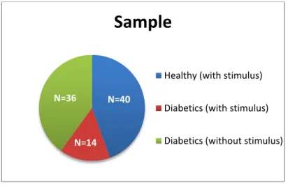
The total sample consists of healthy individuals and diabetics, who were divided into three groups. The reason for this division is that it is not possible to apply a stimulus across the sample of diabetic patients, due the logistic and short time of medical appointment. Thus the groups that constitute the total sample of 90 people, including healthy and diabetic

![Figure 5 - Location of plantar nerves (A) [35]; Location of plantar arteries (B)[36].](https://thumb-eu.123doks.com/thumbv2/123dok_br/15733430.1071728/32.892.123.780.548.823/figure-location-plantar-nerves-location-plantar-arteries-b.webp)
![Figure 7- Illustration of vasodilation and vasoconstriction processes, respectively [50]](https://thumb-eu.123doks.com/thumbv2/123dok_br/15733430.1071728/37.892.195.739.309.601/figure-illustration-vasodilation-vasoconstriction-processes-respectively.webp)
![Figure 8- Wavelength of infrared radiation, divided into short, medium and longe waves [57]](https://thumb-eu.123doks.com/thumbv2/123dok_br/15733430.1071728/39.892.174.751.176.447/figure-wavelength-infrared-radiation-divided-short-medium-longe.webp)