Vol.57, n.2: pp. 223-227, March-April 2014
ISSN 1516-8913 Printed in Brazil BRAZILIAN ARCHIVES OF
BIOLOGY AND TECHNOLOGY
A N I N T E R N A T I O N A L J O U R N A L
Oxidative Stress Parameters as Biomarkers of Risk Factor
for Diabetic Foot among the Patients with Type 2 Diabetes
Ana Carla Pozzi Oliveira
1, Caio Jordão Teixeira
2, Talitha Fernandes Stefanello
1, Marcia
Aparecida Carrara
2, Roberto Barbosa Bazotte
2*, Anacharis Babeto Sá-Nakanishi
3,
Jurandir Fernando Comar
3and Marcia Regina Batista
11Departamento de Análises Clínicas e Biomedicina; Universidade Estadual de Maringá; Maringá - PR - Brasil.
2Departamento de Farmacologia e Terapêutica; Universidade Estadual de Maringá; Maringá - PR - Brasil.
3Departamento de Bioquímica; Universidade Estadual de Maringá; Maringá - PR - Brasil
ABSTRACT
The aim of this study was to determine whether plasma levels of carbonylated proteins, total antioxidant capacity (TAC) and reduced protein thiols could be suitable biomarkers of risk factors for diabetic foot. Individuals with type 2 diabetes with normal protective sensation (normal foot group) vs. loss of protective sensation and/or signs of peripheral arterial disease and/or foot deformities and/or history of ulcers and/or neuropathic fractures and/or amputation (diabetic foot group) were compared. The diabetic foot group showed higher carbonylated protein levels (P = 0.0457) and lower levels of TAC (P = 0.0148) and reduced protein thiols (P = 0.0088), compared with the normal foot group. In general, several other parameters of risk of diabetes complication (blood levels of glycated hemoglobin, glucose and cholesterol, duration of diabetes, body mass index and waist circumference) showed a tendency of higher values in the diabetic foot group. The results suggest that the plasma levels of carbonylated proteins, TAC and reduced protein thiols could furnish information about the risk of diabetic foot, considering that the changes in these biomarkers were associated with the loss of sensitivity and foot ulcerations.
Key words: Diabetic foot, oxidative stress, total antioxidant capacity, reduced protein thiols, protein carbonyls
*Author for correspondence: [email protected]
INTRODUCTION
Combined peripheral neuropathy and ischemia result in a higher risk of foot ulcers in type 1 and type 2 diabetic patients (Singh et al. 2005). The risk of patients with diabetes developing foot ulcers in their lifetime could be as high as 25% (Singh et al. 2005). For this reason, diabetes is the leading cause of amputation worldwide. For example, a global study of lower extremity amputation estimated that 25–90% of all the amputations were associated with diabetes (Global Lower Extremity Amputation Study 2000). Regardless of the high incidence of foot ulcers and
amputations associated with diabetes, the
characterization of risk factors that could prevent foot ulcers and amputations is not well established (Sun et al. 2012). Therefore, the determination of biomarkers for early detection not only of foot ulcers but also nerve damage, infection and gangrene should be investigated.
respectively, (Sun et al. 2012) and that type 2 diabetes represents 95% of diagnosed patients, this study focused on the patients with type 2 diabetes. Accordingly, the blood levels of total antioxidant capacity (TAC), reduced protein thiols and carbonylated proteins in the patients with type 2 diabetes with normal foot vs. diabetic foot were compared.
MATERIALS AND METHODS
Eligibility criteria were confirmed diagnosis of type 2 diabetes and age over 40 years. Exclusion criteria were pregnancy, gestational diabetes, type 1 diabetes and other specific types of diabetes. The study followed the guidelines as described in the Declaration of Helsinki and written consent of the participants was obtained. The study was approved by the Ethics Committee of the State University of Maringá, PR, Brazil (COPEP - CAAE 381/2010). During the consultation, the patients were interviewed using a structured questionnaire, and information about the socio-demographic and disease factors (age, sex, medical history, educational level, marital status, duration of
diabetes, diabetes-related disorders, etc.),
pharmacotherapeutic profile and lifestyle were obtained. After the interview, the measurements
of body mass index (BMI) and waist
circumference and foot examination were
performed.
The foot examination was based on the National Hansen's Disease Program (NHPD) developed by the University of Baton Rouge, LA, USA, which identified those patients who had lost protective sensation (Tan 2010). This diabetic foot screen used a 5.07 monofilament, which delivered 10 g of force on 12 places of application to identify the patients with the risk of developing diabetic foot. The results obtained from this test permitted the classification of the foot into four categories: a) normal protective sensation; b) loss of protective sensation; c) loss of protective sensation plus signs of peripheral arterial disease and/or foot deformities; d) history of ulcers and/or neuropathic fractures and/or amputation. The patients with normal protective sensation were included in the normal foot group, and the other patients were included in the diabetic foot group.
Just before finishing the consultation, the patients received instructions for blood collection. In general, the instructions and procedures for blood
collection were similar to those adopted in a previous study investigating the risk factors of coronary heart disease in the population of northwestern Paraná (Silva et al. 2004). The biochemical parameters investigated were blood glycated hemoglobin A1c (Metus et al. 1999) and serum glucose (Bergmeyer and Bernt 1974), cholesterol (Allain et al. 1974), triacylglycerol (Bucolo and David 1973), creatinine (Bartels et al. 1972), reduced protein thiols (Faure and Lafond 1995), TAC (Erel 2004) and carbonylated proteins (Levine et al. 1990). Part of the results was presented as a percentage (%) and part of the results as mean ± standard deviation (SD). Comparison between the normal foot group and diabetic foot group was carried out using the unpaired Student t-test. P< 0.05 was considered statistically significant.
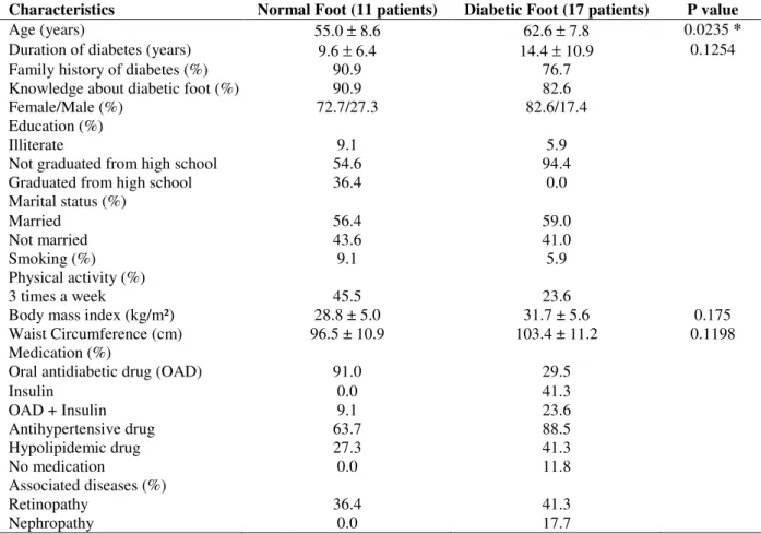
Complete data for 28 patients showed that 11 belonged to normal foot group and 17 to diabetic foot group. Most patients (Table 1) in both the groups (normal, or diabetic foot group) were female (> 70%), were knowledgeable about diabetic foot (> 80%) and had a family history of diabetes (> 75%). The diabetic foot group had a higher age (P = 0.0235) and tendency of longer duration of diabetes, higher BMI and waist
circumference and higher percentage of
nephropathy. In spite of diabetic foot, 11.8% patients in this group were not receiving any medication (Table 1). On the other hand, the normal foot group showed a higher percentage of patients graduated from high school, engaged in physical activity at least three times a week and using oral antidiabetic drugs. Marital status, smoking and presence of retinopathy were similar in the two groups (Table 1). The diabetic foot group revealed a tendency of higher blood
glycated hemoglobin A1c, glucose, total
cholesterol, low-density lipoproteins,
triacylglycerols and creatinine, compared with the normal foot group. Moreover, a tendency for lower high-density lipoproteins was observed in the diabetic foot group (Table 2).
Table 1 - Characteristics of patients in the absence (Normal Foot) or presence (Diabetic Foot) of foot alterations. Part of the results was presented as a percentage (%) and part of the results as mean ± standard deviation. * P < 0.05.
Characteristics Normal Foot (11 patients) Diabetic Foot (17 patients) P value
Age (years) 55.0 ± 8.6 62.6 ± 7.8 0.0235 *
Duration of diabetes (years) 9.6 ± 6.4 14.4 ± 10.9 0.1254
Family history of diabetes (%) 90.9 76.7
Knowledge about diabetic foot (%) 90.9 82.6
Female/Male (%) 72.7/27.3 82.6/17.4
Education (%)
Illiterate 9.1 5.9
Not graduated from high school 54.6 94.4
Graduated from high school 36.4 0.0
Marital status (%)
Married 56.4 59.0
Not married 43.6 41.0
Smoking (%) 9.1 5.9
Physical activity (%)
3 times a week 45.5 23.6
Body mass index (kg/m²) 28.8 ± 5.0 31.7 ± 5.6 0.175
Waist Circumference (cm) 96.5 ± 10.9 103.4 ± 11.2 0.1198
Medication (%)
Oral antidiabetic drug (OAD) 91.0 29.5
Insulin 0.0 41.3
OAD + Insulin 9.1 23.6
Antihypertensive drug 63.7 88.5
Hypolipidemic drug 27.3 41.3
No medication 0.0 11.8
Associated diseases (%)
Retinopathy 36.4 41.3
Nephropathy 0.0 17.7
Table 2 - Biochemical parameters of patients in the absence (Normal Foot) or presence (Diabetic Foot) of foot alterations. The results are presented as mean ± standard deviation. Key: glycated hemoglobin A1c (Hb A1c), fasting glycemia (FG), high-density lipoproteins (HDL), and low-density lipoproteins (LDL).
Parameters Normal Foot (11 patients) Diabetic Foot (17 patients) P value
Hb A1c (%) 7.1 ± 1.2 8.0 ± 1.8 0.2657
FG (mg/dL) 136.7 ± 26.3 175.3 ± 109.7 0.1746
Total cholesterol (mg/dL) 183.8 ± 42.5 214.3 ± 62.3 0.1679
HDL (mg/dL) 53.5 ± 26.7 45.9 ± 14.1 0.3385
LDL (mg/dL) 105.8 ± 26.8 141.6 ± 58.7 0.0708
Triacylglycerols (mg/dL) 122.9 ± 52.3 133.8 ± 54.8 0.6050
Creatinine (mg/dL) 1.1 ± 0.3 1.3 ± 0.3 0.1595
Table 3 - Total antioxidant capacity - TAC (mg/mL), reduced protein thiols (mg/mL) and carbonylated proteins (mg/mg plasma albumin) in the absence (Normal Foot group) or presence (Diabetic Foot group) of foot alterations. The results are presented as mean ± standard deviation. * P < 0.05.
Parameters Normal Foot (11 patients) Diabetic Foot (17 patients) P value
TAC 0.69 ± 0.06 0.61 ± 0.09 0.0148*
Reduced
protein thiols 413.41 ± 28.71 369.22 ± 46.13 0.0088* Carbonylated
proteins 6.86 ± 1.13 8.14 ± 1.81 0.0457*
DISCUSSION
accelerates chemical modification of proteins and function of tissue proteins, precipitating the development of diabetic complications. In this context, there are several hypotheses on the origin of complications, including mitochondrial damage, mitochondrial defect in oxidative phosphorylation, increased oxidative and reductive stress, increased formation of advanced glycation end products (AGES), increased activity of the polyol pathway, hypoxia, altered lipoprotein metabolism, increased protein kinase C activity, altered growth factors and cytokine activities (Baynes and Thorpe 1999; Arya et al. 2011; Papanas and Ziegler 2011). Increased levels of reactive carbonyl compounds derived from proteins by both oxidative and non-oxidative reactions lead to increased chemical modification of proteins, and then, at a later stage, to oxidative stress and tissue damage (Baynes and Thorpe 1999). Therefore, this study evaluated plasma carbonyl proteins as a biomarker of diabetic foot. In addition, two parameters of defense against oxidative stress were assessed, i.e., plasma reduced protein thiols and TAC. The results showed increased plasma levels of carbonylated proteins (P = 0.0457) in patients with diabetic foot.
Carbonyl stress is the result of a higher level of reactive carbonyl species and may be the consequence of an increased substrate stress
and/or a decrease in the efficiency of
detoxification of carbonyl compounds, which leads to increased chemical modification of biomolecules and thereby to a series of tissue dysfunction (Noeman et al. 2011). Therefore, the clinical severity of diabetic foot could be related to the development of carbonyl stress. The present results also showed lower plasma levels of reduced protein thiols (P = 0.0088) and TAC (P = 0.0148) in the patients with diabetic foot. In agreement with these findings, other studies (Bolajoko et al. 2008; Yang et al. 2011) have suggested that elevated oxidative stress could be associated with increased risk of diabetic foot.
Interestingly, the diabetic foot group showed a higher percentage of patients not on insulin therapy, or on antihypertensive, or hypolipidemic therapy. In agreement with these observations, the diabetic foot group showed a tendency of higher glycated hemoglobin level and plasma levels of glucose, triacylglycerol, and cholesterol and its fractions. This study did not include the localization of tissue-specific change in oxidative
stress in the foot, nor did it establish a clear role for blood levels of altered protein thiols, TAC and carbonyl protein in the pathogenesis of diabetic foot. Despite these limitations, results provided evidence that reduced protein thiols, TAC and carbonyl proteins in plasma could be useful biomarkers in the early detection of diabetic foot in the patients with type 2 diabetes.
CONCLUSION
Results showed a decreased defense against oxidative stress in the patients with diabetic foot. Therefore, the plasma levels of TAC, protein thiols and carbonyl proteins could provide additional information about the risk of diabetic foot, considering that the alteration of these biomarkers was associated with the loss of sensitivity and foot ulcerations.
ACKNOWLEDGMENTS
This study was supported by CNPq grant 563870/2010-9 and PRONEX/CNPq/Fundação Araucária. The authors are grateful to C.E. Oliveira, M. Guilhermetti and S. C. Oliveira for their technical assistance during the experiments and Carlos A. dos Santos for data management. Dr. A. Leyva helped with English editing of the manuscript.
REFERENCES
Allain CC, Poon LS, Chan CS, Richmond W, Fu PC. Enzymatic determination of total serum cholesterol.
Clin Chem. 1974; 20(4): 470-475.
Arya AK, Pokharia D, Tripathi K. Relationship between oxidative stress and apoptotic markers in lymphocytes of diabetic patients with chronic non healing wound. Diabetes Res Clin Pract. 2011; 94(3):377-384.
Bartels H, Böhmer M, Heierli C. Serum creatinine determination without protein precipitation. Clin Chim Acta. 1972; 37: 193-197.
Baynes JW, Thorpe SR. Role of oxidative stress in diabetic complications: a new perspective on an old paradigm. Diabetes. 1999; 48(1):1-9.
Bolajoko EB, Mossanda KS, Adeniyi F, Akinosun O, Fasanmade A, Moropane M. Antioxidant and oxidative stress status in type 2 diabetes and diabetic foot ulcer. S Afr Med J. 2008; 98(8):614-617.
Bucolo G, David H. Quantitative determination of serum triglycerides by the enzymes. Clin Chem. 1973; 19(5):476-482.
Erel O. A novel automated direct measurement method for total antioxidant capacity using a new generation, more stable ABTS radical cation. Clin Biochem.
2004; 37(4):277-285.
Faure P, Lafond JL. Measurement of plasma sulphydryl and carbonyl groups as a possible indicator of protein oxidation. In: Analysis of free radicals in biological systems. Boston: Birkhauser Verlag, Basel; 1995. p. 238-247.
Global Lower Extremity Amputation Study Group. Epidemiology of lower extremity amputations in centres in Europe, North America and East Asia. The Global Lower Extremity Amputation Study Group.
Br J Surg. 2000; 87(3):328-337.
Levine RL, Garland D, Oliver CN, Amici A, Climent I, Lenz AG, et al. Determination of carbonyl content in oxidatively modified proteins. Methods Enzymol. 1990; 186: 464-478.
Metus P, Ruzzante N, Bonvicini P, Meneghetti M, Zaninotto M, Plebani M. Immunoturbidimetric assay of glycated hemoglobin. J Clin Lab Anal. 1999; 13(1):5-8.
Monnier L, Colette C, Owens D. The glycemic triumvirate and diabetic complications: is the whole greater than the sum of its component parts?.
Diabetes Res Clin Pract. 2012; 95(3):303-311.
Noeman SA, Hamooda HE, Baalash AA. Biochemical study of oxidative stress markers in the liver, kidney and heart of high fat diet induced obesity in rats.
Diabetol Metab Syndr. 2011; 3:1-8.
Papanas N, Ziegler D. New diagnostic tests for diabetic distal symmetric polyneuropathy. J Diabetes Complications. 2011; 25(1)44-51.
Silva GEC, Bazotte RB, Curi R, Silva MARCP. Investigation of risk factors to coronary heart disease in two countryside villages. Braz Arch Biol Technol.
2004:47(3); 387-390.
Singh N, Armstrong DG, Lipsky BA. Preventing foot ulcers in patients with diabetes. JAMA. 2005; 293(2): 217-228.
Sun JH, Tsai JS, Huang CH, Lin CH, Yang HM, Chan YS, Hsieh SH, Hsu BR, Huang YY. Risk factors for lower extremity amputation in diabetic foot disease categorized by Wagner classification. Diabetes Res Clin Pract. 2012; 95(3): 358-363.
Tan LS. The clinical use of the 10g monofilament and its limitations: a review. Diabetes Res Clin Pract.
2010;90(1):1-7.
Yang XY, Sun L, Xu P, Gong LL, Qiang GF, Zhang L, Du GH. Effects of salvianolic scid A on plantar microcirculation and peripheral nerve function in diabetic rats. Eur J Pharmacol. 2011; 665(1-3):40-46.