Development of a clinical model to predict the likelihood of
identification of the Adamkiewicz artery by angiotomography
Desenvolvimento de modelo clínico para predição da possibilidade de identificação da
artéria de Adamkiewicz por angiotomografia
Alexandre Campos Moraes Amato1,2
*, José Rodrigues Parga Filho
3, Noedir Antônio Groppo Stolf4
Abstract
Background: There are clinically important morphological differences in the Adamkiewicz artery (AKA) between populations that do and do not have aortic disease and they have an influence on the neuroischemic complications involving the spinal cord during surgical operations. It is not yet known whether clinical parameters correlate with the predictability of identification of the artery using angiotomography. Objective: To develop a mathematical model that by correlating clinical parameters with atherosclerosis enables prediction of the probability of identification of the AKA in patients examined with angiotomography.
Method: This is a cross-sectional, observational study using a patient database and image bank. A multivariate statistical analysis was conducted and a logit mathematical model was constructed to predict AKA identification. Significant variables were used to build a formula for calculation of the probability of identification. This model was calibrated and its power of discrimination was assessed using receiver operating characteristic (ROC) curves. Selection of explanatory variables was based on largest area under the ROC curve (p = 0.041) and combined significance of variables. Results: A total of 110 cases were analyzed (54.5% were male, mean age was 60.97 years, and ethnicity coefficients were white -2.471, brown -1.297, and black -0.971) and the AKA was identified in 60.9%. Body mass index: 27.06 ± 0.98 (coef. -0.101); smokers: 55.5% (coef. -1.614/-1.439); diabetes: 13.6%; hypertension: 65.5% (coef. -1.469); dyslipidemia: 58.2%; aortic aneurysm: 38.2%; aortic dissection: 12.7%; and mural thrombus: 24.5%. The constant was 6.262. The formula for calculating the probability of detection is as follows:
( )
( . . . ker . . tan) 1
(e−Coef Etnicity+Coef BMI BMI× +Coef smo +Coef SAH Coef dyslip Cons+ + t +1)− . The prediction model was constructed and made available at: https://vascular.pro/aka-model. Conclusions: Using the covariates ethnicity, body mass index, smoking, arterial hypertension, and dyslipidemia, it proved possible to create a mathematical model for predicting identification of the AKA with a combined significance of nine coefficients (p = 0.042).
Keywords: spinal marrow; spinal column; aorta; Adamkiewicz.
Resumo
Contexto: Diferenças morfológicas da artéria de Adamkiewicz (AKA) entre a população portadora e não portadora de doença aórtica têm importância clínica, influenciando as complicações neuroisquêmicas da medula espinhal em procedimentos operatórios. Ainda não é conhecida a correlação entre parâmetros clínicos e a previsibilidade da identificação dessa artéria pela angiotomografia. Objetivo: Desenvolver um modelo matemático que, através de parâmetros clínicos correlacionados com aterosclerose, possa prever a probabilidade de identificação da AKA em pacientes submetidos a angiotomografias. Método: Estudo observacional transversal utilizando banco de imagens e dados de pacientes. Foi feita análise estatística multivariada e criado modelo matemático logit de predição para identificação da AKA. Variáveis significativas foram utilizadas na montagem da fórmula para cálculo da probabilidade de identificação. O modelo foi calibrado, e a discriminação foi avaliada pela curva receiver operating characteristic (ROC). A seleção das variáveis explanatórias foi guiada pela maior área na curva ROC (p = 0,041) e pela significância combinada das variáveis.
Resultados: Foram avaliados 110 casos (54,5% do sexo masculino, com idade média de 60,97 anos e etnia com coeficiente B -2,471, M -1,297, N -0,971), com AKA identificada em 60,9%. Índice de massa corporal: 27,06 ± 0,98 (coef. -0,101); fumantes: 55,5% (coef. -1,614/-1,439); diabéticos: 13,6%; hipertensos: 65,5% (coef. -1,469); dislipidêmicos: 58,2%; aneurisma aórtico: 38,2%; dissecção aórtica: 12,7%; e trombo mural: 24,5%. Constante de 6,262. Fórmula para cálculo da probabilidade de detecção: ( . . ( ) . . . tan ) 1
(e−Coef Etnia+Coef IMC IMC× +Coef fumante Coef HAS Coef dislip Cons+ + + te +1)− . O modelo de
predição foi criado e disponibilizado no link https://vascular.pro/aka-model. Conclusão: Com as covariáveis etnia, índice de massa corporal, tabagismo, hipertensão arterial e dislipidemia, foi possível criar um modelo matemático de predição de identificação da AKA com significância combinada de nove coeficientes (p = 0,042).
Palavras-chave: medula espinhal; coluna vertebral; aorta; Adamkiewicz.
1 Instituto de Medicina Avançada – AMATO, Cirurgia Vascular e Endovascular, São Paulo, SP, Brasil. 2 Universidade de Santo Amaro – UNISA, Disciplina de Cirurgia Vascular, São Paulo, SP, Brasil.
3 Universidade de São Paulo – USP, Departamento de Radiologia, Faculdade de Medicina, Hospital das Clínicas, Instituto do Coração, São Paulo, SP, Brasil. 4 Universidade de São Paulo – USP, Cirurgia Torácica e Cardiovascular, São Paulo, SP, Brasil.
Financial support: None.
Conflicts of interest: No conflicts of interest declared concerning the publication of this article. Submitted: August 01, 2017. Accepted: December 13, 2017.
INTRODUCTION
Studying morphological differences of the Adamkiewicz artery (AKA) between populations with and without diseases is of clinical importance, because they can influence neuroischemic complications of the spinal cord during surgical operations. The findings could also help to establish the importance of preserving this artery in aortic and neurological surgery, reducing
the risk of spinal cord ischemia.1-3
The intercostal and lumbar arteries that feed the spinal cord originate from the aorta. The intercostal and lumbar arteries divide three times before reaching the spinal cord. The last bifurcation of the spinal branch is constant for anterior and posterior supply of the vertebral canal, at some levels only. Generally, one of the anterior radicular arteries is dominant over the others, in terms of caliber, and is called the great
radicular artery or the AKA.2
Knowledge of the blood supply to the spinal cord is important when planning treatment for aortic diseases, but the vasculature is complex and difficult to study because of the small caliber of the arteries involved, which form an intricate three-dimensional
network with a large degree of anatomic variability2,4
(Figure 1). The fact that there is no consensus on a gold standard imaging exam also makes it difficult to compare existing imaging methods.
Differences in the AKA’s importance to spinal blood supply between different diseases, such as aneurysms, dissections, mural thrombi, and others,
are also unknown, even though this is the population that is most likely to need surgical treatment and, consequently, most exposed to the risk of spinal cord ischemia. It has been demonstrated that aortic disease itself can change the appearance of the spinal blood
supply.5 While it is known which factors influence
identification of the vessels of the spinal blood supply using angiotomography, their effects in combination are unknown.
OBJECTIVE
To construct a mathematical model using clinical variables to predict whether or not the AKA will be identifiable with angiotomography.
METHOD Patients
We conducted a cross-sectional observational study of a database and archived images from a prior
study5 of patients with or without aortic disease who
had been examined with angiotomography between October 2011 and July 2012.
During this period, the Instituto do Coração do Hospital das Clínicas da Faculdade de Medicina da Universidade de São Paulo conducted a total of 128 angiotomographies of the aorta in the diagnostic imaging department, using an Aquilion One scanner (Toshiba™, Medical Systems, Otawara, Japan). One hundred and ten of these examinations were
considered eligible for the study, on the basis of the inclusion and exclusion criteria.
The inclusion criteria were as follows: patients who had undergone angiotomography of the aorta using an Aquilion One scanner and following a predefined protocol, irrespective of the reasons for ordering the examination; patients who accepted an aortic angiotomography as a supplementary test, in cases in which the treating physician had not ordered the examination.
The exclusion criteria employed were as follows: patients who had previously undergone surgery on the descending aorta; patients with rare diseases that could compromise the results because of collateral circulation (Takayasu’s disease, aortic coarction); paraplegic or tetraplegic patients; known allergy to radiology contrast; concurrent enrollment on another study that would interfere with the protocol; patients with intramural hematoma and penetrating aortic ulcer; age less than 25 years, to ensure sample homogeneity; technical failure of opacification of the aorta.
Routine outpatients angiotomographies were analyzed prospectively, using the open source software package
OsiriX (Pixmeo, Geneva, Switzerland).6 Images were
blindly analyzed; the evaluator was unaware of why the examination had been conducted and assessments of whether aortic disease was present were made in a second round of analyses of the images, during which the evaluator was unaware of whether or not the AKA had been identified.
Angiotomography examinations were conducted according to a preestablished routine: images were all acquired using the same Aquilion One, 320-detector scanner, with software for helical scanning with 80 to 160 detectors, following the protocol for arterial contrast studies with 100 to 110 mL of non-ionic contrast administered intravenously over 30 s with an injection pump at a velocity of 4 to 5 mL/s, with the trigger configured for a greater contrast concentration in the descending aorta and a threshold of 150 HU. The technical parameters of the angiotomographic method used follow the recommendations contained
in aortic disease diagnosis and treatment guidelines.7
Patients were stratified according to the following characteristics: sex, age, ethnic group, weight, height, and body mass index (BMI). Presence or absence of the anterior spinal artery, presence and location of the AKA, and presence or absence of aortic disease, thrombus, and dissection were evaluated by a single, experienced observer directly on the highest quality raw DICOM images from the patient’s examination. Additionally, dichotomous risk factors related to aortic diseases were also assessed: diabetes mellitus, metabolic syndrome, hyperlipidemia, and arterial hypertension. The variable smoking was categorized as smoker, ex-smoker, or non-smoker.
Sociodemographic data were obtained from the patients’ medical records and directly from patients and their relatives, as follows: age, sex, and ethnicity. Ethnicity was defined according to the skin color reported by the participant, i.e. self-assessed. Self-reported multiracial variations were coded as brown skin. Patients were also told they could ignore this question.
Data were collected when taking patients’ histories, from medical records, and by telephone from relatives when necessary. The following parameters were evaluated: weight, height, and BMI. Weight and height were measured using an anthropometric balance; BMI was calculated with the formula: weight (in kilograms) divided by the square of height (in meters). Smoking: patients were defined as current smokers if they had smoked one or more cigarettes during the preceding 30 days and as ex-smokers if they had ever smoked for more than 30 days. Diabetes mellitus (DM): patients were classified as diabetics if they had been taking oral hypoglycemic drugs and/or insulin, or had two fasting glycemia results ≥ 126 mg/dL. Systemic arterial hypertension (SAH): patients were considered hypertensive if they were taking antihypertensives and/or had systolic arterial blood pressure (SBP) greater than 140 mmHg and/or diastolic arterial blood pressure (DBP) greater than 90 mmHg. Dyslipidemia: patients were classified as having dyslipidemia if they were taking antihyperlipidemics and/or had elevated low density lipoprotein cholesterol (LDL-C ≥ 160 mg/dL) and/or elevated triglycerides (TG ≥ 150 mg/dL) and/or high density lipoprotein cholesterol (HDL-C) < 40 mg/dL
for men and < 50 mg/dL for women.8 Metabolic
syndrome: diagnoses of metabolic syndrome were made according to International Diabetes Federation criteria. Presence of abdominal obesity is an essential criterion, combined with two or more of the following criteria: triglycerides ≥ 150 mg/dL; HDL-C < 40 mg/dL for men and < 50 mg/dL for women; SBP ≥ 130 mmHg and/or DBP ≥ 85 mmHg, or treatment for hypertension; fasting glycemia ≥ 100 mg/dL or treatment for
diabetes mellitus.8
The method used to identify AKA has been used by the author, described and validated in a previous
publication,3,6 and an explanatory video illustrating the
technique is available at http://vascular.cc/aka.html.
Statistical analysis
The data obtained from patient histories and medical records were input on a secure online database
(GoogleDocs). After verification of the consistency
of data, descriptive analyses were conducted for each group of patients.
US). Models were defined as good if the area under the receiver operating characteristic (ROC) curve was greater than 0.7 and as excellent if the area under the ROC curve exceeded 0.85. The logit model was chosen since the outcome was binary and significance was defined as p < 0.05. Once the model had been constructed, a formula was derived to predict the outcome variable. The prediction formula calculates the expected value of the outcome (identification or failure to identify the AKA) based on the initial variables selected as meeting the criterion of area under the ROC curve greater than 0.7.
Considering the entire sample, Table 1 lists the variables collected and the preliminary analyses of
the database that have been published previously.3,5
The outcome selected was identification or failure to identify the AKA using the method proposed. The initial explanatory co-variables were chosen on the basis of prior knowledge of the influence of body mass, arterial disease, and atherosclerosis. Covariates with coefficients significantly different
from zero to the 10% level were maintained. Thus, ethnicity, smoking, BMI, hypertension, and dyslipidemia were chosen to construct the predictive model (Table 2). Selection of explanatory variables
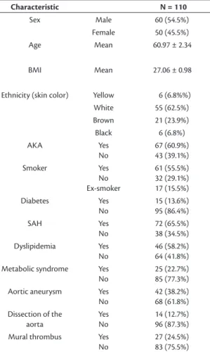
Table 1. Sociodemographic data on the study population.
Characteristic N = 110
Sex Male 60 (54.5%)
Female 50 (45.5%)
Age Mean 60.97 ± 2.34
BMI Mean 27.06 ± 0.98
Ethnicity (skin color) Yellow 6 (6.8%%)
White 55 (62.5%)
Brown 21 (23.9%)
Black 6 (6.8%)
AKA Yes
No
67 (60.9%) 43 (39.1%)
Smoker Yes
No Ex-smoker
61 (55.5%) 32 (29.1%) 17 (15.5%)
Diabetes Yes
No
15 (13.6%) 95 (86.4%)
SAH Yes
No
72 (65.5%) 38 (34.5%)
Dyslipidemia Yes
No
46 (58.2%) 64 (41.8%) Metabolic syndrome Yes
No
25 (22.7%) 85 (77.3%) Aortic aneurysm Yes
No
42 (38.2%) 68 (61.8%) Dissection of the
aorta
Yes No
14 (12.7%) 96 (87.3%)
Mural thrombus Yes
No
27 (24.5%) 83 (75.5%)
AKA, Adamkiewicz artery; SAH, systemic arterial hypertension; BMI, body mass index.
Table 2. Significance of variables in the model studied and
coefficients for the influence of variables on the outcome.
Exp
lan
at
or
y
var
iab
le
C
o
effi
ci
ent
St
an
d
ar
d
e
rr
or
p Signific
an
ce
Ethnicity (skin color) 0.0677 *
White -2.471 (1.264) 0.0505 *
Brown -1.297 (1.329) 0.3291 Black -0.971 (1.597) 0.5433
BMI -0.101 (0.055) 0.0664 *
Smoker 0.0196 **
Yes -1.614 (0.801) 0.0439 **
Ex-smoker -1.439 (0.595) 0.0156 **
SAH -1.469 (0.604) 0.0150 **
Dyslipidemia 0.97 (0.555) 0.0806 *
Constant 6.262 (2.018) 0.0019 ***
*Coefficient significantly different from zero to the 10% level; **Coefficient significantly different from zero to the 5% level; ***Coefficient significantly different from zero to the 1% level. SAH, systemic arterial hypertension; BMI, body mass index.
Figure 2. (a) Receiver operating characteristic (ROC) curve.
was guided by largest area under the ROC curve
(Figure 2A) and by the combined significance of the
variables (Figure 2B), so that a balance was achieved between the number of explanatory variables, without excessive variables, and the greatest influence on area under the ROC curve.
This investigation was conducted in accordance with the principles set out in the Helsinki Declaration. The study was approved by the Scientific Commission at the Instituto do Coração (InCor), Universidade de São Paulo, and the Research Ethics Commission at the HC-FMUSP on 05/05/2010, under reference number 0089/10. It is registered on the Brazilian national human research ethics register (SISNEP) maintained by the National Research Ethics Commission (CONEP - Comissão Nacional de Ética em Pesquisa), under number 0088.0.015.000-10. All patients or their guardians were given explanations about the objectives of the study, agreed to participation, and signed free and informed consent forms, which included the current data analysis in the terms of permission.
Results
The formula for calculating the probability of detecting the AKA
( )
( . . . ker . . tan) 1
(e−Coef Etnicity+Coef BMI BMI× +Coef smo +Coef SAH Coef dyslip Cons+ + t +1)−
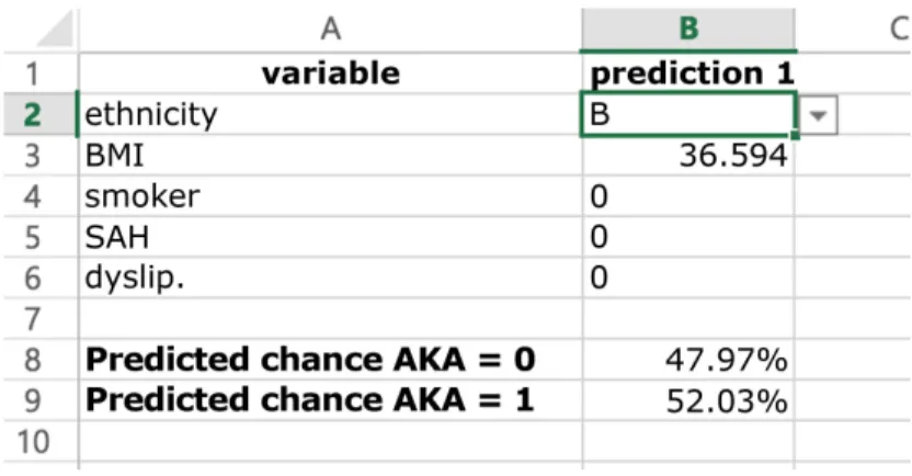
enabled construction of the prediction model, which was exported to an excel spreadsheet (Figure 3).
Table 2 lists the constant and the coefficients for the
influence of the variables selected for calculation of the outcome. The model was formatted and sent to an online service and then published for general use at the web address https://vascular.pro/aka-model. The variables can be changed in real-time and calculate the probability of identification of the AKA.
DISCUSSION
Spinal cord ischemia is a possible complication of aortic surgery and, while rare, it is devastating
and merits study.9 In vivo examination of the AKA
is difficult because the method considered to be the gold standard, angiography, currently has unacceptably high rates of serious complications: 8.2% for localized complications, 3.7% for non-neurological complications,
and 2.2% for neurological complications,10 and does
not achieve detection in 100% of cases (without aortic
disease: 68%;10 with aortic disease: 60%).11
The angiotomographic characteristics of the AKA in populations with and without aortic disease have been studied and reported in an earlier publication
by the author.3,5 Improved knowledge of the spinal
blood supply should affect and provide a foundation for creation of new strategies for prevention of spinal cord ischemia during aortic surgery and other neurological surgical procedures. Boll et al. analyzed 100 examinations of patients with pancreatic cancer and applied a modified version of a graphical algorithm for reconstruction of images of cerebral vessels. The AKA was identified in all cases, but it is unclear whether the high rate of detection was because of the absence of aortic disease or the different method used
to process the images.12
According to the literature, it is possible to identify the AKA using computed tomography in around
70% of cases analyzed,1,6,8-12 although the cause of
non-identification in the remaining cases is unclear. Influence from atherosclerosis, mural thrombus,
dissection, and body mass has been suggested.3,5
The imaging protocol used was as close as possible to habitual angiotomography protocols, so that it represented the routine examinations conducted at many medical centers. Care was taken to minimize
the radiation dose to reduce risks.13
Figure 3. Simplified appearance of the model for prediction of identification of the AKA on a basic electronic spreadsheet. Interface
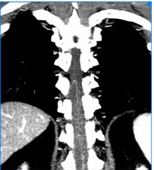
Since angiotomography is a method based on X-rays, it suffers from artifacts close to very dense structures, such as bones. The vessels studied are surrounded by a bony framework, but modern equipment allows this
problem to be reduced to a minimum14-16 (Figure 4).
All of the variables tested representing comorbidities and risk factors for atherosclerosis were associated with lower rates of AKA identification. In the subset of smokers, ex-smokers, and hypertensive patients, the likelihood of non-identification of the AKA was statistically significant. Patients in whom the AKA could not be identified had a higher incidence of metabolic syndrome (31.1%), compared with those in whom the AKA could be evaluated, who had a lower rate of metabolic syndrome (15.7%). This fact may be a consequence of this syndrome being related to
more advanced atherosclerotic vascular damage,17
increasing the likelihood of non-identification of the AKA by 2.42 times (p = 0.054) in comparison with those who did not have metabolic syndrome. All of the binary aortic disease variables tested (mural thrombus, dissection and/or thrombus, and aneurysms with and without dissection) were associated with reductions in identification of AKA with statistical significance, probably because of abnormal blood flow through the aorta and its branches, occlusion of the ostia of intercostal arteries, tortuosity of arteries secondary to anatomic distortion, and/or occlusion
by dissection flap, atherosclerotic plaque, or mural thrombus. When the AKA is not found, it could be occluded, it may not exist, or it may be too narrow to be detected by the method used.
Clinical variables were identified that have significance with relation to identification of the AKA, enabling a mathematical model for prediction of the result to be constructed. The absence of a low-risk gold-standard method for comparison of methods is one factor limiting this study. Setting a maximum limit for acceptable radiation dosage also limited the quality of the images acquired. However, the method employed required a mean of 12 mSv/(mGy·cm), which is considered a safe
dose, is reproducible,18,19 and provides satisfactory
coverage of the aorta.
CONCLUSIONS
It proved possible to use the covariates ethnicity, BMI, smoking, arterial hypertension, and dyslipidemia to create a mathematical model to predict identification of the AKA.
REFERÊNCIAS
1. Melissano G, Bertoglio L, Civelli V, et al. Demonstration of the Adamkiewicz artery by multidetector computed tomography angiography analysed with the open-source software OsiriX. Eur J Vasc Endovasc Surg. 2009;37(4):395-400. http://dx.doi. org/10.1016/j.ejvs.2008.12.022. PMid:19230726.
2. Amato ACM, Stolf NAG. Anatomy of spinal blood supply. J Vasc Bras. 2015;14(3):248-52. http://dx.doi.org/10.1590/1677-5449.0004.
3. Amato ACM, Parga JR Fo, Stolf NAG. Predictors of Adamkiewicz artery and anterior spinal artery detection through computerized tomographic angiography. SAGE Open Med. 2017;5:1-7. http:// dx.doi.org/10.1177/2050312117711599. PMid:28616230.
4. Melissano G, Civilini E, Bertoglio L, Calliari F, Amato ACM, Chiesa R. Angio-CT imaging of the spinal cord vascularisation: a pictorial essay. Eur J Vasc Endovasc Surg. 2010;39(4):436-40. http://dx.doi. org/10.1016/j.ejvs.2009.11.026. PMid:20034815.
5. Amato ACM, Parga JR Fo, Stolf NAG. Influential factors on the evaluation of Adamkiewicz artery using a 320-detector row computed tomography device. Ann Vasc Surg. 2017;44:136-45. http://dx.doi.org/10.1016/j.avsg.2017.02.019. PMid:28501659.
6. Ou P, Schmit P, Layouss W, Sidi D, Bonnet D, Brunelle F. CT angiography of the artery of Adamkiewicz with 64-section technology: first experience in children. AJNR Am J Neuroradiol. 2007;28(2):216-9. PMid:17296982.
7. Hiratzka LF, Bakris GL, Beckman JA, et al. 2010 ACCF/AHA/ AATS/ACR/ASA/SCA/SCAI/SIR/STS/SVM guidelines for the diagnosis and management of patients with thoracic aortic disease - executive summary: a report of the american college of cardiology foundation/american heart association task force on practice guidelines, american association for thoracic surgery, american college of radiology, american stroke association, society of cardiovascular anesthesiologists, society for cardiovascular angiography and interventions, society of interventional radiology, society of thoracic surgeons, and society for vascular medicine.
Figure 4. Final result of the methodology employed for
Anesth Analg. 2010;111(2):279-315. http://dx.doi.org/10.1213/ ANE.0b013e3181dd869b. PMid:20664093.
8. Sposito AC, Caramelli B, Fonseca FAH, et al. IV brazilian guideline for dyslipidemia and atherosclerosis prevention: department of atherosclerosis of Brazilian Society of Cardiology. Arq Bras Cardiol. 2007;88:2-19. http://dx.doi.org/10.1590/S0066-782X2007000700002. PMid:17515982.
9. Gao L, Wang L, Su B, Wang P, Ye J, Shen H. The vascular supply to the spinal cord and its relationship to anterior spine surgical approaches. Spine J. 2013;13(8):966-73. http://dx.doi.org/10.1016/j. spinee.2013.03.017. PMid:23608560.
10. Forbes G, Nichols DA, Jack CR Jr, et al. Complications of spinal cord arteriography: prospective assessment of risk for diagnostic procedures. Radiology. 1988;169(2):479-84. http://dx.doi.org/10.1148/ radiology.169.2.3174997. PMid:3174997.
11. Savader SJ, Williams GM, Trerotola SO, et al. Preoperative spinal artery localization and its relationship to postoperative neurologic complications. Radiology. 1993;189(1):165-71. http://dx.doi. org/10.1148/radiology.189.1.8372189. PMid:8372189.
12. Boll DT, Bulow H, Blackham KA, Aschoff AJ, Schmitz BL. MDCT angiography of the spinal vasculature and the artery of Adamkiewicz. AJR Am J Roentgenol. 2006;187(4):1054-60. http:// dx.doi.org/10.2214/AJR.05.0562. PMid:16985157.
13. Sodickson A, Baeyens PF, Andriole KP, et al. Recurrent CT, cumulative radiation exposure, and associated radiation-induced cancer risks from CT of adults. Radiology. 2009;251(1):175-84. http://dx.doi. org/10.1148/radiol.2511081296. PMid:19332852.
14. Joseph PM, Ruth C. A method for simultaneous correction of spectrum hardening artifacts in CT images containing both bone and iodine. Med Phys. 1997;24(10):1629-34. http://dx.doi. org/10.1118/1.597970. PMid:9350717.
15. Joseph PM, Spital RD. A method for correcting bone induced artifacts in computed tomography scanners. J Comput Assist Tomogr. 1978;2(1):100-8. http://dx.doi.org/10.1097/00004728-197801000-00017. PMid:670461.
16. Goodenough DJ, Weaver KE, Costaridou H, Eerdmans H, Huysmans P. A new software correction approach to volume averaging artifacts in CT. Comput Radiol. 1986;10(2-3):87-98. http://dx.doi. org/10.1016/0730-4862(86)90050-8. PMid:3754803.
17. Olijhoek JK, van der Graaf Y, Banga JD, et al. The metabolic syndrome is associated with advanced vascular damage in patients with coronary heart disease, stroke, peripheral arterial disease or abdominal aortic aneurysm. Eur Heart J. 2004;25(4):342-8. http:// dx.doi.org/10.1016/j.ehj.2003.12.007. PMid:14984924.
18. Huda W, Ogden KM, Khorasani MR. Converting dose-length product to effective dose at CT. Radiology. 2008;248(3):995-1003. http://dx.doi.org/10.1148/radiol.2483071964. PMid:18710988.
19. Van Unnik JG, Broerse JJ, Geleijns J, Jansen JT, Zoetelief J, Zweers D. Survey of CT techniques and absorbed dose in various dutch hospitals. Br J Radiol. 1997;70(832):367-71. http://dx.doi.org/10.1259/ bjr.70.832.9166072. PMid:9166072.
*
Correspondence
Alexandre Campos Moraes Amato Av. Brasil, 2283 - Jardim América CEP 01431-001 - São Paulo (SP), Brasil Tel.: +55 (11) 5053-2222 E-mail: [email protected]
Author information
ACMA - Professor of Vascular Surgery, Universidade de Santo Amaro (UNISA); Full member, Sociedade Brasileira de Angiologia e Cirurgia Vascular; Board-certified in Vascular and Endovascular Surgery by Sociedade Brasileira de Angiologia e Cirurgia Vascular, and in Vascular Doppler Ultrasound by Colégio Brasileiro de Radiologia. JRPF - Assistant Physician and Investigator, Departamento de Radiologia, Instituto do Coração (InCor), Universidade de São Paulo. NAGS - Emeritus Professor of Cardiovascular Surgery, Faculdade de Medicina da Universidade de São Paulo (FAMUSP).
Author contributions
Conception and design: ACMA, JRPF, NAGS Analysis and interpretation: ACMA, JRPF, NAGS Data collection: ACMA, JRPF, NAGS Writing the article: ACMA, NAGS Critical revision of the article: ACMA, JRPF, NAGS Final approval of the article*: ACMA, JRPF, NAGS Statistical analysis: ACMA Overall responsibility: ACMA