This study evaluated the effect of the accelerated artificial aging (AAA) on feldspar ceramic strength and the reinforcing effect promoted by adhesive cementation with resin luting agent. One hundred twenty feldspar ceramic disks were obtained. Sixty disks were acid-etched, silanized, and coated with an experimental resin luting agent simulating the adhesive luting procedures. Four groups were created (n=30): uncoated ceramic (control group), uncoated ceramic submitted to AAA, ceramic coated with resin luting agent, and coated ceramic submitted to AAA. Biaxial flexural testing with ball-on-ring setup was carried out. Biaxial flexural strength (σbf, MPa), characteristic strength (σ0,
MPa), and Weibull modulus (m) were calculated for axial positions z=0 (ceramic surface) and z=−t2 (luting agent surface). Data of σbf at positions z=0 and z=-t2 were separately
submitted to statistical analyses (α=0.05). The uncoated ceramic submitted to AAA had no significant difference in σbf and σ0 compared with the control group. Resin coating of
the ceramic increased σbfand σ0 at z=0. The AAA increased the σbf and σ0 for the
resin-coated ceramic specimens at z=0 and also the σ0 at axial position z=-t2. The structural
reliability at z=0 and z=-t2 was not influenced by the variables tested. In conclusion, resin
coating improved the mechanical strength of the feldspar ceramic. The AAA procedure was not effective in aging the uncoated or resin-coated feldspar ceramic specimens.
Feldspar Ceramic Strength and The
Reinforcing Effect by Adhesive
Cementation Under Accelerated Aging
Fabíola Jardim Barbon1,2, Rafael Ratto Moraes2, Noéli Boscato2, Rodrigo
Alessandretti1, Aloísio Oro Spazzin1
1Department of Restorative
Dentistry, School of Dentistry, IMED - Meridional Faculty, Passo Fundo, RS, Brazil
2Department of Restorative
Dentistry, School of Dentistry, UFPel - Universidade Federal de Pelotas, Pelotas, RS, Brazil
Correspondence: Aloísio Oro Spazzin, St. Senador Pinheiro, 304, Bairro Rodrigues, 99070-220 Passo Fundo, RS, Brasil. Tel/ Fax: +55-54-3045-6100. e-mail: [email protected]
Key Words: ceramic, resin luting agent, mechanical strength, Weibull analysis.
Introduction
Feldspar dental ceramics are used in several rehabilitation treatments due to their biocompatibility, satisfactory functional and esthetic clinical outcomes (1, 2). These ceramics are intrinsically brittle, however they may be strengthened by adhesive cementation to the dental structure using resin luting agents (3-5). The ceramic strengthening provided by resin luting agent is clinically relevant by decreasing crack growth and improving the mechanical strength performance of feldspar ceramics (4, 6). Adhesive cementation is responsible for allowing even very thin veneers to be bonded to tooth structures without immediate fracture, providing clinical success and longevity.
Many mechanisms have been used to explain the ceramic strengthening effect, such as induction of crack closure stress by resin shrinkage upon curing (7) and ceramic crack healing by resin infiltration (8). The role of the resin luting agent in transferring stresses from load-bearing restorations to the underlying dental structures is still not fully elucidated. Recent studies have suggested that the ceramic strengthening is dependent on the formation of a proper interpenetrated resin-ceramic interphase (3, 5). In addition, studies evaluating the effects of elastic properties of resin luting agents on ceramic strengthening indicated that higher elastic moduli are favorable for
ceramic strengthening (4, 9-11).
Studies have used several methods of accelerated artificial aging (AAA) to simulate the effects of intraoral aging and evaluate the mechanical strength and other physical properties of dental materials (12-15). One these methods is the AAA system associating the use of ultraviolet-B light, moisture, and heating cycles applied for short- and long-term analyses of the behavior of non-metallic materials (12, 15). Color changes, degradation of bonded interfaces, and crack growth mechanisms will likely be influenced by intrinsic physical-chemical modifications in the materials during the degradation process induced by AAA (12). The literature presents limited information about the effect of the AAA on the strength of the ceramic and of the ceramic-resin luting agent set. This issue is particularly relevant considering that very thin feldspar ceramic laminate veneers have been extensively used in restorative dentistry.
Feldspar ceramic strength under accelerated aging
Material and Methods
Study DesignOne hundred twenty feldspar ceramic disks were obtained. An experimental resin luting agent, prepared with an elastic modulus of 13.3 GPa, was used to coat half the number of the acid-etched, ceramic disks simulating the adhesive luting procedures. Half the number of specimens obtained was submitted to AAA. In total, four groups were tested: uncoated ceramic disks (control group, C), uncoated ceramic disks submitted to AAA (C-AAA), ceramic coated with resin luting agent (CR), and ceramic coated with resin luting agent submitted to AAA (CR-AAA). Thirty specimens were tested in each group to allow appropriate Weibull analysis. The response variables were biaxial flexural strength (σbf, MPa), characteristic strength (σ0, MPa), and
Weibull modulus (m).
Preparation of the Ceramic Disks
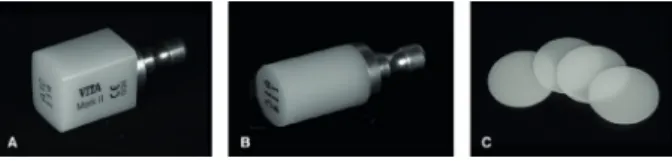
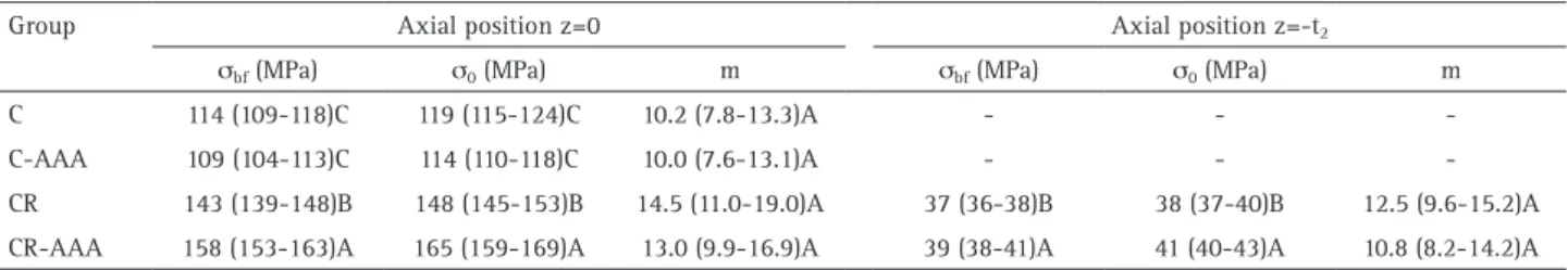
Feldspar ceramic blocks for CAD/CAM (I14 A1C Vitablocs Mark II; Vita Zahnfabrik, Bad Säckingen, Germany) were milled under water-cooling obtaining a cylindrical shape (12 mm of diameter) and then they were sectioned using a diamond saw (Isomet 1000; Buehler, Lake Bluff, IL, USA) obtaining ceramic disks (0.8 ± 0.1 mm thickness) (Fig. 1). This thickness was used due to be similar the thickness of ceramic laminate veneers restorations under clinical situations. The ceramic disks were wet-polished using 600 and 1200-grit SiC abrasive papers (Norton S.A., São Paulo, SP, Brazil), and observed using ×40 magnification to exclude disks with visible flaws or cracks. The thickness and diameter of the ceramic disks were measured using a digital caliper (Mitutoyo, Tokyo, Japan). One hundred and twenty ceramic disks were randomly divided into the four groups tested (n=30): C, C-AAA, CR, and CR-AAA.
Formulation of the Experimental Resin Luting Agent
The experimental resin luting agent was formulated using a 55% mass fraction of urethane dimethacrylate (UDMA), 5% mass fraction of 2,2-bis[4-(2-hydroxy-3-methacryloyloxypropyl)phenyl]-propane (BisGMA), and 40% mass fraction of methyl methacrylate (MMA), all from Esstech Inc. (Essington, PA, USA). A 0.8% mass fraction of ethyl 4-dimethylamino benzoate (Sigma-Aldrich, St.
Louis, MO, USA) was used as coinitiator, and a 0.4% mass fraction of camphorquinone (Sigma-Aldrich) was used as photosensitizer. Barium-borosilicate glass particles (2 μm average size coated with 1% mass fraction of silane coupling agent; V-119-4120; Esstech) were added to the resin matrix at 67% mass fraction. The components of the resin luting agent were mechanically mixed using a centrifugal mixer (SpeedMixer DAC150; FlackTek, Landrum, SC, USA) to produce a homogeneous paste.
Five disks of the resin luting agent were made using a cylindrical silicone mold with 5 mm diameter and 2 mm thickness. After the filling of the mold with the resin luting agent, the top surface was covered with a Mylar strip, a ceramic disk was positioned over the mold, and light-activation was carried out through the ceramic for 60 s using an LED unit (Radii; SDI, Bayswater, Victoria, Australia) with irradiance of 1200 mW/cm2 monitored throughout
the experiment. The disks were embedded in poly(methyl methacrylate) resin and the top surface was wet-polished using 400, 1200, 1500, 2000, and 2500-grit SiC abrasive papers (Norton S.A.). Final polishing was accomplished using felt disk (Buehler) with diamond suspensions of 3, 1, and 0.25 μm particles (Buehler). Five Knoop hardness indentations were performed on the top surface of each disk, under a load of 50 kgf for 15 s, using a microindenter (FM-700; Futuretech, Tokyo, Japan). In this method, the elastic recovery of the material is considered to calculate the elastic modulus. Then, the readings of the indentation diagonals were performed 60 s after the indention.
The decrease in the length of the indentation diagonals caused by the elastic recovery of the material is related to the hardness and elastic modulus ratio (H/E), thus the
E was calculated using the Equation 1 (16):
where a is the minor and b the major diagonal of the Knoop indentation in the fully loaded state, so the ratio a/b is given by a constant 0.140647, in the other words, this constant is given by Knoop diamond shape; a’ and b’
are the altered dimensions when fully recovered, which were measured using the microscopy and functions of the microindenter; and α1= 0.45 is a proportionality constant.
The five readings for each specimen were averaged. The mean of elastic modulus of the resin luting agent used was 13.3 GPa (95% confidence interval: 11.5–15.0).
Ceramic-Resin Specimens (Resin-Coating)
The ceramic disks had one of the surfaces etched with 10% hydrofluoric acid for 90 s (Dentsply Caulk, Milford, DE, USA), washed for 60 s, and dried with water- and oil-free
F
. J. Barbon et al.
compressed air for 30 s. Two layers of a silane-coupling agent (Dentsply) were applied and dried after 60 s with water- and oil-free compressed air for 30 s. A standard volume of resin luting agent was applied on the center of the disk, and a Mylar strip was manually pressed to extrude the resin luting agent; then this assembly was placed on top of a polished disk (14 mm diameter, 2 mm thickness) of resin composite (Filtek Z350 XT; 3M ESPE, St. Paul, MN, USA) used to simulate dentin. The disk was centralized on a leveled loading platform. The top ceramic surface was loaded with a 5 N load for 60 s using a 10-mm-diameter ball indenter with an intermediary 2-mm-thick silicone layer to reduce contact-induced damage (17). The extruded excess of the resin luting agent was removed using a microbrush. The resin luting agent was light-cured through the ceramic for 60 s, and then the thickness of the luting agent layer was measured. Ceramic-resin specimens with luting agent layer thickness outside the range between 100 μm and 150 μm were discarded and replaced by new specimens.
Accelerated Artificial Aging (AAA)
Half the number of ceramic disks and ceramic-resin specimens (groups C and CR, respectively) were dry-stored at 37°C for 24 h in lightproof containers before of the biaxial flexural test. The other half (groups C-AAA and CR-AAA) was submitted to AAA (Equilam, Diadema, SP, Brazil). The specimens were exposed to ultraviolet-B irradiation from 40 W UV-B fluorescent tubes with an irradiation peak of 0.89 W/m2/mm, in wavelength of 340 nm, at a temperature
of 60oC for 4 h, followed by condensation at a temperature
of 50oC for 4 h, and 15 min of continuous water spray
between each cycle. The specimens were submitted to an aging time of 120 h.
Biaxial Flexural Test
The σbf of the specimens was evaluated using a
ball-on-ring setup on a mechanical testing machine (DL500; EMIC, São José dos Pinhais, PR, Brazil). The specimens were positioned on a 10-mm-diameter knife-edged support and then centrally loaded using a spherical indenter with 4 mm diameter at a crosshead speed of 1 mm/min. A thin rubber dam sheet was placed between the disk and support to accommodate slight irregularities in specimen geometry. The σbf (MPa) of the ceramic disks was calculated using
the Equation 2 (18):
where P is the fracture load (N), ν the Poisson’s ratio (0.25) of ceramic (19), t the disk thickness (mm), a the
radius of the knife-edged support (mm), R the radius of the disk-shaped specimen (mm), and b the radius of the loading contact area at the specimen center (mm), determined using the Equation 3 (20):
The σbf of the resin-coated specimens was calculated
according to the analytical solutions described in previous studies (3-5,17,21). The elastic modulus of the ceramic (E*1)
and resin luting agent (E*2) were calculated as a function
of the Poisson’s ratio of the ceramic and resin luting agent, according to the Equation 4:
where E1 is the elastic modulus of the ceramic (19), E2
is the measured elastic modulus of the resin luting agent, and ν1 and ν2 the Poisson’s ratios of the ceramic (0.25) (19)
and resin luting agent (0.27) (22). The neutral plane (tn) of the ceramic-resin specimens was calculated as a function of the ceramic and resin luting agent thicknesses (t1 and t2,
respectively) and the calculated and , using the Equation 5:
The σbf of the ceramic-resin disks was calculated at
z-axial positions at the specimen center, where the ceramic surface at the bonded interface is located (position z=0) and the resin luting agent surface above the ring of the ball-on-ring setup is located (position z=-t2), using the
Feldspar ceramic strength under accelerated aging
Statistical Analysis
The confidence intervals for the means (95% CI) were calculated for σbf of the ceramic disks and ceramic-resin
specimens at axial positions z=0 and z=-t2. The one-way
Analysis of Variance (ANOVA) followed by the Student-Newman-Keuls’ test was performed for σbf data at axial
position z=0; while the Student’s t-test was performed for σbf data at axial position z=-t2 (α=0.05). A Weibull analysis
was accomplished for σbf data using the software Minitab
v.14 (Minitab Inc., State College, PA, USA), Weibull modulus (m), characteristic strength (σ0), and 95% upper and lower
confidence bounds were calculated using the maximum likelihood method.
Results
Table 1 presents the results for σbf, σ0, and m for the
uncoated ceramic disks and resin-coated ceramic and the different axial positions of the ceramic-resin specimens (z=0 and z=-t2). The AAA had no significant influence on the σbf
(p=0.129) and σ0 of the ceramic disks. Resin coating of the
ceramic significantly increased σbf(p<0.001) and σ0 at z=0
as compared to uncoated ceramic disks. The AAA increased the σbf (p<0.001) and σ0 for the ceramic-resin specimens
at z=0 and also the σbf (p=0.019) and σ0 at axial position
z=-t2. The Weibull plots for all groups at axial position z=0
(all groups) and z=-t2 (ceramic-resin specimens) are shown
in Figures 2 and 3, respectively. The structural reliability at z=0 and z=-t2 was not influenced by variables tested, with
no significant differences in m across groups.
Discussion
Results of the present study provide evidence of the strengthening effect that resin luting agents have on feldspar ceramic. This reinforcing effect is in agreement with previous studies (3,4,23) and is likely explained by the formation of a ceramic-resin hybrid layer resulting from the interpenetration of the resin luting agent in the etched ceramic surface (3, 5). It has been shown that the reinforcing effect of ceramic by resin-coating is impacted by the elastic properties of the resin luting agent and that the use of resin luting agents with higher elastic modulus seems beneficial for the adhesive cementation of feldspar ceramics (4, 9, 10). A recent study have further explained the ceramic strengthening mechanism by showing that the use of resin luting agents with higher elastic moduli lead to increased stress concentration within the luting agent
Table 1. Estimates (95% confidence intervals) for mean biaxial flexural strength (σbf), characteristic strength (σ0), and Weibull modulus (m), n=30
Group Axial position z=0 Axial position z=-t2
σbf (MPa) σ0 (MPa) m σbf (MPa) σ0 (MPa) m C 114 (109-118)C 119 (115-124)C 10.2 (7.8-13.3)A - -
-C-AAA 109 (104-113)C 114 (110-118)C 10.0 (7.6-13.1)A - -
-CR 143 (139-148)B 148 (145-153)B 14.5 (11.0-19.0)A 37 (36-38)B 38 (37-40)B 12.5 (9.6-15.2)A
CR-AAA 158 (153-163)A 165 (159-169)A 13.0 (9.9-16.9)A 39 (38-41)A 41 (40-43)A 10.8 (8.2-14.2)A
C: Uncoated ceramic; CR: Resin-coated ceramic; AAA: Accelerated artificial aging. Distinct letters in the same column indicate significant differences between groups (p<0.05). ANOVA followed by the Student-Newman-Keuls’ test (z=0) and Student’s t-test (z=-t2) were used for σbf data (α=0.05). Figure 2. Weibull plot showing the probability of failure (%) vs. stress
(MPa) for all groups at axial position z=0.
F
. J. Barbon et al.
layer, reducing the magnitude of stresses reaching the ceramic (11). In that scenario, in addition the higher loads required to fracture the ceramic, the stresses navigating toward the ceramic also have higher difficulty in finding flaws to initiate failure (11). The fractographic analysis additionally showed that luting agents with higher elastic moduli had failure origins sometimes not located at the ceramic structure of ceramic-resin interface but rather at the luting agent free surface (11). It has been also shown a decreased ceramic strength after acid-etching, indicating that the adhesive cementation is able to overcome the strength degradation caused by acid-etching and further improve the mechanical strength of the bonded ceramic assembly (3,11).
The specimens were exposed to harsh storage conditions in order to simulate a physical-chemical aging, which was accomplished by AAA. The AAA method used here was chosen because it simulates the UV-B irradiation, moisture, and heating together. Theoretically, these aging conditions may occur in laminate veneer restorations, particularly in maxillary anterior teeth. Studies have shown degradation effects of dental materials by similar AAA methods compared to the method used in the present study (12, 13, 15). However, the AAA had no significant effect on the σbf and σ0 of the ceramic disks, rejecting the first study
hypothesis. This finding might be accounted to the higher chemical stability of the ceramic compared with the other dental restorative materials. By contrast, studies that used thermal-mechanical methods to age ceramic specimens have shown significant degradation of ceramic materials (24, 25). Therefore, thermal-mechanical aging methods are likely more appropriate than AAA to evaluate the degradation of ceramics.
In contrast, the AAA increased the σbf and σ0 of the
ceramic in the bonded interface (axial position z=0), thus the second study hypothesis was also rejected. This result could be associated with the AAA process, which consists the cycles application of heating and light, thus it may have increased the degree of C=C conversion of the resin luting agent, and hence its stiffness. Studies previous showed improved mechanical proprieties for some restorative composites after AAA (15, 26), suggesting that the aging could have increased the modulus of the elasticity of the resin luting agent, consequently improving the ceramic strengthening as explained before. In addition, the σ0 of
the resin luting agent (axial position z=-t2) for the
ceramic-resin specimens also was higher after AAA. Further studies evaluating the degree of C=C conversion, flexural strength, and elastic modulus of the resin luting agents after AAA may confirm this assumption. One of the limitations of the methods used here is that tooth abutments are not employed in the biaxial flexural test, and the interaction
of the resin luting agent with dental structures is also important for the mechanical performance of bonded ceramic.
In conclusion, resin coating improved the mechanical strength of the feldspar ceramic. The AAA procedure was not effective in aging the uncoated or resin-coated feldspar ceramic specimens.
Resumo
Este estudo avaliou o efeito do envelhecimento artificial acelerado (EAA) na resistência da cerâmica feldspática e o reforço promovido pela cimentação adesiva com cimento resinoso. Cento e vinte discos de cerâmica feldspática foram obtidos. Sessenta discos foram condicionados com ácido, silanizados, e recobertos com um cimento resinoso experimental simulando os procedimentos de cimentação adesiva. Quatro grupos foram criados (n=30): cerâmica sem recobrimento (grupo controle), cerâmica sem recobrimento submetida ao EAA, cerâmica recoberta com cimento resinoso, cerâmica recoberta com cimento resinoso submetida ao EAA. O teste de resistência à flexão biaxial foi realizado utilizando o dispositivo pistão-anel. Resistência à flexão biaxial (σfb, MPa), resistência característica (σ0, MPa), e módulo de Weibull (m) foram calculados para as posições axiais z=0 (superfície da cerâmica) e z=−t2 (superfície do cimento). Os
dados de σfb em z=0 e z=−t2 foram submetidos a análises estatísticas
separadamente (α=0,05). A cerâmica não recoberta submetida ao EAA não teve diferença significante na σfb e σ0 comparada com o grupo controle. O recobrimento com cimento resinoso da cerâmica aumentou a σfb e σ0 em z=0. O EAA aumentou a σfb e σ0 para os espécimes de cerâmica recobertos com cimento resinoso em z=0 e também a σ0 em z=−t2. A
confiabilidade em z=0 e z=−t2 não foi influenciada pelas variáveis testadas.
Concluindo, o recobrimento com cimento resinoso melhorou a resistência mecânica da cerâmica feldspática. O procedimento de EAA não foi efetivo em envelhecer os espécimes de cerâmica feldspática recobertos ou não com cimento resinoso.
Acknowledgements
The study was supported by CNPq/Brazil (grant 503897/2012-4). Authors thank Esstech Inc. for donation of the reagents used in the study.
References
1. Federizzi L, Gomes EA, Baratro SS, Baratto-Filho F, Bacchi A, Spazzin AO. Use of Feldspathic Porcelain Veneers to Improve Smile Harmony: A 3-Year Follow-up Report. Braz Dent J 2016;27:767-774.
2. Layton DM, Walton TR. The up to 21-year clinical outcome and survival of feldspathic porcelain veneers: accounting for clustering. Int J Prosthodont 2012;25:604-612.
3. Soares LD, Basso GR, Spazzin AO, Griggs J, Moraes RR. Mechanical reliability of air-abraded and acid-etched bonded feldspar ceramic. Dent Mater 2016;32:433-441.
4. Spazzin AO, Guarda GB, Oliveira-Ogliari A, Leal FB, Correr-Sobrinho L, Moraes RR. Strengthening of Porcelain Provided by Resin Cements and Flowable Composites. Oper Dent 2016;41:179-188.
5. Fleming GJ, Maguire FR, Bhamra G, Burke FM, Marquis PM. The strengthening mechanism of resin cements on porcelain surfaces. J Dent Res 2006;85:272-276.
6. Cao X, Fleming GJP, Addison O. The impact of resin-coating on sub-critical crack extension in a porcelain laminate veneer material. Dent Mater 2017;33:498-504.
7. Nathanson D. Principles of porcelain use as an inlay/onlay material. In: Gaber GAG, R. E., editor. Porcelain and composite inlays and onlays Chicago: Quintessence; 1993. p. 23-32.
Feldspar ceramic strength under accelerated aging
9. Addison O, Marquis PM, Fleming GJ. Resin elasticity and the strengthening of all-ceramic restorations. J Dent Res 2007;86:519-523. 10. Fleming GJ, Hooi P, Addison O. The influence of resin flexural modulus on the magnitude of ceramic strengthening. Dent Mater 2012;28:769-776.
11. Spazzin AO, Bacchi A, Alessandretti R, Santos MB, Basso GR, Griggs J, et al. Ceramic strengthening by tuning the elastic moduli of resin-based luting agents. Dent Mater 2017;33:358-366.
12. dos Reis AC, de Castro DT, Schiavon MA, da Silva LJ, Agnelli JA. Microstructure and mechanical properties of composite resins subjected to accelerated artificial aging. Braz Dent J 2013;24:599-604. 13. Takahashi JM, Consani RL, Henriques GE, Nobilo MA, Mesquita MF.
Effect of accelerated aging on permanent deformation and tensile bond strength of autopolymerizing soft denture liners. J Prosthodont 2011;20:200-204.
14. Andre M, Kou W, Sjogren G, Sundh A. Effects of pretreatments and hydrothermal aging on biaxial flexural strength of lithium di-silicate and Mg-PSZ ceramics. J Dent 2016;55:25-32.
15. Gomes PN, Dias SC, Moyses MR, Pereira LJ, Negrillo BG, Ribeiro JC. Effect of artificial accelerated aging on Vickers microhardness of composite resins. Gen Dent 2008;56:695-699.
16. Marshall DB, Noma T, Evans AG. A simple method for determining elastic-modulus-to-hardness ratios using Knoop indentation measurements. J Am Ceram Soc 1982;65:C175-C176.
17. Addison O, Sodhi A, Fleming GJ. Seating load parameters impact on dental ceramic reinforcement conferred by cementation with resin-cements. Dent Mater 2010;26:915-921.
18. Pagniano RP, Seghi RR, Rosenstiel SF, Wang R, Katsube N. The effect
of a layer of resin luting agent on the biaxial flexure strength of two all-ceramic systems. J Prosthet Dent 2005;93:459-466.
19. Zeng K, Oden A, Rowcliffe D. Evaluation of mechanical properties of dental ceramic core materials in combination with porcelains. Int J Prosthodont 1998;11:183-189.
20. Hooi P, Addison O, Fleming GJ. Testing rate and cementation seating load effects on resin-strengthening of a dental porcelain analogue. J Dent 2013;41:514-520.
21. Hsueh CH, Lance MJ, Ferber MK. Stress distributions in thin bilayer discs subjected to ball-on-ring tests. J Am Ceram Soc 2005;88:1687-1690. 22. De Jager N, Pallav P, Feilzer AJ. The apparent increase of the Young’s
modulus in thin cement layers. Dent Mater 2004;20:457-462. 23. Addison O, Marquis PM, Fleming GJ. Quantifying the strength of a
resin-coated dental ceramic. J Dent Res 2008;87:542-547.
24. Borba M, Cesar PF, Griggs JA, Della Bona A. Step-stress analysis for predicting dental ceramic reliability. Dent Mater 2013;29:913-918. 25. Cotes C, Arata A, Melo RM, Bottino MA, Machado JP, Souza RO. Effects
of aging procedures on the topographic surface, structural stability, and mechanical strength of a ZrO2-based dental ceramic. Dent Mater 2014;30:396-404.
26. Chaves FO, Farias NC, Medeiros LM, Alonso RC, Di Hipolito V, D’Alpino PH. Mechanical properties of composites as functions of the syringe storage temperature and energy dose. J Appl Oral Sci 2015;23:120-128.