Júri:
Presidente: Prof. Doutora Maria Alexandra Núncio de Carvalho Ramos Fernandes
Arguente: Prof. Doutor Pedro Miguel Ribeiro Viana Baptista
Sara Melo Dias
Licenciada em Ciências Biomédicas
Characterization of a chromosome rearrangement
associated with cardiopathy and autism
Dissertação para obtenção do Grau de Mestre em
Genética Molecular e Biomedicina
Orientador: Doutor Dezsö David, Investigador Auxiliar, Departamento de
Genética Humana do Instituto Nacional de Saúde Doutor Ricardo
Jorge, I.P.
iii
Júri:Presidente: Prof. Doutora Maria Alexandra Núncio de Carvalho Ramos Fernandes
Arguente: Prof. Doutor Pedro Viana Baptista
Júri:
Presidente: Prof. Doutora Maria Alexandra Núncio de Carvalho Ramos Fernandes
Arguente: Prof. Doutor Pedro Miguel Ribeiro Viana Baptista
Sara Melo Dias
Licenciada em Ciências Biomédicas
Characterization of a chromosome rearrangement
associated with cardiopathy and autism
Dissertação para obtenção do Grau de Mestre em
Genética Molecular e Biomedicina
Orientador: Doutor Dezsö David, Investigador Auxiliar, Departamento de
Genética Humana do Instituto Nacional de Saúde Doutor Ricardo
Jorge, I.P.
v
I
dentification of candidate gene for Cognitive Deficiencies through characterization of chromosome rearrangementsCopyright © Sara Melo Dias, Faculdade de Ciências e Tecnologia, Universidade Nova de Lisboa.
vii
Acknowlegments
This Project would not have been possible without appropriate authorizations from Doctor Fernando de Almeida and Doctor José Albuquerque, of the Directive Board of Instituto Nacional de Saúde Doutor Ricardo Jorge (INSA, I.P.), and from Doctor Glória Isidro, Coordinator of the Human Genetics Department of INSA, I.P.,for which I am extremely grateful.
I would like to thank to Doctor David Dezsö to give me the opportunity to preform my master’s thesis in his research group, Genomic Diseases group at Human Genetic Department of INSA, I.P., integrated in the Harvard Medical School-Portugal translational research program project entitled “Citogenética de Próxima Geração Irrompe nos Cuidados de Saúde e contribui para a anotação do Genoma Humano”. And also for all the support, help and advices without which this project would not exist.
I would also like to thank to Mariana Marques, Master of Science, and Manuela Cardoso, Master of Science, my “mentors”, for everything. You both guided me through this project, helped when was necessary and for integrating myself in the routines of the laboratory having the patience to teach me the techniques and the places where the things were, without you this would be chaotic.
To Joana Fino, Master of Science, for performing the bioinformatics analyses associated to this study, without you none of this would be possible.
To Doctor João Freixo for the recruitment of the family in study and for providing all the clinical information needed.
To my friends and family for the constant support, for not letting me down, and for helping me to see the light even in the darkest moments. I also want to thank my boyfriend who has been my safe haven at the most difficult times.
Thank you all,
ix
Abstract
Chromosomal rearrangements have been associated with multiple congenital abnormalities, including malformative syndromes and global developmental delay.
The aim of this study was identification of candidate genes for a complex phenotype characterized by cardiopathy and autism, identified in an individual with a chromosome translocation t(4;7)(q21.1;p21.2). Since classical and molecular cytogenetic analyses have low resolutions, large-insert whole-genome sequencing (liWGS) was applied for identification and mapping of structural chromosomal alterations. By this approach, the 4q21.1 breakpoint was identified between genomic positions chr4:73,918,924-74,049,529 on 4q13.3, whereas the 7p21.2 breakpoint between chr7:13,184,731-14,536,001 [GRCh38/hg38]; suggesting the occurrence of deletions at both breakpoints. Additionally, a 473Kb deletion on 2p16.3 was also identified in the proband. Nucleotide-level resolution of the breakpoints and familial segregation analysis were carried out by amplification of the junction fragments and Sanger sequencing. At the 4q13.3 breakpoint, the 130Kb deletion erases four genes PF4, PPBP, CXCL5 and CXCL3, whereas at 7p21.2, the 1351Kb deletion removes the entire ETV1 and disrupts DGKB and the long non-coding intergenic (Linc) RNA AC011288.2. Furthermore, at this breakpoint region, genomic array analysis identified in the proband’s father a 742Kb deletion comprising DGKB and ETV but not the LincRNA AC011288.2. The maternally inherited 473Kb deletion on 2p16.3 removes the first 5 exons of NRXN1, a gene associated with Pitt-Hopkins like syndrome (OMIM #614325), susceptibility to schizophrenia and chromosome 2p16.3 deletion (OMIM #614332). Similar deletions have been reported with incomplete penetrance and variable expressivity. Several genes from the 7p21.2 breakpoint region and especially those affected by the deletion, DGKB, ETV1 and LincRNA AC011288.2, have been linked with cognitive, speech, language and auditory disorders. In conclusion, coinheritance of the maternally derived deletion on 2p16.3 and the deletion at the breakpoint of the der(7) on 7p21.2 appear to be the most contributively alterations for the proband’s phenotype. At the time, NRXN1, DGKB, ETV1 and LincRNA AC011288.2 are the most likely genes to be responsible for the proband’s phenotype, being those mainly characterised by cardiopathy and autism.
xi
Resumo
Os rearranjos cromossómicos estruturais têm sido associados a anomalias congénitas incluindo síndromes malformativas e atraso global do desenvolvimento. O objetivo deste estudo foi identificar os genes candidatos para um fenótipo caracterizado por cardiopatia e autismo, presente num individuo com o rearranjo cromossómico t(4;7)(q21.1;p21.2). Visto que as análises de citogenética clássica e molecular têm baixa resolução, a sequenciação pangenómica de grandes insertos foi a metodologia usada para mapear as alterações cromossómicas estruturais. Através desta abordagem determinou-se que o ponto de quebra previamente identificado em 4q21.1, está na região chr4:73.918.924-74.049.529 em 4q13.3, e que o ponto de quebra de 7p21.2 está na região chr7:13.184.731-14.536.001, [GRCh38/hg38] sugerindo a ocorrência de deleções nos pontos de quebra. Adicionalmente, foi identificada uma deleção de 473Kb em 2p16.3 no probando. A determinação dos pontos de quebra com resolução nucleotídica e os estudos de segregação familiar foram feitos por amplificação e sequenciação Sanger dos fragmentos de junção. No ponto de quebra em 4q13.3 a deleção de 130Kb elimina 4 genes, PF4, PPBP, CXCL5 e CXCL3, enquanto que em 7p21.2 a deleção de 1351Kb remove ETV1 e interrompe DGKB e o LincRNA AC011288.2. em 7.21.2 o array genómico identificou uma deleção de 742Kb no pai do probando, envolvendo apenas DGKB e ETV1. A deleção de 473Kb, de origem materna, elimina os 5 exões iniciais de NRXN1, um gene associado a síndrome de Pitt-Hopkins (OMIM #614325), suscetibilidade para esquizofrenia e a deleções em 2p16.3 (OMIM #614332). Deleções semelhantes foram reportadas como tendo penetrância incompleta e expressividade variável. Vários genes em 7p21.1 têm sido associados a desordens cognitivas, de linguagem e auditivas. Em suma, A co-herança da deleção materna em 2p16.3 e a deleção associada ao ponto de quebra do der(7) em 7p21.2, aparentam ser as alterações cromossómicas com maior contribuição para o fenótipo. Assim, NRXN1, DGKB, ETV1 e LincRNA AC011288.2 foram identificados como sendo os genes mais prováveis de serem causadores do fenótipo de cardiopatia e autismo apresentado pelo probando.
xiii
Table of Contents
Acknowlegments ... vii
Abstract... ix
Resumo ... xi
Table of Contents ... xiii
List of Figures ... xvii
List of Abbreviations ... xxi
1. Introduction ... 1
1.1. Considerations about the Human Genome and its organization ... 1
1.2. Chromosomal abnormalities ... 2
1.2.1. Numerical Chromosomal abnormalities... 3
1.2.2. Structural Chromosomal abnormalities ... 3
1.3. Initial considerations about Congenital Anomalies ... 5
1.3.1. Congenital Heart diseases ... 6
1.3.2. Neurocognitive and Neurodevelopmental Deficiencies ... 10
1.4. Methods of identification of Chromosomal abnormalities ... 13
1.4.1. Classical Cytogenetics ... 14
1.4.2. Molecular Cytogenetics ... 14
1.4.3. Next Generation Cytogenetics ... 16
1.5. C-technologies and Studies of 3D organization of Human Genome ... 16
2. Objectives ... 19
3. Analyses carried out prior to this study ... 21
3.1. Classical cytogenetic analysis ... 21
3.2. Molecular cytogenetic analysis ... 21
4. Material and Methods ... 23
4.1. Blood Samples, RNA extraction and production of Lymphoblastoid Cell Lines (LCLs) ... 23
4.2. DNA and RNA Extractions from LCL ... 24
4.3. Large Insert Whole Genome Sequencing (liWGS) and Bioinformatic Analysis ... 24
xiv
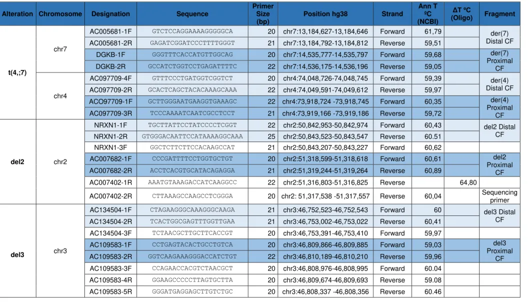
4.4.1. Sequence-specific oligonucleotide design for the control and junction fragments of eachchromosomal rearrangement ... 25
4.4.2. PCR amplification of Control and Junction Fragments... 28
4.4.3. PCR fragments purification and Automated direct dideoxy sequencing - Sanger sequencing adaptation ... 33
4.5. Electroelution, a method for DNA fragment purification ... 34
4.6. Culture, Extraction and Characterization of plasmids for Establishment of Urinary Cell Lines……….…….35
4.7. Hi-C – Studies about Chromatin Conformation ... 37
5. Results and Discussion ... 39
5.1. Clinical description ... 39
5.2. Cytogenetic analyses ... 39
5.3. Identification of structural chromosome rearrangements by large insert whole genome sequencing (liWGS) ... 41
5.4. Determination of the chromosomal breakpoints with nucleotide resolution ... 42
5.4.1. Translocation between 4q13.3 and 7p21.2 ... 42
5.4.2. Deletion on 2p16.3 ... 45
5.4.3. Deletion on 3p21.1 ... 47
5.4.4. Deletions on chromosomes 9 and 11 ... 49
5.5. Identification and characterization of the candidate genes ... 50
5.5.1. Identification of the candidate genes from 4q13.3 breakpoint region... 50
5.5.2. Identification of the candidate genes from 7p21.2 breakpoint region... 53
5.5.3. Identification of the candidate genes from 2p16.3 breakpoint region... 55
5.5.4. Identification of the candidate genes from 3p21.1 breakpoint region... 58
5.6. Molecular pathogenesis – Protein coding Genes located on TADs interrupted by the Chromosomal Rearrangements and their relation with the clinical phenotype ... 58
5.7. Mechanisms underlying the formation of t(4;7)(q13.3;p21.2) ... 65
5.8. Electroelution assays for isolation of DNA fragments ... 67
6. Conclusion ... 69
7. Future work ... 73
8. References ... 75
xv
Appendix 1 – Establishment of permanent growing lymphoblastoid cell lines (Adapted from Neitzel,1986)………...………..81
Appendix 2 –“PAXgene® Blood RNA Kit” RNA extraction from whole blood Workflow (Adapted from PAXgene® Blood RNA Kit Handbook (PreAnalytiX 2015)) ... 84
Appendix 3 –“QIAamp® DNA blood midi kit” DNA extraction Workflow (Adapted from QIAamp® DNA Blood Midi/Maxi Handbook (QIAGEN 2015)) ... 85
Appendix 4 – “QIAamp® RNA Blood Mini” RNA extraction from LCL Workflow (Adapted from QIAamp® RNA Blood Mini Handbook (QIAGEN 2010)) ... 86
Appendix 5 – “Amicon® Ultra-0.5 Centrifugal Filter Devices” PCR product purification Workflow (Adapted from Amicon® Ultra-0.5 Centrifugal Filter Devices user guide (Millipore 2009)) ... 87
Appendix 6 –“ZR Plasmid MiniprepTM - Classic” Plasmid extraction from Miniprep Cultured Bacteria Workflow (Adapted from ZR Plasmid Miniprep™-Classic (Zymo Research Corporation 2013)) ... 88
Appendix 7 –“HiSpeed® Plasmid Purification” Plasmid extraction from Midiprep Cultured Bacteria Workflow (Adapted from HiSpeed® Plasmid Purification Handbook, (QIAGEN 2012)) ... 89
Appendix 8 – Complete protocol for Plasmid Culture and Isolation (Adapted from Addgene and HiSpeed® Plasmid Purification Handbook, 2012) ... 90
Appendix 9 – in situ Hi-C Nuclei Isolation and Staining protocol for light microscopy (based on Belaghzal et al. 2016; Rao et al. 2014 and Ramani et al. 2016 protocols) ... 94
Appendix 10 – in situ Hi-C Protocol Developed for the First partial Experimental Assay (based on Belaghzal et al. 2016; Rao et al. 2014 and Ramani et al. 2016 protocols) ... 95
xvii
List of Figures
Figure 1.1 – Classification of Chromosomal Aberrations. ... 2
Figure 1.2 – Map of genic overlap for neurodevelopmental disorders obtained by replication process and combination of CNV morbidity maps. (adapted from “Genetic studies in intellectual disability and related disorders” - Vissers et al. 2015) ... 13
Figure 1.3 – Summary of the strategies used nowadays to define the genetic architecture of chromosomal rearrangement after Karyotyping. (adapted from “Genetics of Congenital Heart Disease: The Glass Half Empty” - Akl C. Fahed et al. 2014.) ... 14
Figure 1.4 – Overview of Hi-C method. (adapted from “Hi‐C 2.0: An Optimized Hi-C procedure for High Resolution Genome Wide Mapping of chromosome conformation” - Belaghzal et al. 2016) ... 18
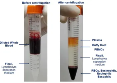
Figure 4.1 – Representation of Ficoll Gradient before and after centrifugation, for isolation of PBMCs to obtain LCLs. ... 23
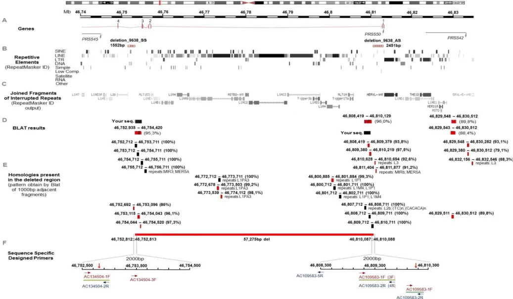
Figure 4.2 – Overview of the genomic region deleted on chromosome 3. ... 26
Figure 4.3 – Diagrammatic representation of cycle sequencing reaction, where the fluorescent ddNTPs were incorporated into the sequencing products. (adapted from Applied Biosystems chemistry guide – DNA sequencing by Capillary Electrophoresis) ... 33
Figure 4.4 – Bacterial growth plates after overnight incubation. ... 35
Figure 5.1 – Pedigree of the family in study with indication of the cytogenetic analyses results for each individual. T ... 40
Figure 5.2 – PCR confirmation of t(4;7)(q13.3;p21,2) of the proband. ... 43
Figure 5.3 – PCR confirmation of the familial study performed about t(4;7)(q13.3;p21,2). PCR of control and junction fragments was carried out from index and his mother DNA samples... 43
Figure 5.4 – Ideograms illustrating the derivate chromosomes resulted from t(4;7)(q13.3;p21.2). ... 44
Figure 5.5 – Nucleotide sequences of der(4) breakpoint aligned against the reference sequence. ... 45
Figure 5.6 – Nucleotide sequences of der(7) breakpoint aligned against the reference sequence. ... 45
Figure 5.7 – PCR confirmation of the familial study performed about del2p16.3. PCR of control and junction fragments was carried out from index and his mother DNA samples. ... 46
Figure 5.8 – Ideogram illustrating chromosome 2 with a deletion on 2p16.3. ... 46
Figure 5.9 – Nucleotide sequences of del2 breakpoint aligned against the reference sequence... 47
Figure 5.10 – PCR results from the amplification of control and junction fragments of del3p21.1 from a proband sample. ... 48
Figure 5.11 – PCR results from the amplification of control and junction fragments of del3p21.1 from a control sample. ... 49
Figure 5.12 – Structural variants present on the genomic region chr9:25,258,355-25,352,944 ... 49
Figure 5.13 – Structural variants present on the genomic region chr11:25,680,453-25,699,601. ... 50
Figure 5.14 – Overview of der(4) breakpoints genomic region. ... 51
Figure 5.15 – Overview of der(7) breakpoints genomic region. ... 54
xix
List of Tables
Table 1.1 – List of most common structural chromosomal abnormalities. ... 4
Table 1.2 – Genes causative of congenital heart defects. (adapted from “Genetics of Congenital Heart Disease” - Richards and Garg 2010) ... 9
Table 1.3 – Most recurrent CNVs associated with non-syndromic congenital Heart Defects. (adapted from “Genetics of Congenital Heart Disease: The Glass Half Empty” - Akl C. Fahed et al. 2014.) ... 10
Table 4.1 – Summary of the designed primers for this case study. Specifications of each primer, like its sequence, size, strand, position and annealing temperature are enumerated. ... 27
Table 4.2 – Composition of PCR master-mix made to this study... 28
Table 4.3 – Primer combination for amplification of control and junction fragments of der(4). ... 28
Table 4.4 – Primer combination for amplification of control and junction fragments of der(7). ... 29
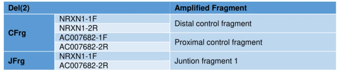
Table 4.5 – Primer combination for amplification of control and junction fragments of del2. ... 30
Table 4.6 – New Junction fragment amplification option with a new reverse primer only designed for reverse sanger sequencing of del2 junction fragment. ... 31
Table 4.7 – Primer combination for amplification of control and junction fragments of del3. ... 32
Table 5.1 – Summary of the results from the bioinformatic analysis carried out on the data received from liWGS of proband's genome. (reference sequence: human genome assembly [GRCh38/hg38]) ... 41
Table 5.2 – Protein coding genes located on the two TADs interrupted by t(4;7)(q13.3;p21.2) breakpoints. Genome version [hg38]. TADs are according to Dixon et al. 2012. ... 60
Table 5.3 – Protein coding genes located on the two TADs interrupted by del2(p16.3;p16.3) breakpoints. Genome version [hg38]. TADs are according to Dixon et al. 2012. ... 61
xxi
List of Abbreviations
3C – Chromosome Conformation Capture
3D – Three Dimensional
aCGH – Comparative genomic hybridization array
ACTC1 – Actin alpha cardiac muscle
AFP – Alpha-fetoprotein
AIFM1 – Apoptosis inducing factor mitochondria associated 1
ALB – Albumin
ASD – Autism Spectrum Disorder
BACs – Bacterial artificial chromosomes
BCR – Balanced Chromosomal Rearrangements
CCDC88C – Coiled-coil domain containing 88C
CCR – Complex Chromosomal Rearrangement
CD36 – CD36 molecule
CGH – Comparative Genomic Hybridization
CHD – Congenital Heart Defect
chr – Chromosome
CIT – Citron Rho interacting serine/threonine kinase
CITED2 – Cbp/P300 interacting transactivator with Glu/Asp rich carboxy terminal domain 2
CMV – Cytomegalovirus
CNV – Copy Number Variation
COL1A1 – Collagen type I alpha 1 chain
COL1A2 – Collagen type I alpha 2 chain
CRTAP – Cartilage associated protein
CXCL3 – Chemokine, C-X-C motif, ligand 3
CXCL5 – Chemokine, C-X-C motif, ligand 5
CXCL8 – Chemokine, C-X-C motif, ligand 5
del – Deletion
xxii
DGKB – Diacylglycerol beta, 90kDDMSO – Dimethyl sulfoxide
DNA – Deoxyribonucleic Acid
dNTPs – Deoxynucleotides
dup – Duplication
EBV – Epstein-Barr virus
EDTA – Ethylenediamine tetraacetic acid
ELN – Elastin
ETV1 – E-twenty-six variant gene 1
EUROCAT – European surveillance of congenital anomalies
FISH – Fluorescence in situ Hybridization FLNA – Filamin A
FMR1 – Fragile X mental retardation 1
FSHR – Follicle stimulating hormone receptor
GABRB3 – Gamma-aminobutyric acid type A receptor beta 3 sebunit
GATA4 – GATA binding protein 4
GTL – Giemsa-Trypsin-leishman
Hi – Haploinsufficiency index
hTERT – Telomerase catalytic subunit gene
ID – Intellectual Disability
ins – Insertion
INSA, I.P. – Instituto Nacional de Saúde Doutor Ricardo Jorge, Instituto Público
inv – Inversion
IQ – Intelligence Quotient
JAG1 – Jagged 1
KRAS – KRAS proto oncogene, GTPase
LB – Lysogeny broth
LCL – Lymphoblastoid Cell Line
LHGCR – Luteinizing hormone/ choriogonadotropin receptor
xxiii
LOH – Loss of heterozygositylow-TE – Low-Tris- Ethylenediamine tetraacetic acid
MECP2 – Methyl CpG binding protein 2
MEF2C – Myocyte enhancer factor 2C
MYH6 – Myosin heavy chain 6
NCBI – National Center for biotechnology information
NDD – Neurodevelopmental Disorder
NF1 – Neurofibromin 1
NGS – Next Generation Sequencing
NLGN2 – Neuroligin 2
NLGN3 – Neuroligin 3
NOTCH1 – Notch 1
NOTCH2 – Notch 2
NRAS – NRAS proto-oncogene, GTPase
NRXN1 – Neurexin 1
NX2-5 – NK2 Homeobox 5
PACs – P1 artificial chromosomes
PBMCs – Peripheral Blood Mononuclear Cells
PBS – Phosphate Buffered Saline
PF4 – Platelet factor 4
PMSE – Proteasome activator subunit 4
PNPT1 – Polyribonucleotide nucleotidyltransferase 1
PPBP – Pro-platelet basic protein
PRSS50 – Protease serine 50
PTEN – Phosphatase and tensin homolog
PTH1R – Parathyroid hormone 1 receptor
RPMI – Roswell ParK memorial institute
RPS27A – Ribosomal protein S27A
RTN4 – Neurite outgrowth inhibitor (reticulon 4) clathrin heavy chain linker domain containing I
xxiv
SHANK3 – SH3 and multiple Ankyrin repeat domains 3SNP – Single Nucleotide Polymorphism
SNV – Single Nucleotide Variant
SOS1 – SOS RAS/RAC guanine nucleotide exchange factor 1
SOX3 – SRY-box 3
SV40 – Simian virus 40
TAD – Topological association domain
TAE – Tris-Acetate-EDTA
TBE – Tris-Borate-EDTA
TBX20 – T-box 20
TBX5 – T-box 5
TEAD1 – TEA domain transcription factor 1
TLL1 – Tolloid like 1
TNFSF11 – TNF superfamily member 11
TPP2 – Tripeptidyl Peptidase 2
TSC1 – Tuberous sclerosis 1
TSC2 – Tuberous sclerosis 2
TUBA1A – Tubulin alpha 1 a
TUBB3 – Tubulin beta 3 class III
UBE3A – Ubiquitin protein ligase E3A
UCSC – University of California, Santa Cruz
UV – Ultraviolet
WGS – Whole genome sequencing
xxv
List of Symbols
~ – Approximately
% – Percentage
%(v/v) – Volume per volume percentage
%(wt/v) – Weight per volume percentage
bp – Base pair
F – Forward
Kb – Kilo-base pair
M – Molar
Mb – Mega-base pair
mM – Millimolar
n – Haploid number, refers to the number of chromosomes (n=23)
OD – Optical density
p – Chromosome short arm
q – Chromosome long arm
R – Reverse
rpm – Rotations per minute
t – chromosomal translocation
U – Enzyme units
X – Chromosome X
xg – Relative centrifugal force
1
1. Introduction
1.1.
Considerations about the Human Genome and its organization
The Human Genome is the set of all the genetic information related to Homo Sapiens specie, codified on more or less 3 billion DNA base pairs allotted in 46 chromosomes. (Gardner, R.J. Mckinlay; Sutherland, GranT R.; Shaffer 2012; National Library of Medicine 2017). These 46 chromosomes are organized in 22 matching pairs and two sex chromosomes. One homolog of each pair is inherited from the mother and the other one is inherited from the father. (Gardner, R.J. Mckinlay; Sutherland, GranT R.; Shaffer 2012)
The 22 matching pairs correspond to Autosomes and the remaining ones correspond to the Sex Chromosomes or Genosome. Although autosomes’ homologs have the same morphology in both sexes, sex chromosomes are different in males and females. Females have a pair of X while males have a X and a Y chromosome. A single set of homologs, one of each autosome, plus one of the sex chromosomes, is the haploid set (n=23). That only exists, as such, in gametocytes. All the other cells in the organism, somatic cells, have the diploid set (2n=46). (Gardner, R.J. Mckinlay; Sutherland, GranT R.; Shaffer 2012)
Chromosomes are formed by chromatin, a complex of DNA and proteins, in its condensed state. There are different condensed forms of chromatin: euchromatin corresponds to the less condensed form, and constitutes coding DNA – genes; on the other hand, heterochromatin is the most condensed form, and constitutes the non-coding DNA present in centromeres and telomeres. DNA associated proteins, histones and non-histones, provide scaffolding of chromosomes. (Gardner, R.J. Mckinlay; Sutherland, GranT R.; Shaffer 2012) Histones are strongly conserved alkaline proteins, their positive charges allow them to associate with DNA, which is negatively charged, and help condense it into chromatin fibres. Those fibres loop around some histones that function like spools and form nucleosomes. (Nature Education 2014)
Each chromosome has 2 arms, capped at terminal extremities by telomeres, and divided by the centromere. The shorter arms are designated as p while the longer arms are named as q. The centromere is a specialized region of non-coding DNA that, at mitosis, provides the site at which the spindle apparatus can be anchored and draw each separated chromatid to the opposite poles of the dividing cell. Telomeres are specialized non-coding DNA sequences comprising many repeats of the sequence “TTAGGG” that confers stable ends to chromosomes preventing its fusion with the chromatin of other chromosomes. (Gardner, R.J. Mckinlay; Sutherland, GranT R.; Shaffer 2012; Nature Education 2014)
2
1.2.
Chromosomal abnormalities
Since chromosomes contain the genetic information essential to form an healthy human being, any change in the number or structure of chromosomes may lead to death, mental retardation, a variety of genetic syndromes, infertility and spontaneous abortions. (Barbara H. Czepulkowski 2001; Baht and Wani 2017)
The structural features and the architecture of the human genome can induce genomic instability due to the presence of specific regions with high susceptibility to mutations and rearrangements. (Lupski and Stankiewicz 2005)
Is important to note that certain parts of some chromosomes may show variation in population, a phenomenon known as heteromorphism. Watson-Crick base pair changes, that originate single nucleotide polymorphisms (SNPs), and by copy number variations (CNVs) are causes of Heteromorphism. (Lupski and Stankiewicz 2005; Zhang et al. 2009)
Chromosomal rearrangements occur by both homologous and non-homologous recombination mechanisms. Homologous mechanisms appear to be the predominant pathway for the formation of genomic rearrangements. (Lupski and Stankiewicz 2005)
Chromosomal abnormalities, in the form of deviations from the normal state of diploidy or in the form of structural rearrangements may lead to genetic imbalance, ending up in a variety of syndromes. (Baht and Wani 2017) Chromosomal abnormalities are classified as numerical or structural and each one of them be constitutional or acquired. An abnormality is considered constitutional if it arises during the embryonary development, being present in cells throughout the organism, and it is considered acquired if the individual was born without any cytogenetic alteration, having a normal karyotype and the chromosomal alteration appears in a subset of cells or tissues. Acquired chromosomal alterations are characteristic from processes like tumorigenesis. (Barbara H. Czepulkowski 2001; Gardner, R.J. Mckinlay; Sutherland, GranT R.; Shaffer 2012)
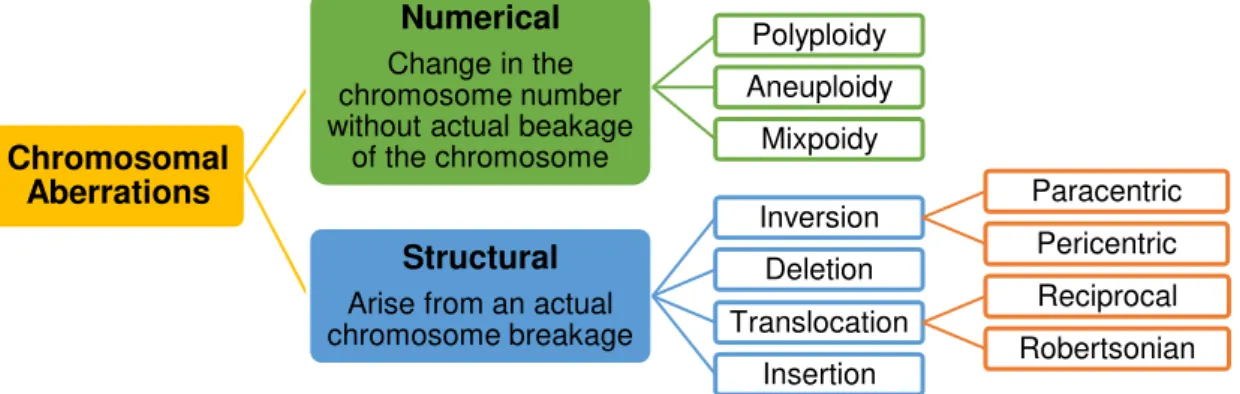
If an abnormality is large enough to be visible using the light microscope, being bigger than 4Mb in length, it is termed a cytogenetically visible. (Barbara H. Czepulkowski 2001) The different forms of observed chromosomal alterations, numerical and structural, are summarized in figure 1.1.
Figure 1.1 – Classification of Chromosomal Aberrations.
Chromosomal
Aberrations
Numerical
Change in the chromosome number without actual beakage
of the chromosome
Polyploidy Aneuploidy
Mixpoidy
Structural
3
Like previously said chromosomal rearrangements may lead to genomic imbalance. The pathogenic mechanisms that arise from chromosomal abnormalities, are:1. Gene dosage effect, caused by a lack (deletion, disruption of a gene) or excess (duplication) of chromosomal material, whether for a whole chromosome or a part of a chromosome;
2. Meiotic impairment, due to incongruent paternal origin of a chromosome or chromosomal segment;
3. A position effect, whereby a gene loss its normal functions when placed in a new chromosomal environment, this is caused for example by a translocation;
4. Combinations of the aforementioned. (Gardner, R.J. Mckinlay; Sutherland, GranT R.; Shaffer 2012; Baht and Wani 2017)
Genomic disorders are a class of human conditions that result from rearrangements of the human genome rather than from DNA sequence base changes. Those rearrangements convey traits or susceptibility to traits. (Lupski 2009)
1.2.1. Numerical Chromosomal abnormalities
Numerical chromosomal changes involve an alteration of chromosome number without actual breakage of the chromosome. These are subdivided in three different classes of alterations:
- Polyploidy (more than two paired homologous sets, this condition is not compatible with life);
- Aneuploidy (extra copies of a single chromosome or loss of one of both homologous);
- Mixploidy (someone who possesses two or more genetically different cell lines from a single zygote or occasionally because of chimerism). (Barbara H. Czepulkowski 2001)
1.2.2. Structural Chromosomal abnormalities
Structural chromosomal alterations are the result of chromosomal breakage in one or more chromosomes. There are lots of ways in which a chromosome can be altered after a breakage, so there are a large variety of structural chromosome changes. Breaks near the centromere are rapidly repaired by reparation enzymes. When the chromosome breaks in distal fragments terminal deletions are formed, because repair enzymes are not able to fix the breakage. (Barbara H. Czepulkowski 2001)
Breaks in more than one chromosome tend to disrupt the correct function of repair enzymes, which tend to join the wrong ends of chromosomes, being that the basis of the remaining structural chromosome anomalies. (Barbara H. Czepulkowski 2001) There are four types of alterations in chromosome structure, such as, deletions (del), duplications (dup), inversions (inv) and translocations (t). (Baht and Wani 2017)
4
under homozygous condition, while heterozygotes often have pairing difficulties in meiosis, leading to formation of inversion loops. (Barbara H. Czepulkowski 2001; Baht and Wani 2017)A deletion represents a missing segment on a chromosome. It occurs when chromosomal arms split, forming a chromosomal fragment and the remaining chromosome re-joins. The chromosomal fragment formed from the breakage is posteriorly degraded. (Barbara H. Czepulkowski 2001; Baht and Wani 2017)
A ring chromosome is formed in a situation of breakage occurs in either side of the centromere, and the two ends of the segment are joined together, forming a circular chromosomal fragment. (Barbara H. Czepulkowski 2001)
Translocation phenomenon takes place in a situation of breakage in two or more chromosomes, at the same time, and fragments from different chromosomes are relocated and re-joined to other chromosomes, resulting in abnormal chromosomes with genetic material from more than one chromosome. Translocations are subdivided in distinct classes: reciprocal translocations (translocations where the broken acentric fragments, distal to the two breakpoints, are exchanged) and centric or Robertsonian translocations (two acrocentric chromosomes break near the centromere and the large fragments of these chromosomes fuse together.). (Barbara H. Czepulkowski 2001; Baht and Wani 2017)
Insertions are the result of three breaks, two in one chromosome, producing an acentric fragment, which is then, inserted at a break formed in another chromosome. (Barbara H. Czepulkowski 2001). Table 1.1 summarizes and illustrates the different types of structural chromosomal aberrations.
Table 1.1 – List of most common structural chromosomal abnormalities.
Two Breaks in a single chromosome
Two or more breaks in different
chromosomes
Inversion
Paracentric
Pericentric
Reciprocal Translocation
Interstitial Deletion
Centric (Robertsonian) Translocation
5
Complex chromosomal rearrangements (CCRs) are rare, structural chromosomal abnormalities involving three or more cytogenetic breakpoints located on two or more chromosomes. (Hemmat et al. 2014; Pan et al. 2016)CCRs can be balanced (if there is no gain or loss of chromosomal material) or unbalanced (if there is gain or loss of chromosomal material), and are usually associated with significant risk of mental retardation and phenotypic anomalies attributable to gene disruption, cryptic imbalances, or position effects. (Hemmat et al. 2014)
Balanced chromosomal rearrangements (BCR) occur sporadically in the population or are segregate within families, with a frequency of 1 in every 2000 live births. The risk of developing congenital anomalies or neurodevelopmental disorders (NDDs) has been estimated to be 6.1 percent (%) for a the novo BCR. (Utami et al. 2014)
Up to 5% of reference haploid human genome is copied in DNA fragments bigger than 1 kilo-base pair (Kb) in size and with higher than 90% sequence identity to the human genome reference sequence. These fragments are termed Low copy repeats (LCRs). LCRs longer than 10Kb and of over approximately (∼) 97% sequence identity can lead to local genomic instability, and stimulate CNV formation. (Stankiewicz and Lupski 2010)
A CNV is a structural alteration that implies an imbalance on human genome. Its length may vary between 1Kb to several mega-base pair (Mb). CNVs perturb the normal biological balance of the diploid state at any given locus. Deletions, duplications, triplications, insertions, or translocations can result in CNVs. CNVs can be inherited or sporadic, and large de novo CNVs are thought more likely to be disease causative than the small ones. Phenotypic effects of CNVs are sometimes unclear and depend mainly on whether dosage-sensitive genes or regulatory sequences that are affected by the genomic rearrangement. (Stankiewicz and Lupski 2010)
1.3.
Initial considerations about Congenital Anomalies
Congenital anomalies, also known as birth defects, congenital disorders or congenital malformations, can be defined as structural or functional anomalies that occur during intrauterine life and that can be detected prenatally, at birth or sometimes only later in infancy. (World Health Organization 2017) Major Congenital Anomalies are those which are lethal, carry high mortality or have other serious medical functional consequences. Congenital malformations are a major cause of early spontaneous abortions, because some foetus malformations are incompatible with intrauterine survival. (Dolk et al. 2011; World Health Organization 2017)
Approximately 50% of all congenital anomalies cannot be linked to a specific cause, although there are some identified causes or risk factors associated with the development of these defects. (World Health Organization 2017)Both genetic and environmental factors are involved in the causation of congenital anomalies. (Dolk et al. 2011)
6
Structural chromosomal rearrangements, including CCRs, have been associated with multiple congenital abnormalities, including malformative syndromes and global developmental delay. (Houge et al. 2003; Kloosterman and Hochstenbach 2014). Usually there is a correlation between the number of chromosomal breakpoints and the severity of the phenotype, although is the genomic location of these breakpoints that accounts most for the phenotypical consequences. (Houge et al. 2003; Kloosterman and Hochstenbach 2014)A genetic abnormality is diagnosed by the clinician with the help of genetic tests and family history, where the results of the genetic tests explain why the children is malformed. Genetic syndromes, associated with chromosomal anomalies, microdeletions, single gene mutations or by genetic imprinting, account for less than one fifth of the total reported cases of congenital anomalies. (Dolk et al. 2011)
The Maternal exposure to certain pesticides and other chemicals, as well as certain medications, alcohol, tobacco, radiation and some pathogens during pregnancy, may increase the risk of having a foetus or neonate affected by congenital anomalies. Advanced maternal age is also a risk factor for abnormal intrauterine fetal development. (World Health Organization 2017)
In the vast majority of cases, the cause cannot be identified as one single factor, these include congenital anomalies of multifactorial origin. In these cases many genetic and environmental factors contribute additively, such that, the individual embryo/foetus surpasses a “threshold” beyond which it can no longer self-regulate to follow the normal developmental pattern. (Dolk et al. 2011)
The main challenge for genetic research is the interaction between genetic factors and environmental factors, such that, specific environmental exposures are only teratogenic in the presence of specific predisposing genetic factors. (Dolk et al. 2011)
European surveillance of congenital anomalies (EUROCAT) is the network of population-based registers of congenital anomalies in Europe, with a common protocol and data quality review, covering 1.5 million annual births in 22 countries. (Dolk et al. 2011) EUROCAT recorded a total prevalence of major congenital anomalies of 23.9 per 1000 births for 2003 to 2007, in which 80% were live births. The prevalence of chromosomal anomalies was 3.6 per 1000 births. Congenital heart defects (CHD) were the most common non-chromosomal subgroup, at 6.5 per 1000 births, followed by limb defects (3.8 per 1000) anomalies of urinary system (3.1 per 1000) and nervous system defects (2.3 per 1000). (Dolk et al. 2011)
1.3.1. Congenital Heart diseases
Congenital heart defects (CHD) are problems linked to the heart's structure that are present at birth. These defects can involve the interior walls of the heart, the valves inside the heart or the arteries and veins that carry blood to the body or to the heart. CHD might change the normal flow of blood through the heart. (National Heart)
7
many types of CHD that range from simple defects (with no symptoms) to complex defects (with severe, life-threatening symptoms). (National Heart) The incidence of these type of malformation is 19-75 per 1000 live births, and are also present in an even greater proportion of miscarriages. (Richards and Garg 2010)Although medical and surgical techniques for diagnosis and treatment of CHD have advanced through time, CHD aetiology is not completely understood. (National Heart; Richards and Garg 2010). Classical studies have found that CHD is multifactorial, accounting with genetic predisposition and environmental influence. (Richards and Garg 2010) However, sequencing the human genome and advances in molecular techniques led to increasing evidence of a stronger role of the genetic factors. (Richards and Garg 2010)
Non-genetic aetiologies of CHD include environmental teratogens (dioxins, polychlorinated biphenyls, pesticides), maternal exposures to alcohol, isotretinoin, thalidomide or anti-seizure medications, and infectious agents like rubella. Anti-retroviral and the epidemic of obesity with associated phenotypes of diabetes and hypercholesterolemia are nowadays recognized as emerging risk factors for CHD. (Akl C. Fahed et al. 2014)
Initial human genetics methodologies, like G-banding karyotyping, had poor resolution, which restricted analyses to inherited forms of CHD, and lead to a defective identification of the genes that might be linked to the presence of heart defects. (Akl C. Fahed et al. 2014; American Heart Association 2017)
Most children with CHD don’t have other birth defects. 25 to 40% of cases of CHD are associated with other anomalies or as part of an identified genetic syndrome. About 30% of children with chromosomal abnormalities have CHD. (Richards and Garg 2010)
Interatrial communications are the most common of the congenital heart defects, accounting for 6-8% of malformed hearts. (Orphanet 2017) Having a communication between atrial chambers does not imply having a deficiency on atrial septum 3/4 of all cases are the ostium secundum defect, characterized by a large aperture on foramen ovale site. Ostium primum defect accounts for 1/6 of defects, and constitutes an atrioventricular septal defect with shunting only at atrial level. 1/10 of the cases are classified as sinus venosus defect, that is a hole outside the confines of the oval fossa, being frequently associated with anomalous connection of the right pulmonary veins. Coronary sinus defect, is the rarest defect. It is due to absence of the walls that separate the sinus from the left atrium, being found at the mouth of the coronary sinus. (Orphanet 2017)
8
1.3.1.1.
Chromosomal rearrangements and Genes associated with CHD
In the past couple of decades, there has been a greater understanding on molecular pathways regulating cardiac development. Through gene target technology mouse models of a multitude of CHD were created, which led to the identification of transcriptional regulators, signalling molecules and structural genes critical for normal cardiac morphogenesis. Multiple genes were also identified as being controlled by these highly conserved molecular pathways. These investigations have assisted in the identification of the genetic aetiology of CHD, and provide evidence that many genes may have etiologic roles in CHD. (Richards and Garg 2010)
Nowadays, there are several methods available for genetic research of CHDs, including cytogenetic analysis, linkage and association studies, CNV and DNA micro-array analysis, and whole exome sequencing. The use of new technologies has increased the possibility of identification of new genes and chromosomal loci in syndromic and non-syndromic CHDs. (Grollmuss et al. 2016)
Each chromosomal anomaly is preferentially associated with specific types of CHDs. (Grollmuss et al. 2016) Numerical alterations in chromosomes account for a significant portion of CHD cases. Chromosomal aneuploidy, the first recognized genetic cause of CHD, continues to be a major aetiology today. 50% of individuals born with trisomy 21 have CHD, ranging from atrial and ventricular septal defects to atrioventricular canal lesions; 80% of individuals born with trisomy 13 have CHD, with heterotaxy and laterality defects becoming more common; nearly all individuals born with trisomy 18 have CHD; ~1/3 of females with monosomy X (Turner syndrome) have CHD, most common diagnoses include bicuspid aortic valve, aortic stenosis, hypoplastic left heart syndrome and coarctation of the aorta; ~1/5 of males 47, XXY (Klienfelter syndrome) have CHD, with patent ductus arteriosus and atrial septal defects prevailing. (Richards and Garg 2010)
Submicroscopic chromosomal anomalies associated with syndromic CHDs are the 22q11.2 deletions, causative of DiGeorge/velocardiofacial with conotruncal heart defects, the deletions on 7q11.23 causative of Williams syndromes with supravalvular aortic stenosis, the 5p deletions causative of cri-du-cha syndrome, with atrial and ventricular septal defects and patent ducts arteriosus, the 4p deletions causative of Wolf-Hirschhorn syndrome, the 8p deletion syndrome, the 10p deletions, the 11q deletions causative of Jacobsen syndrome, the deletion and duplication on 1q21.1, the 1p36 deletion syndrome and the deletions on 20p12 causative of Alagille syndrome, all with different combination of various cardiac defects. (Pierpont et al. 2007; Grollmuss et al. 2016)
It is important to note that interatrial communications, are the cardiac defect most shared between the previous identified genomic syndromes.
9
These developments on the technologies genomic technologies turned possible to identify that single-gene defects can lead to isolated CHD and reveal more about molecular pathways important for cardiac morphogenesis. (Richards and Garg 2010) On table 1.2 are summarized the genes associated to single-gene mutation induced CHD and the respective phenotype observed on patients.Table 1.2 – Genes causative of congenital heart defects. (adapted from “Genetics of Congenital Heart Disease” - Richards and Garg 2010)
Phenotypes Gene
Atrial septal defect, atrioventricular conduction delay, tetralogy of
Fallot, tricuspid valve abnormalities NKX2.5 Atrial septal defect, ventricular septal defect GATA4 Atrial septal defect, hypertrophic cardiomyopathy MYH6 Cardiac septation defects associated with pulmonary hypertension BMPR2
Endocardial cushion defects CRELD1, ALK2
Bicuspid aortic valve, early valve calcification NOTCH1 D – transposition of the great arteries PROSIT-240
It is not easy to define precisely the genetic defects underlying non-syndromic CHDs, due to the genetic and clinical heterogeneity of these malformations. (Grollmuss et al. 2016)
Through genotype-phenotype observations were concluded that CHD are not due to a global change in genomic content but rather from altered dose of specific genes. This concept gained more clarity with the development of methodologies, like array comparative genomic hybridization (aCGH) and whole genome sequencing, to define sub- chromosomal changes in genome structure, like CNVs. (Akl C. Fahed et al. 2014) Although is not easy to distinguish between pathogenetic CNVs and benign polymorphic variants, it was observed that the vast majority of individuals with CHD don’t have only single gene defects, but have unique CNVs associated with cardiac malformations. (Richards and Garg 2010; Grollmuss et al. 2016)
10
Table 1.3 – Most recurrent CNVs associated with non-syndromic congenital Heart Defects. (adapted from “Geneticsof Congenital Heart Disease: The Glass Half Empty” - Akl C. Fahed et al. 2014.)
Locus Size Range (Kb) Genes
1q21.1 418-3,981 PRKAB2, FM05, CHD1L, BCL9, ACP6, GJA5, CD160, PDZK1, NBPF11, FMO5, GJA8
3p25.1 175-12,380 RAF J, TMEM40
3q22.1-3q26.1 680-32,134 FOXL2, NPHP3,FAM62C, CEP70, FAIM, PIK3CB, FOXL2, BPESC1
4q22.1 45 PPM1K
5q14.1-5q14.3 4,937-5,454 EDIL3, VCAN, SSBP2, TMEM167A
5q35.3 264-1,777 CNOT6, GFPT2, FLT4, ZNF879, ZNF345C, ADAMTS2, NSD1
7q11.23 330-348 FKBP6
8p23.1 67-12,000 GATA4, NEIL2, FDFT1, CSTB, SOX7
9q34.3 190-263 NOTCH1, EHMT1
11p15.5 256-271 HRAS
13q14.11 555-1,430 TNFSF11
15q11.2 238-2,285 TUBGCP5, CYFIP1, NIPA2, NIPA1
16p13.11 1,414-2,903 MYH11
18q11.1-18q11.2 308-6,118 GATA6
19p13.3 52-805 MIER2, CNN2, FSTL3, PTBP1, WDR18, GNA11, S1PR4
Xp22.2 509-615 MID1
1.3.2. Neurocognitive and Neurodevelopmental Deficiencies
Cognition refers to the mental processes or thinking skills that allow people to learn and function in daily life, in society. These are the skills needed to process information, think, read, understand and solve problems. The main types of cognition include: attention, memory, processing information, solving problems, and planning/organizing. (Health)
NDDs are a group of conditions with onset on developmental period, with typical manifestation in early stages of development, and are characterized by developmental deficits that traduce impairments on personal, social, educational and occupational functioning. The range of these deficits varies from specific limitations, of learning, control or executive functions to global impairments of social skills or intelligence (American Psychiatric Association 2013)
11
It affects about 1-2% of the general population worldwide. ID is a broad diagnosis encompassing a wide variety of overlapping phenotypes and severities, it can range from mild to profound and can occur as an isolates phenotype or can be associated with either clinical symptoms or as part of a syndrome. (Vaidutis et al. 2016) Usually individuals with autism spectrum disorders also present intellectual disability. (American Psychiatric Association 2013)The causes of ID vary with the severity of the disorder: - moderate to severe cases (Intelligence Quotient (IQ) score <50) are much more likely to be due to a single pathological cause than are mild cases (IQ score 50-70) which are thought to be multifactorial in origin. Chromosomal and genetic disorders account for 30-40% of causes of moderate to severe mental retardation, environmental factors explain a further 10-30% and 40% of the cases have unknown causes. (Knight et al. 1999)
ID is often overlooked because of its association with other neuropsychiatric disorders such as autism, epilepsy and schizophrenia, a fact that also contributes to its near invisibility in global health programs. (Ropers 2010)
It is estimated that ∼1% of the human population has an autism spectrum disorder (ASD), being diagnosed in a 4/1 ratio male/female. (Zoghbi and Bear 2012; Samsam et al. 2014) ASD is characterized by persistent deficits in social communication and interaction across multiple contexts, excessively repetitive behaviors, restricted interests and insistence on sameness. (American Psychiatric Association 2013) The molecular pathogenesis of ASD is not already fully understood, since it has the contribution of a multitude of factors, constituting a multifactorial disease. (Samsam et al. 2014)
The evaluation of genetic causes of neurodevelopmental disorders remains challenging because these conditions are generally heterogeneous with many different genetic alterations resulting in clinically indistinguishable phenotypes. (Vaidutis et al. 2016)
1.3.2.1. Chromosomal Rearrangements and genes associated with Neurodevelopmental
Disorders
Cytogenetically visible chromosome aberrations account for ∼15% of all patients with severe ID. Submicroscopic deletions and duplications have recently emerged as an equally important, novel cause of ID. These are too small to be detectable under the microscope, and their identification is possible nowadays thanks to evolution of Molecular Cytogenetic Techniques and Next Generation Sequencing (NGS). (Ropers 2010) The usage of NGS methodologies like extensive gene panels, exome sequencing and the whole genome sequencing, allowed new ID genes to be identified in rapid succession. (Vissers et al. 2015)
There is some evidence that small chromosomal rearrangements involving the subtelomeric regions of chromosomes are also important causative of ID, being identified mostly on ID sporadic forms. (Rafati et al. 2012)
12
ligase E3A(UBE3A), methyl CpG binding protein 2(MECP2), phosphatase and tensin homolog(PTEN) and SH3 and multiple Ankyrin repeat domains 3 (SHANK3)genes, being those genes associated to several syndromes. (Zoghbi and Bear 2012) Rare mutations on neuroligins, neuroligin 3 (NLGN3), neuroligin 2 (NLGN2) and on neurexin 1, neurexin 1 (NRXN1) were also described as being a cause of autism. (Zoghbi and Bear 2012)Banerjee and his co-workers decribed the de novo translocation, t(1;2)(q31.3;p16.3)dn and the paternally inherited insertion, ins(16;2)(q22.1;p16.1p16.3)pat was being associated with ASD.
The presence of CNVs encompassing a variety of different genomic regions has been shown to be strongly associated with diseases such as autism, schizophrenia and developmental delay, along with being identified in individuals with multiple anomalies. (Richards and Garg 2010)
Performing comparative genomic hybridization array (aCGH) to scan the genome of autists and their healthy siblings’, has shown de novo, but individually rare, CNVs are significantly associated with autism. (Richards and Garg 2010) And more recently the microdeletion or reciprocal microduplication of 16p11.2 and 15q13 were identified to be also significantly associated with autism, being present in more than 1% of the cases. (Richards and Garg 2010; Banerjee et al. 2014)
Similar studies made about schizophrenia found out that the CNVs detected in schizophrenic patients modify genes involved in neurodevelopment. Multiple studies detected that abnormal chromosomal copy number is also associated with mental retardation associated with dysmorphism. (Richards and Garg 2010)
Proving pathogenicity of mutations observed in patients with intellectual disability and establishing the candidate ID genes (ID-causing gene when mutated) is still complicated, even in this post-NGS era, since it is a polygenic disability maybe with hundreds of genetic loci giving small contributions to the phenotype. (American Psychiatric Association 2013) Replication, which consist in the observation of multiple unrelated but phenotypically similar patients with mutations that are predicted to be damaging in the same gene, is still a key requirement in the process of identifying the candidate ID genes. (Vissers et al. 2015)
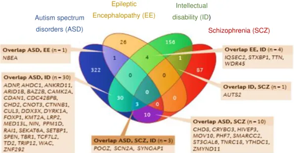
13
Figure 1.2 – Map of genic overlap for neurodevelopmental disorders obtained by replication process andcombination of CNV morbidity maps. (adapted from “Genetic studies in intellectual disability and related disorders”
- Vissers et al. 2015)
1.4.
Methods of identification of Chromosomal abnormalities
The characterization of constitutional chromosomal rearrangements is important for identification of the genetic aetiology of congenital abnormalities, and it is also fundamental for functional annotation of human genome. (David et al. 2013)
The combination of several molecular cytogenetic techniques with NGS constitutes a powerful methodology for characterization of chromosomal rearrangements. (Pan et al. 2016)
The advances on genomic technologies allows precise delineation of structural chromosomal rearrangements at nucleotide level, and since its results are becoming increasingly feasible, those techniques provide major improvements on genetic counselling. (Ordulu et al. 2014; Pan et al. 2016)
Karyotyping, aCGH and single nucleotide polymorphism array (array SNP) are the first-tier diagnostic tools to investigate chromosomal rearrangements. (Utami et al. 2014) BCRs are typically unidentified by low resolution methods, such as karyotyping. And since BCRs do not result in large gains or losses of genetic material at the breakpoint, molecular cytogenetic techniques, like microarrays, which detect CNVs at a considerable resolution fail in the identification on copy number neutral rearrangements. (Talkowski et al. 2011; Utami et al. 2014)
Most of times, interpretation of a rearrangement’s clinical impact is based upon a broad chromosome region, insensitive to the specific gene(s) disrupted or deregulated and neglecting the sequence complexity that might underlie these rearrangements. (Talkowski et al. 2011) Nowadays, with the development of molecular cytogenetics techniques and the advent of NGS, it is possible to define chromosomal breakpoints at nucleotide resolution, and different approaches can be combined as shown in figure 1.3 in order to better understand the architecture of chromosomal rearrangements.
Autism spectrum disorders (ASD)
Epileptic Encephalopathy (EE)
Intellectual disability (ID)
14
Figure 1.3 – Summary of the strategies used nowadays to define the genetic architecture of chromosomal rearrangement after Karyotyping. (adapted from “Genetics of Congenital Heart Disease: The Glass Half Empty” - Akl C. Fahed et al. 2014.)1.4.1. Classical Cytogenetics
Conventional cytogenetics consists in the analysis of chromosomes through microscopy techniques. It allows scanning the genome in the pursuit for aberrations that involve both gains and losses of portions of the genome, as well as rearrangements within and among chromosomes. (Pinkel and LI 2005)
Karyotyping, is one of the most common and easily accessible assays used to determine the chromosomal constitution of human cells. (Bates 2011) However it is unable to resolve structural rearrangements to the nucleotide level, since its overall resolution is greater than 5Mb, and its breakpoint determination resolution varies to 5 to 15Mb, which makes it difficult to analyse the actual pathological burden of the rearrangement. (Ordulu et al. 2014)
The analysis by karyotyping is only possible recurring to banding techniques, which consists in staining of metaphasic or pro-metaphasic chromosomes with a suitable dye like Giemsa. Staining patterns produced on the chromosomes are sometimes ambiguous, and the resolution is limited, both by the optical characteristics of microscopes and the complex manner in which DNA is packaged into chromosomes. (Pinkel and LI 2005)
Although banding techniques represent the central theme in every cytogenetics laboratory it is mandatory to recur to methodologies of genomic analysis with better resolution and less limitation, since specific identification of disrupted genomic region(s) is critical in diagnosis and management of constitutional and acquired rearrangements, especially as annotation of the human genome accelerates. (Wan 2010; Ordulu et al. 2014)
1.4.2. Molecular Cytogenetics
15
FISH analysis was managed to overcome many of the limitations of karyotyping, it is sensitive rapid and a critical complement to conventional cytogenetics. It is based on the capacity of single stranded DNA sequences hybridizes with a complementary DNA sequence, and consists in the use of fluorescent specific probes for specific genetic loci, such as telomeres, centromeres or sites of known gain or loss of DNA sequences. It turns possible the detection of both numerical and structural chromosome alterations. (Pinkel and LI 2005; Wan 2010)In diagnosis laboratory, the most useful FISH probes system is: centromere probes used for chromosomic enumeration (useful for the detection of numerical chromosomal alterations), chromosome painting probes (useful in deciphering major chromosomal rearrangements) and locus specific gene probes for gene fusions. (Wan 2010)
Although, FISH is an improvement to classical cytogenetic analysis, the complexity of the staining pattern that is produced is limited by the number of FISH probes that can be distinguished, and it has the same limitations of optical microscopy and chromosome structure that affect chromosome banding. (Pinkel and LI 2005)
Aberrations that involve gain or loss of chromosome segments are particularly important in medical cytogenetics. (Pinkel and LI 2005) Comparative genomic hybridization (CGH), is a technique based on quantitative tow colours fluorescence in-situ hybridization, and it allows the detection of genetic imbalances throughout the screening of the entire genome. (Pinkel and LI 2005; Wan 2010)
Using microarrays, the resolution of CGH can be much greater than with standard cytogenetics, by substitution of the hybridization targets on metaphasic chromosomes with genomic segments. These genomic segments can be bacterial artificial chromosome (BACs) or P1 artificial chromosome (PACs) clones for hybridization targets. Array CGH is a technology for “molecular karyotyping” with a resolution of 100Kb to 1Mb. This resolution can be improved by overlapping clones, turning possible to detect gains or losses as small as 40-80Kb. (Wan 2010)
The resolution and coverage of array CGH are dependent on the density of the array used. Microarrays with dense coverage of the entire genome allow screening for aberrations at any location without the need to have prior knowledge about where to test. (Pinkel and LI 2005; Wan 2010)
Thus, array CGH offers the ability to screen genomes of affected individuals to discover new aberrations, determine if apparently balanced translocations also involve gains or losses of DNA, establish phenotype-genotype relationships for conditions with variable aberrations, and provide comprehensive clinical diagnostic information. In combination with techniques such as chromosome microdissection or chromosome sorting, array CGH can map translocation breakpoints with high precision. (Pinkel and LI 2005)
16
FISH and/or High resolution chromosomal microarray studies have identified cryptic CCRs as a cause of abnormal phenotype in a significant number of patients with apparently balanced chromosomal rearrangements. (Hemmat et al. 2014)1.4.3. Next Generation Cytogenetics
“Next generation cytogenetics” is an integration of traditional cytogenetic techniques and the NGS. (Ordulu et al. 2014)
Sequencing breakpoints of structural chromosome rearrangements has been possible since 1980s, however, precise localization of these aberrations to the nucleotide level in a genome wide context only became feasible with recent improvements of sequencing techniques. (Ordulu et al. 2014)
NGS is a powerful tool for genomics research. It has allowed rapid characterization of genomes, exomes and transcriptomes. Whole-genome sequencing (WGS) is the most comprehensive method for analysing the genome. (Liang et al. 2014; illumina 2017)
Although the cost of performing WGS has decreased in recent years, it remains costly compared with other methods like exome and ribonucleic acid (RNA) sequencing (RNAseq). This fact triggeredfor the need of identifying an alternative WGS strategy for identifying the breakpoints that characterize structural variants and copy number changes. (Wan 2010)
Analysing the whole genome using NGS delivers a base-by-base view of all genomic alterations, including single nucleotide variants (SNV), insertions and deletions, copy number changes, and structural variations. (illumina 2017)
Several different approaches were described to perform massively parallel sequencing of large fragments, such as large-insert sequencing, mate-pair sequencing, and jumping libraries, which all represent similar approaches to sequencing large genomic inserts. (Talkowski et al. 2011)
Next generation paired-end whole-genome sequencing involves sequencing both ends of a DNA fragment with predetermined size and yields millions of paired short reads from the ends of these fragments. This increases the likelihood of alignment to the reference genome and facilitates detection of genomic rearrangements, repetitive sequences, and gene fusions. (Talkowski et al. 2011; Liang et al. 2014)
The application of this methodology requires the preparation of is the use of Illumina’s mate pair library. The standard protocol requires 10 milligrams (mg) of genomic DNA and supports the evaluation of regions spanning up to ~2–5Kb. (Liang et al. 2014)
1.5.
C-technologies and Studies of 3D organization of Human Genome
17
expression, regulation and other genomic activities. (Belaghzal et al. 2017) Unfortunately, the understanding of higher order genomic structure is coarse, fragmented or incomplete. (Dixon et al. 2012)Preforming studies about three-dimensional (3D) conformation and organization of chromatin allow the better understanding of spatial aspects of gene regulation, chromosome morphogenesis and genome stability and transmission. Also enhance the understanding of the biophysics of chromatin and further enable the investigation of pathologies related to genome instability or nuclear morphology. (Belton et al. 2013)
Recent Studies have revealed the existence of millions of potential cis-regulatory elements in human genome, with a great number of them residing in intergenic regions and away from their target gene promoters. (Schmitt et al. 2016) These elements, which largely consist in enhancers, influence the transcription of target genes through looping of chromatin fibbers during animal development, which has been described for many enhancers. (Schmitt et al. 2016)
There are many techniques available to observe the spatial organization of chromatin and these are classified into microscopic and molecular assays. (Belton et al. 2013)
Electron microscopy is used to analyse the architecture of the nucleus in nanometre (nm) scale detail, while fluorescent (light) microscopy provides information on the shape and distribution of specific chromosomes and chromosomal loci, as well as the co-association of specific loci with sub-nuclear compartments, with resolution of 50-100nm. (Belton et al. 2013)
Microscopic techniques have the major disadvantage of lack of connectivity to the genomic sequence, i.e. incredible fine resolution architecture cannot be assigned to a specific location in the genome. (Belton et al. 2013). The use of sequence specific fluorescent probes in FISH assays allows to go through limitations of microscopy, but these methods are still limited in throughput, allowing the analysis of only a few loci simultaneously. (Belton et al. 2013)
Molecular assays are based in the concept that dense matrices of chromatin interactions could be used to determine the spatial organization of chromatin domains, chromosomes and ultimately entire genomes. (Belaghzal et al. 2017) This concept was explained with the development of the methodology of chromosome conformation capture (3C) where the genomic sequence itself is the output, completely connecting chromosome structure and genomic sequence. (Belton et al. 2013; Belaghzal et al. 2017)
3C and its derived 3C-based technologies are commonly used for studying chromatin interactions in eukaryotic cells. These techniques have uncovered general features of genome organization, which include the existence of hierarchical chromatin structures such as compartments, topologically associating domains (TADs), sub-TADs, insulted domains and chromatin loops. (Schmitt et al. 2016)
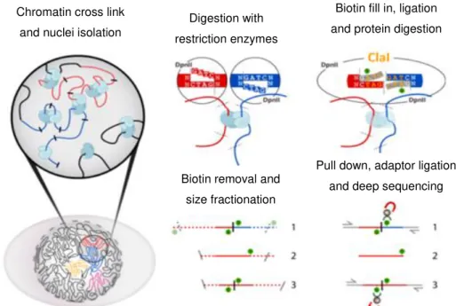
18
crosslinked DNA is then digested using restriction enzymes and the ends of the obtained DNA fragments are re-ligated in diluted conditions that strongly favour ligation of the juxtaposed DNA fragments. The frequency of ligation between two genomic loci is then assed using polymerase chain reaction (PCR) or direct DNA sequencing. (Schmitt et al. 2016) On figure 1.4 is a schematic representation of the whole process of Hi-C method. The overhangs left after digestion will depend on the chosen restriction enzyme.Figure 1.4 – Overview of Hi-C method. (adapted from “Hi‐C 2.0: An Optimized Hi-C procedure for High Resolution
Genome Wide Mapping of chromosome conformation” - Belaghzal et al. 2016) Digestion with
restriction enzymes
Biotin fill in, ligation and protein digestion
Biotin removal and size fractionation
Pull down, adaptor ligation and deep sequencing Chromatin cross link