0 1987 by The American Society for Biochemistry and Molecular Biology, Inc. Printed in U.S.A.
Mutant and Chimeric
Recombinant Plasminogen Activators
PRODUCTION IN EUKARYOTIC CELLS AND PRELIMINARY CHARACTERIZATION*
(Received for publication, December 23, 1986)
Laurent PierardS, Paul Jacobs$, Dirk Gheyseng, Marc HoylaertsS, Bruno Andre$,
Ljubica Topisirovic$, Alfred0 CravadorS, FranCoise de Forestat, Albert Herzog$, Desire CollenTl, Michel De Wildet, and Alex Bollen$
From the $Department of Applied Genetics, Uniuersiti Libre de Bruxelles, 24 rue de I’Zndustrie, B-1400 NiveUes, Belgium, the
§Molecular Genetics Department, Smith Kline RZT, 89 rue de I’Zmtitut, B-1330 Rixensart, Belgium, and the
TCenter for Thrombosis and Vascular Research, Department of Medical Research, University of Leuven, Campus Gasthuisberg,
49 Herestraat, B-3000 Leuven, Belgium
Mutant urokinase-type plasminogen activator (u-
PA) genes and hybrid genes between tissue-type plas-
minogen activator (t-PA) and u-PA have been designed
to direct the synthesis of new plasminogen activators and to investigate the structure-function relationship in these molecules. The following classes of constructs
were made starting from cDNA encoding human t-PA
or u-PA: 1) u-PA mutants in which the Arg16’ and LyslSs were substituted with threonine, thus prevent-
ing cleavage by thrombin and plasmin; 2) hybrid mol-
ecules in which the NH2-terminal regions of t-PA
(amino acid residues 1-67, 1-262, or 1-313) were
fused with the COOH-terminal region of u-PA (amino
acids 136-411, 139-411, or 195-411, respectively);
and 3) a hybrid molecule in which the second kringle
of t-PA (amino acids 173-262) was inserted between
amino acids 130 and 139 of u-PA. In all cases but one,
the recombinant proteins, produced by transfected eu- karyotic cells, were efficiently secreted in the culture
medium. The translation products have been tested for
their ability to activate plasminogen after in situ bind-
ing to an insolubilized monoclonal antibody directed
against urokinase. All recombinant enzymes were
shown to be active, except those in which LyslaS of u-
P A was substituted with threonine. Recombination of
structural regions derived from t-PA, such as the fin-
ger, the kringle 2, or most of the A-chain sequences,
with the protease part or the complete u-PA molecule
did not impair the catalytic activity of the hybrid poly-
peptides. This observation supports the hypothesis that
structural domains in t-PA and u-PA fold independ-
ently from one to another.
The fibrinolytic system plays a major role in the removal of insoluble fibrin from the vascular bed. It is triggered by the conversion of an inactive proenzyme, plasminogen, into the active enzyme, plasmin, which will degrade fibrin clots into
soluble components (1).
Among several plasminogen activators, two immunologi-
cally distinct enzymes, tissue-type plasminogen activator (t-
* This work has been funded through a research contract between the Walloon Region (Service des Technologies Nouvelles), the Uni- versity of Brussels, and Smith Kline RIT, Belgium. The costs of publication of this article were defrayed in part by the payment of page charges. This article must therefore be hereby marked “adver-
tisement” in accordance with 18 U.S.C. Section 1734 solely to indicate
this fact.
PA)’ and urokinase-type plasminogen activator (u-PA), have been extensively studied (for a review, see Ref. 2). The first one, t-PA, found to be identical to blood plasminogen acti-
vator (3), has been isolated from human uterus (4). The
second enzyme, u-PA, has been identified in human urine and kidney cells (5, 6).
Both proteins are serine proteases of 70,000 and 54,000
daltons, respectively, synthesized as single-chain polypeptides including a signal sequence involved in secretion (7-9). Single- chain plasminogen activators are processed by plasmin to form active enzymes composed of two disulfide-linked poly-
peptides. t-PA is cleaved at the Ar$78-Ile279 bond and single-
chain u-PA (scu-PA), primarily, between Lyslm and Ile’” residues. Secondary cleavages in the u-PA molecule occur at
the Arg’56-Phe’57 (with thrombin) (10) and L y ~ ’ ~ ~ - L y s ’ ~ ~
bonds, the latter event producing the low molecular size form of the enzyme (33,000 daltons) which has similar properties as the 54,000-dalton species (11).
Although both enzymes activate plasminogen, t-PA and u-
P A present different fibrinolytic properties. Indeed, plasmin- ogen activation by t-PA is highly fibrin-specific because the activator binds to the fibrin clot. Plasminogen then binds to both t-PA and fibrin, thus forming a cyclic ternary complex with increased stability (12). Both single-chain and two-chain
t-PA have very similar fibrinolytic efficacy; this implies that
the conversion of single-chain to two-chain t-PA at the sur-
face of the fibrin clot (13) has no physiological significance. On the contrary, two-chain u-PA displays little affinity for fibrin and activates free and fibrin-bound plasminogen equally well. Single-chain urokinase (scu-PA), which has been isolated recently by several groups (14-17), is a plasminogen activator with better fibrin specificity than u-PA (18-20). scu-
P A thus displays intrinsic plasminogen activator properties (21,22).
A comparison of the amino acid and nucleotide sequences of t-PA and u-PA reveals extensive homology between their B-chains (COOH-terminal regions) which carry the active site. The A-chains (NH2-terminal regions), however, differ in
some significant aspects (7-9, 23, 24). t-PA contains two
kringle domains, whereas u-PA has only one. These kringle
The abbreviations used are: t-PA, tissue-type plasminogen acti- vator; u-PA, urokinase-type plasminogen activator; scu-PA, single- chain u-PA ELISA, enzyme-linked immunosorbent assay; PBS, phosphate-buffered saline; SV40, simian virus 40; BGH, bovine growth hormone; bp, base pairs; kringle, triple loop disulfide-bonded structures occurring in t-PA (twice), u-PA (once), and plasminogen (five times); finger, amino-terminal region of t-PA, homologous to the finger-like domains in fibronectin; aa, amino acid(s).
11772
New
RecombinantPlasminogen Activators
domains are highly homologous to equivalent structures of plasminogen involved in fibrin-binding (25, 26). In addition,
the NH2-terminal region of t-PA contains a finger-like do-
main similar in structure to the fibrin-binding regions of
fibronectin (27, 28). The high affinity of t-PA for fibrin has
been attributed to the presence of both the finger and kringle
domains in the enzyme (29, 30). Because plasminogen acti-
vation by t-PA occurs with a low catalytic rate constant (k2
-
0.5 s-') (31), new plasminogen activators with both a highfibrin specificity and a high turnover rate constant might
constitute improved thrombolytic agents. Therefore, we have constructed, using recombinant DNA techniques, sequences coding for t-PA/scu-PA hybrids and for mutant scu-PA mol- ecules resistant to the cleavage by plasmin. In the present study, we found that the recombinant proteins, produced by transfected eukaryotic cells, are efficiently secreted in the culture medium and, in most instances, display specific activ- ities for the activation of plasminogen comparable to that of natural u-PA.
EXPERIMENTAL PROCEDURES~ RESULTS
Construction of Mutant u-PA and of Chimeric t-PA/u-PA
Coding Sequences-In an effort to improve thrombolytic se-
lectivity and fibrin specificity of plasminogen activators, a family of vectors carrying sequences encoding new plasmino- gen activators have been created through recombinant DNA
technology. As outlined in the Miniprint Section, the starting
material for the new constructs is carried by three plasmids: pULBlOOO and pULB1135 carry a preprourokinase cDNA (9)
and pDSPl.lTPA25.BGH carries a t-PA precursor cDNA.
The recombinant molecules derived from the manipulation of these DNAs share common features: they were all obtained
as HindIII-Sac1 cassettes carrying a 5'-terminal sequence
coding for a signal peptide and a 3'-terminal sequence corre-
sponding to the whole or partial B-chain of u-PA. In all cases,
the u-PA catalytic site has been maintained. The new con- structs, however, differed either in the nature of the A-chain or in the sequence coding for the activation site of the proen- zymes.
Full-length recombinant DNA molecules were obtained by subcloning various DNA fragments into the Hind111 and Sac1 sites of plasmid pULB1221 (42). When necessary, sequences joining DNA fragments of different origins were synthesized
chemically and added to the ligation mixtures. The conformity
of the recombinant DNAs to the expected sequences was then
checked by DNA sequencing before proceeding to the inser- tion of HindIII-Sac1 coding sequences into the eukaryotic transient expression vector pDSP1.1BGH (35), between the SV40 early promoter and the BGH polyadenylation signal (see Miniprint Section). Upon transfection in Chinese ham-
ster R1610 or/and Cos I monkey cells, recombinant plasmin-
ogen activators were produced and secreted in the culture medium.
Characteristics of recombinant plasminogen activators are shown in Table 2 and the new enzymes schematically repre-
sented in Fig. 2. The products can be classified in three main
*
Portions of this paper (including "Experimental Procedures," Fig.1, and Table 1) are presented in miniprint at the end of this paper. Miniprint is easily read with the aid of a standard magnifying glass. Full size photocopies are available from the Journal of Biological Chemistry, 9650 Rockville Pike, Bethesda, MD 20814. Request DOC- ument No. 86M 4391, cite the authors, and include a check or money order for $3.20 per set of photocopies. Full size photocopies are also included in the microfilm edition of the Journal that is available from Waverly Press.
groups. The first one consists of modified preprourokinase molecules (Table 2). Two of these enzymes, ppUK.410 and ppUK.410/366, coded for by pULB9122 and pULB9134, carry
amino acid substitutions in the B-chain as compared to
pULBlOOO and pULB1135. These modifications were intro- duced to assess their effect on enzymatic activity, in view of the reported discrepancies between the deduced amino acid sequence of cloned preprourokinase (9) and the sequence of the purified natural enzyme (23,24). Another set of constructs from the same group, Scupa n.c.410 and Scupa n.c.410/366 (coded for by pULB9129 and pULB9135), derives from the former molecules; additional amino acid substitutions have been introduced at the physiological activation site in the
proenzyme (Arg15'j and L Y S ' ~ ~ are respectively replaced by
threonine). The purpose of these constructions was to obtain prourokinase molecules with similar enzymatic properties as the natural single-chain species (scu-PA) (18-20), but resist- ant to cleavage by plasmin. The last constructs belonging
to the first group, pULB9139 and pULB9152 (coding for
ppUK.(410/366/13l)del and Scupa n.c.(410/366/131)del), were designed to eliminate the secondary cleavage site of urokinase ( L ~ s ' ~ ~ - L y s ' ~ ' j ) and to replace amino acid 131, tryp- tophan in pULBlOOO (9), by the cysteine residue found in the natural protein (23). This was achieved by deleting a stretch of amino acids (132-147) and replacing it with a shorter link (Ser-Thr) identical to the one found in t-PA at comparable positions of the enzyme. The product ppUK.(410/366/131)del, coded for by pULB9139, consists thus of a deleted but activ- ableprourokinase, whereas Scupa n.c.(410/366/131)del, coded for by pULB9152, is deleted and non-activable. Finally, for comparison purposes, we constructed a recombinant preprou-
rokinase DNA (pULB9154) identical to that described by
Heyneker et al. (8).
The second group of constructions comprises four chimeric molecules. Taking into account the hypothesis of exon shuf- fling as a mechanism for protein evolution (48), we tried to
recombine cDNA fragments, derived from t-PA and u-PA,
corresponding as precisely as possible to exons in the genes
and to structural domains in the corresponding proteins.
Fg.t-PA/UK.410 and Fg.t-PA/UK.410/366 (coded for by pULB9120 and pULB9124) result from the fusion of the
finger domain of t-PA to the COOH-terminus of scu-PA. The
two species are identical, except for the amino acid at position 366 in urokinase (glycine in pULB9124 and cysteine in
pULB9120). Both molecules were designed to explore the
potential role of the t-PA finger domain (29) in fibrin binding
when associated with scu-PA. Another molecule, tPPUK.410/ 366, encoded by plasmidpULB9151, combines a larger portion of t-PA, the A-chain, to the B-chain of u-PA; it is designed to confer to scu-PA the fibrin specificity of t-PA, which
appears to be associated to the A-chain moeity (49, 50). A
similar product, tPKUK.410 (coded for by pULB9125), con-
sists of the NH2-terminal part of t-PA containing the activa-
tion site, up to amino acid 313 in the B-chain, fused to the remaining COOH-terminal part of the B-chain of u-PA.
The single representative of the third group, UK-K2.410/ 366, coded for by plasmid pULB9137, is a nearly complete
scu-PA polypeptide wherein the kringle 2 region of t-PA has
been inserted between the single kringle domain and the B- chain. It was designed to test the hypothesis that the kringle
2 region of t-PA behaves as an autonomous domain conferring
fibrin binding ability to the enzyme (30).
Expression of Recombinant Plasminogen Activators in Cell
Cultures-Eukaryotic cells transfected with the recombinant plasmids described above were cultivated for 3-5 days in the presence of aprotinin to prevent conversion of the recombi-
TABLE I1
General description of recombinant plasminogen activators
Amino acid residue
Plasmid Product denomination position" Description
131 366 410 pULBlOOO pULB1135 pDSPl.lTPA25BGH pULB9122 pULB9134 pULB9154 PPUK PPUK t-PA ppUK.410 ppUK.410/366 ppUK.410/366/131 pULB9139 pULB9129 pULB9135 pULB9152 pULB9120 pULB9124 pULB9151 pULB9125 pULB9137 Scupa n.c.410 Scupa n.c.410/366 Scupa n.c.(410/366/131)del Fg.t-PA/UK.410 Fg.t-PA/UK.410/366 tPPUK.410/366 tPKUK.410 UK-K2.410/366 Val Val Ala Ala Ala Ala Ala Ala Ala Ala Ala Ala Ala Ala
Preprourokinase cDNA clone (9) Preprourokinase cDNA clone (9) t-PA cDNA clone
ppUK where Val"' has been re- placed by Ala
ppUK.410 where Cysm has been replaced with Gly
ppUK.410/366 where Trp131 has been replaced with Cys; iden- tical to preprourokinase previ- ously published (8, 23, 24) ppUK.410/366/131 where aa
132-147 have been replaced by Ser-Thr
ppUK.410 where aa 156 and 158 have been replaced by Thr in order to produce an uncleava- ble ppUK
Scupa n.c.410 where Cysm has been replaced by Gly
Scupa n.c. where aa 132 and 147 have been replaced by Ser-Thr t-PA sequence from the ATG co-
don to aa 67 followed by the sequence coding for aa 136 to
the stop codon of prouroki- Fg.t-PANK.410 where aa corre-
nase
sponding to position 366 of prourokinase has been re- placed by Gly
t-PA sequence from the ATG co- don to aa 262 followed by the sequence coding for aa 139 to the stop codon of prouroki- nase
t-PA sequence from the ATG co- don to aa 313 followed by the sequence coding for aa 195 to the stop codon of prouroki- nase
Sequence coding for kringle 2 of
t-PA (aa 173-262) inserted be- tween aa 130 and 139 of pre- Drourokinase
Amino acid (aa) positions relative to preprourokinase sequence; aa 131 belongs to the A-chain and aa 366 and
aa 410 to the B-chain.
nant plasminogen activators secreted in the medium. Dosage of the recombinant polypeptides by ELISA using two mono- clonal antibodies, AAU2 and AAU6 (46), reveals that all recombinant plasmids, except pULB9125, direct the transi- tory expression of urokinase-like material (Table 3). Culture
supernatants and extracts of cells transfected with pULB9125
(tPKUK.410) were consistently negative when assayed with anti-urokinase or anti-t-PA antibodies (data not shown).
Plasminogen Activation by Immobilized Recombinant Acti-
vators-The assay for plasminogen activation consisted of a
two-step procedure. First, standard urokinase or cell culture
supernatants were incubated with matrix-bound monoclonal antibody AAU2. Specific complexes were then exposed to plasminogen and to a plasmin-specific chromogenic substrate,
D-Ile-Pro-Arg-p-nitroanilide. Any plasmin resulting from the
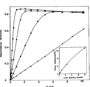
activation of plasminogen will thus react with the substrate and release the paranitroaniline chromophor which can be monitored at 405 nm. In all cases, typical sigmoidal curves
were observed when plotting absorbance as a function of time
( t ) (data not shown). As previously described by Drapier et al.
(47), plotting of AdO5 as a function of squared time
(e)
enablesus to linearize the assay as long as initial conditions are valid
(see Miniprint Section) (Fig. 3). The slope of these straight
lines is almost proportional to the total u-PA concentration present in the experimental standard incubation mixtures at
the moment of enzyme immobilization onto matrix-bound
antibody (Fig. 3, inset). From this relationship it is concluded
that, within the experimental range tested (0-8 IU/ml), the
amount of immobilized enzyme is proportional to the u-PA concentration in the upstanding solution. The dose-depend- ent plasminogen activation thus enables us to evaluate the enzymatic activity present in initial incubation mixtures.
Compared to the curves obtained with control u-PA (Fig. 3), those for all recombinant plasminogen activators and for
purified natural scu-PA appeared biphasic. As shown for three
11774
New Recombinant Plasminogen Activators
3 4 t I B 6 #-I I 6.-.
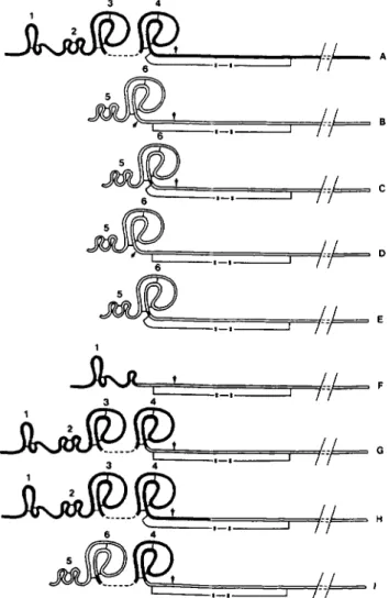
FIG. 2. Schematic representation of the A-chain structural
domains in recombinant plasminogen activators. Open and solid
lines correspond to sequences originating from scu-PA and t-PA,
respectively. The different recombinant plasminogen activators are
indicated by the following letters: A, t-PA; B, u-PA, ppUK.410,
ppUK/410/366, or ppUK/410/366/131; C, ppUK.(410/366/13l)de1;
D, Scupa n.c.410 or Scupa n.c.410/366; E, Scupa n.c.(410/366/13l)del;
F, Fg.t-PA/UK.410 or Fg.t-PA/UK.410/366; G, tPPUK.410/366; H,
tPKUK.410; I , UK-K2.410/366. Arrows point to cleavage sites. Rel-
evant disulfide bridges, in the A-chain and between the A- and B-
chains, are shown. For simplification purposes, B-chains are only
partially represented. Numbers refer to structural domains of A-
chains: I , finger domain of t-PA; 2, epidermal growth factor domain
of t-PA; 3, kringle 1 of t-PA; 4, kringle 2 domain of t-PA; 5, epidermal
growth factor domain of u-PA; 6, kringle domain of u-PA.
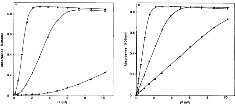
9134), the linear phase was preceeded by an exponential lag
phase (Fig. 4A). Pretreatment of immobilized recombinant
plasminogen activators with plasmin (or with trypsin) and careful elimination of the converting enzyme completely abol- ished this initial lag phase in subsequent plasminogen acti- vation experiments (Fig. 4B). Slopes measured in both exper- imental conditions were identical, considering the steep part of the curves only. The data thus indicate that the initial
phases, as observed in Fig. 4 A , correspond to the activation
of the immobilized recombinant activators which, under the experimental conditions, are harvested essentially as single-
chain molecules. This was confirmed by the fact that the same
lag phase was observed with standard one-chain urokinase purified from Calu-3 cell line (data not shown). Therefore, the enzymatic activity present in cell culture supernatants was determined by comparing the slopes of the linear part of
the curves obtained for recombinant enzymes (Fig. 4A) to the
standard u-PA system (Fig. 3). The enzyme activities are presented in Table 3 for all recombinant plasminogen acti- vators tested; activities ranged from 0 to 4.5 IU/ml of culture supernatant. Apparent specific activities of the recombinant plasminogen activators were obtained by the ratio of measured
activities to the amount of antigen (assuming that they display
similar affinities for the monoclonal antibodies AAU2 and AAU6 as the standard 54,000-dalton u-PA). As seen in Table 3, values range from 35,000 and 100,000 IU per mg of 54,000- dalton activator, except for non-activable scu-PA molecules. From these data, it can be concluded that two-chain recom- binant enzymes activate plasminogen with catalytic efficien- cies comparable to that of u-PA and, thus, that they have maintained a correct three-dimensional active site. On the other hand, no or only very weak activity has been found in
the supernatants of respectively R1610 and Cos I cells al-
though they expressed efficiently the non-activable scu-PA
molecules. As expected, the modification of the activation site
resulted in a single-chain product which cannot be trans- formed into the two-chain active species in the presence of plasminogen and chromogenic substrate or by plasmin (data not shown). Whether non-convertible scu-PA-like and natu- ral scu-PA will be able to activate plasminogen directly in a freely diffusing system remains to be determined.
DISCUSSION
Thrombolytic agents lacking fibrin specificity, such as u-
PA or streptokinase, induce thrombolysis but in association
with generalized plasminogen activation and fibrinogen
breakdown. t-PA induces thrombolysis with a high degree of
clot selectivity due to a markedly higher rate of plasminogen activation at the surface of the fibrin clot, as compared to rates observed in the absence of fibrin (12). Efficient and fibrin-selective thrombolysis has also been obtained with scu- PA, the single-chain precursor of u-PA (22). The mechanism of this selectivity is not fully understood but appears distinct
from that of t-PA.
One way to design improved thrombolytic agents would consist of the combination, in a single molecule, of two essential characteristics: high fibrin-mediated plasminogen activation and low fibrin-independent plasminogen activa-
tion. Such agents would be expected to display, in uiuo, a
fibrinolysis/fibrinogenolysis ratio at least equal or superior to
that of t-PA or scu-PA.
In the present study, three main groups of plasminogen activators have been produced; first, we constructed scu-PA- like molecules (Scupa n.c.410 and Scupa n.c.410/366) wherein conversion to two-chain urokinase was prevented by substi- tuting two amino acids involved in the cleavage of the natural scu-PA molecule. This approach finds its rationale in the fact
that the conversion of scu-PA into u-PA, in uiuo, is not a
prerequisite for thrombolysis, but leads to a loss in clot selectivity.
In a second approach, we recombined several domains de-
rived from the A-chain of t-PA with u-PA (in part or in toto).
Fg.t-PA/UK.410 and Fg.t-PA/UK.410/366 consist of the low
molecular weight scu-PA carrying on its NH2 terminus the
finger domain of t-PA; tPPUK.410/366 contains the A-chain
of t-PA fused to the low molecular weight scu-PA, and
tPKUK.410 is similar to tPPUK.410/366 but contains the cleavage site of t-PA. We hypothesized that some of these molecules might not only induce clot selectivity for plasmin- ogen activation, via the mechanism of scu-PA, but also en-
hance fibrin specificity by binding to the clot via their t-PA
structures. Another hybrid product, UK-K2.410/366, carries
Plasmid
TABLE I11
Expression levels and activity determinations of recombinant plasminogen activators Cell type R1610 cos I Gene product ELISA” supernatant Activity in Ib pULB9122 pULB9134 pULB9139 pULB9120 pULB9124 pULB9151 pULB9125 pULB9137 IIb pULB9134 pULB9129 pULB9135 pULB9151 pULB9137 IIIb pULB9122 pULB9154 pULB9135 ~ULB9152 ppUK.410 ppUK.410/366 ppUK.(410/366/13l)del Fg.t-PA/UK.410 Fg.t-PA/UK.410/366 tPPUK.410/366 tPKUK.410 UK-K2.410/366 ppUK.410/366 Scupa n.c.410 Scupa n.c.410/366 tPPUK.410/366 UK-K2.410/366 ppUK.410 ppUK.410/366/131 Scupa n.c.410/366 ScuDa n.c.(410/366/131)del 3.6 3 3.2 12 11.4 0 1.9 1.1 74.3 18 29 6.6 13.8 7.5 7.5 9 3.8 IU/ml 0.23 0.2 0.22 0.43 0.49 0.21 0 0.08 2.64 0 0 4.44 0.43 0.48 0.36 0 0 Appar- ent Activity in specific supernatant activity” IU/W w l m l IU/ml 62,400 66,000 68,900 35,300 43,000 109,900 0 69,000 35,500 48 0 2.28 40 0 0.12 96 0.57 66,300 21.5 1.34 31,250 7.3 0.38 63,500 8.4 0.62 48.000 6.9 0.52 Appar- ent activity” specific IU/mg 47,000 2,900 5,900 62,000 52,200 73,700 75,000 0 6.5 0.02 3,100 0 4 0.01 2,500 . . I .
Concentrations and apparent specific activities are given in 54,000-dalton urokinase equivalents. I, 11, and I11 refer to three independent transfection experiments.
FIG. 3. Kinetic analysis of plasminogen activation by puri- fied u-PA. Formation of paranitroanilide was recorded at 405 nm. The system contains purified u-PA linked to the monoclonal antibody AAU2, plasminogen (1 PM) and the plasmin-specific chromogenic substrate o-Ile-Pro-Arg-p-nitroanilide (0.5 mM). Data were plotted versus squared time. Slopes of the linear phases represent the accel- eration of paranitroanilide formation. Four concentrations of purified u-PA were assayed 0, 7.9 IU/ml; 0, 2.5 IU/ml; B, 0.79 IU/ml; A,
0.25 IU/ml. Inset, acceleration of pNA formation plotted versus u- PA activities. The curve can be used to evaluate the activity present in cell culture supernatants assayed on microtiter plates.
into the nearly complete scu-PA molecule; this form is ex-
pected to yield a potent urokinase-like plasminogen activator showing high fibrin affinity if, indeed, the kringle 2 behaves as an autonomous domain.
All recombinant plasminogen activators, except tPKUK.410,
were efficiently produced in cell cultures. In addition, specific
activities of recombinant two-chain u-PA and of chimeric polypeptides were comparable to that of natural u-PA, indi-
cating that the catalytic site carried by the urokinase moiety
of the molecules has been maintained and is fully functional for plasminogen activation.
The recombinant uncleavable scu-PA molecules (Scupa
n.c.410 and Scupa n.c.410/366), derived from transfected
R1610 cells, did not show any activity in our assay system.
However, the supernatants derived from transfected Cos I
cells exhibited a slightly higher level of activity than the
control. This is apparently due to the secretion by the cells of
an endogenous plasminogen activator. Indeed, pretreatment
of Cos I cell supernatant with plasmin confirmed this hypoth-
esis (data not shown). We showed also that plasmin was
unable to convert uncleavable scu-PA derived from trans-
fected R1610 cells into an amidolytically active species. This observation supports the conclusion that Scupa n.c. proteins are effectively stable one-chain molecules. Whether the ab-
sence of the activation site in the Scupa n.c. molecule has any
decisive influence on its biological in vitro and in vivo activi-
ties will be investigated in more detail once the recombinant product is obtained in large amounts and purified.
The scu-PA molecule encoded by the cDNA described in
Jacobs et al. (9) differs at three positions from the amino acid
sequence of the natural protein (23,24) and from the deduced
sequence derived from an independently isolated cDNA clone
(8). We showed that none of these differences had a significant
New Recombinant Plasminogen Activators
2 4 6 8 10 0.8-
0.6 In 0E,
3 P)c
9
SI
f 0.4 0.2 0FIG. 4. Kinetic analysis of plasminogen activation by recombinant ppUK.410/366 secreted in culture medium of transfected Cos I cells. In A , procedure was as for Fig. 3. Three dilutions of the cell culture
supernatant were tested 0, undiluted supernatant; 0, 3-fold dilution; W, 10-fold dilution. Slopes of linear phases were used to evaluate the activity of the supernatants by comparison to the standard curve shown in the inset of
Fig. 3. In E , procedure and dilutions of cell culture supernatant were as for A except that ppUK.410/366 linked to
the monoclonal antibody AAU2 was converted to its two-chain form, prior to the assay, by exposure to plasmin as described under “Experimental Procedures.” The data show that the lag phases observed in A are due to the conversion of one-chain ppUK.410/366 to its two-chain form.
vation capability. In addition, deleting the cleavage site re-
sponsible for conversion of 54,000-dalton u-PA to 33,000-
dalton u-PA ( L ~ s ’ ~ ~ - L y s ’ ~ ) was equally without effect.
A number of hypotheses might be tested once these mutant
and chimeric plasminogen activators have been produced in efficient host/vector systems, extensively purified, and char-
acterized. A first step in this direction has been already
achieved for two of our constructs, pULB9122 (ppUK.410)
and pULB9120 (Fg.t-PA/UK.410), and the data are presented
in the accompanying paper (51).
Acknowledgments-We are grateful to M. Massaer, A. Weyens, B.
Lambert, C. De Buyl, and J. Jottard for their help in the course of this work. We are endebted to Dr. M. E. Reff (Smith Kline & French, Molecular Genetics Department, 709 Swedeland Road, Swedeland, PA 19479) for providing the t-PA cDNA clone pDSPl.lTPA25BGH.
REFERENCES
1. Collen, D. (1980) Thromb. Haemostasis 43, 77-89
2. Collen, D., and Lijnen, H. R. (1986) Crit. Reu. Oncol. Hematol. 4,
3. Mullertz, S. (1953) Proc. SOC. Exp. Biol. Med. 82,291-297
4. Rijken, D. C., Wijngaards, G., Zaal-de-Jong, M., and Welbergen, 5. Mac Farlane, R. C., and Pilling, J. (1947) Nature 159, 779-784 6. Nolan, C., Hall, L. S., Barlow, G. H., and Tribby, I. I. E. (1977)
Bwchim. Bwphys. Acta 496,384-400
7. Pennica, D., Holmes, W. E., Kohr, W. J., Harkins, R. N., Vehar, G. A., Ward, C. A., Bennett, W. F., Yelverton, E., Seeburg, P. H., Heyneker, H. L., Goeddel, D. V., and Collen, D. (1983)
Nature 301,214-221
8. Heyneker, H. L., Holmes, W. E., and Vehar, G. A. (1983) Euro- pean Patent No. 0092182/A2, Bulletin 83/42, October 26, 1983 9. Jacobs, P., Cravador, A., Loriau, R., Brockly, F., Colau, B.,
Chuchana, P., Van Elsen, A., Herzog, A., and Bollen, A. (1985) 10. Ichinose, A., Fujikawa, K., and Suyama, T. (1986) J . BioZ. Chem. 11. Murano, G., and Aronson, D. L. (1979) Thromb. Haemostasis 42,
249-301
J. (1979) Biochim. Biophys. Acta 580, 140-153
D N A ( N Y ) 4, 139-145
26 1,3486-3489 1066-1068
12. Hoylaerts, M., Rijken, D. C., Lijnen, H. R., and Collen, D. (1982)
J. Biol. Chem. 257,2912-2919
13. Wallen, P., Bergsdorf, N., RHnby, M. (1982) Biochim. Biophys.
Acta 719,318-328
14. Nielsen, L. S., Hansen, J. G., Skriver, L., Wilson, E. L., Kaltoft, 6415
K., Zeuthen, J., and Dan@, K. (1982) Biochemistry 21, 6410- 15. Husain, S. S., Gurewich, V., and Lipinksi, B. (1983) Arch.
Bwchem. Biophys. 220,31-38
16. Wun, T.-C., Ossowski, L., and Reich, E. (1982) J. Bwl. Chem.
17. Stump, D. C., Lijnen, H. R., and Collen, D. (1986) J . Bwl. Chem.
18. Lijnen, H. R., Zamarron, C., Blaber, M., Winkler, M. E., and 19. Collen, D., Zamarron, C., Lijnen, H. R., and Hoylaerts, M. (1986) 20. Pannell, R., and Gurewich, V. (1986) Blood 67,1215-1223
21. Collen, D., Stassen, J. M., Blaber, M., Winkler, M., and Verstaete,
22. Van de Werf. F.. Masahiro. N., and Collen, D. (1986) Ann. Intern. 257,7262-7268
261,1274-1278
Collen, D. (1986) J . Biol. Chem. 261, 1253-1258 J. Bwl. Chem. 261, 1259-1266
M. (1984) Thromb. Haemostasis 52,27-30
Med. 104; 345-348
23. Guntzler, W. A., Steffens, G. J., Otting, F., Kim, S. M. A., Frankus, E., and Flohe, L. (1982) Hoppe-Seyler’s Z. Physwl.
. .
.~
Chem. 363,.1155-1165
24. Steffens, G. J., Guntzler, W. A., Otting, F., Frankus, E., and Flohe, L. (1982) Hoppe-Seyler’s Z. Physwl. Chem. 363, 1043-
1058
25. Lucas, M. A., Fretto, L. J., and McKee, P. A. (1983) J . BWZ.
Chem. 258,4249-4256
26. Vali, Z., and Patthy, L. (1984) J. Biol. Chem. 259, 13690-13694
27. Sekiguchi, K., Fukuda, M., and Hakamori, S. (1981) J . BwZ. Chem. 256,6452-6462
28. Petersen, T. E., Thogersen, H. C., Skorstengaard, K., Vibe- Pedersen, K., Sahl, P., Sottrup-Jensen, L., and Magnusson, S.
(1983) Proc. Natl. Acad. Sci. U. S. A. 80, 137-141
29. Banyai, L., Varadi, A., and Patthy, L. (1983) FEES Lett. 163, 30. Zonneveld, A.-J., Veerman, H., and Pannekoek, H. (1986) Proc. 31. Lijnen, H. R., Van Hoef, B., and Collen, D. (1984) Eur. J.
32. Deutsch, D. G., and Mertz, E. T. (1970) Science 170,1095-1096 37-41
Natl. Acad. Sci. U. S. A . 83,4670-4674
33. 34. 35. 36. 37. 38. 39. 40. 41. 42.
Heusterspreute, M., Vinh Ha Thi, E. S., Tournis-Gamble, S., Kennedy, N., and Davison, J. (1985) Gene ( A m t . ) 39,299-304 Davison, J., Heusterspreute, M., Merchez, M., and Brunel, F.
(1984) Gene (Amst.) 28,311-318
Pfarr, D. S., Sathe, G., and Reff, M. E. (1985) DNA (NY) 4,461- 467
Maniatis, T., Fritsch, E. F., and Sambrook, J. (1982) Molecular Cloning:A Laboratory Manual, Cold Spring Harbor Laboratory, Cold Spring Harbor, NY
Sinha, N. D., Biernat, J., and Koster, H. (1984) Nucleic Acids
Res. 12,4539-4557
Matteucci, M. D., and Caruthers, M. H. (1981) J. Am. Chem. SOC.
Froehler, B. C., and Matteucci, M. D. (1983) Tetrahedron Lett. Maxam, A. M., and Gilbert, W. (1977) Proc. Natl. Acad. Sci. U. Sanger, F., Nicklen, S., and Coulson, A. R. (1977) Proc. Natl. Jacobs, P., Cravador, A., Loriau, R., Herzog, A., and Bollen, A.
103,3185-3191 24,3171-3174 S. A. 74,560-564
Acad. Sci. U. S. A . 74,5463-5467
(1984) Belgian Patent Application Number 0/214048
43. Thirion, J.-P., Banville, D., and Noel, H. (1976) Genetics 83, 44. Gluzman, Y. (1981) Cell 2 3 , 175-182
45. Wigler, M., Sweet, R., Sim, G. K., Wold, B., Pellicer, A., Lacy,
E., Maniatis, T., Silverstein, S., and Axel, R. (1979) Cell 1 6 , 46. Herion, P., Portetelle, D., Franssen, J.-D., Urbain, J., and Bollen,
47. Drapier, J. C., Tenu, J. P., Lemaire, G., and Petit, J. F. (1979) 48. Gilbert, W., Marchionni, M., and McKnight, G. (1986) Cell 4 6 , 49. Rijken, C., and Groeneveld, E. (1986) J. Bwl. Chem. 2 6 1 , 3098-
137-147 777-785 A. (1983) Biosci. Rep. 3, 381-388 Biochimie (Paris) 61,463-471 151-154 3102
50. Holvoet, P., Lijnen, H. R., and Collen, D. (1986) Eur. J . Biochem. 158.173-177
51. Gheysen, D., Lijnen, H. R., Pierard, L., de Foresta, F., Demarsin,
E., Jacobs, P., De Wilde, M., Bollen, A., and Collen, D. (1987)
- , - ~
J. Biol. Chem. 262, 11714-11719
SUPPLEUENTARY MATERIAL
L O
M u t a n t and chimaeric recombrnant p l a s m i n o g e n a c f l v a f a r s
-Production i n e u k e r y o i i c cells and prellmlnarv charscterirsllon
by
P i e r a r d . L.. Jacobs. P., G h e y s e n , D . , Hoylaerto. M . .
Andre. B.. Topisirovic. L.. C r a v a d o r , A , . n e F o r e s t a . F..
Herzog. A , . C o l l e n . D . . De W i l d e , M. and Bollen. A.
EXPERIMENTAL PROCEDURES
uareria1s:
Restriction endonucleases. T4 DNA p ~ l y m e r a s e . T 4 D N A l i g a s e . Eocherichia
c o l i DNA polymerase I ( K l e n o u fragment) and T4 polynucleotide kinase w e r e
Biolsbo. Apiorlnln was f r o m Sigma. f e t a l c a l f s e r u m from F l o w laboraforiee purchased from Boehrlnger-Mannhelm Biochemxcals. Amersham o r N e w England and D - V a l - L e u - L y s - p - n i f r o a n i l i d e (S2251) f r o m Kablvltrum. Human ~ l a s n i n o ~ e n was purified 8 s dercrlbed (32). O n e and two-chaln urokinase w e r e prepared f G o m
human l u n g adenocarcinoma line Calu-I (ATCC, HTB-551 a s deecrlbed by S t u m p e t
al.(l7). Thelr a c f i v l t i e s w e r e determined u s i n g the r e f e r e n c e standard uraklnase o f Calblochem-Behring C a r p . T h e r e a c f i v l f l e s w e r e 7 0 , 0 0 0 IUImg and 7 9 , 1 0 0 I U I m g respectluely f o r one-chain and two-chain urokinase.
been described e a r l l e r . Plasmid p D S P l . l T P A Z 5 B G H , p r o v i d e d by D r . Reff. i s a
P l a s m i d s p U L B I000 (9). pULB 1118 (9), pJRDl84 ( 3 3 1 and pJRD158 ( 3 4 1 have pDSPl.1BGH-like plasmid wherein a f-PA cDNA has been inserted. Plasmid cloned ~n rhla plasmid a r e flanked by the S Y 4 0 e a r l y promoter end the B G H
pDSP1.lBGH (38) i s e transient expression v e c t o r f o r e u k a r y o t i c cells. G e n e s
polyadenylation s i t e . A11 recombinant g e n e s described in this p a p e r have been introduced i n pDSP1.lBGH a s Hind 1 1 1 - S a c I c a s s e L t e 9 ( f z g . I ) . The procedures
e l . ( 3 6 1 . O l i g o d e o x y n u c l e o t i d e s w e r e synthesized on a Applied B l o s y s f e m f o r DNA preparsrion and r e s r r l c t i o n a n a l y s i s w e r e as published by H a n l a f i s e t
S y n t h e s i z e r m o d e l 3 8 0 A v i a the solid-phase phosphorarnldite method 8 s
previously described (37,38,39). Llgaflon e n d bacterial & r a n s f o r m a r i o n o f e c c o r d i n g t o Uenlatio e t a 1 (16). The methods of M a x a m s o h G l l b e r f ( 4 0 ) and Escherichia coli K12 s t r a i n MH 2 9 4 (end*, thr .hsr-. hrm ) w e r e performed S e n g e r e t 8 1 . (41) w e r e used f o r DNA S e q u e n c e analysis.
p U L B 9 1 2 2
T h i s r e c o m b i n a n t p l e s m d c o d e s f o r p r e p r o u r o k i n a s e . If h a s been constructed a s f a l l o w s : B 1 4 4 0 bp 8 x 1 I - a 1 I DNA fragment v a s derived f r o m
plasmid pULB 1000. If encompasses the urokinase =DNA m o l e c u l e coding f o r a s 16
in the s l g n a l s e q u e n c e t o the stop codon and lncludes in its 3 ' terminus. the
poly C extension and 124 bp of pBRl22 s e q u e n c e s . T h i s f r a g m e n t was treated with T4 DNA polymerase (I61 and llgated on i t s 5 ' end L o a 21 bp Hind I I I - T a b l e 1 1 t m C O O S L I U L T t h e s e q u e n c e corresponding t o t h e ATG initiating coda; and the urokinase signal peptide U P t o a 8 -16. T h e m o l e c u l e coding f o r preprourokinese. ppUK. v a s then ligated (16) T O t h e i n t e r m e d i a t e E. C O I L
x 8 8 further manipulated firstly to r e m o v e exceeding I' p o l y C and pBR322 plasmid. pULB 1 2 2 1 (42). c u t with and u. The resulting plasmid
s e q u e n c e s , and secondly t o substitute the codon for V a l 410 present in pULB
1000 o r pULB 1 1 3 5 by a codon specifying Alanine. This was done by replacing a
2 1 1 bp BemH I-Sac I D N A fragment by a 21 bp double-stranded synthetic DNA
adaptor (adaptor 2.Table 1 ) coding f o r amino acld 398 t o the s t o p codon followed by B protruding end. A 1309 bp H i n d 1 1 1 - S a c I c a s s e t t e coding
for ppUK.410 was excised f r o m t h e r e s u l t i n g recombinant plasmld pULB 9117 and PULB 9122.
inserted i n f o the Y e C f o ~ pDSPl.1BGH. t o produce the f r n a l recomblnanf plasmid (blunt ended s i t e 1 double-stranded synthetic DNA adaptor (adaptor I
PULB 9129
This plasmid c a r r i e s t h e s e q u e n c e c o d l n g f o r preprauroklnase i n which the activation s x t e (Arg 156-Phe 157-Lyr 158) i s replaced b v Thr 156-Phe 157-Thr recovered. This fragment c a r r i e s o n i t s I' e n d , t h e ATG i n i r i a f n g c o d o n . t h e
1 5 8 . S r a r t l n g from plasmid pULB 9117. B 1 1 7 5 b p M - B a r n B I f r a g m e n t was s e q u e n c e coding f r o m aa-19 t o a a 1 4 9 o f p r e p r o u r a k ~ n a o e and. o n 1 f 8 5 ' end. the
s e q u e n c e coding for a a 1 9 8 t o the stop codon. those t w o r e g i o n s b e i n g
separated by the s e q u e n c e 8 o f pULB 1221 p r e s e n t i n the plaenid pULB 9117. On
the other hand. a 2 9 2 bp EcoR I - X h o I1 f r a g m e n t coding f o r a e 163 t o a * 260 o f
preprourokinase was derived f r o m PULB 9117. A thlrd f r a g m e n t , B 4 1 4 bp
-- piece f r o m PULB 9117. encoding aa 2 6 0 t o a a 3 9 8 o f t h e adaptor coding f o r as 149 t o o a 161 including the t w o a m i n o acid substit-
preprourokinase. was purlfled. At l a s t , a double-stranded 4 1 b p Bsl I-EcoR I
described above (adaptor 4. T a b l e I 1 w a s synfheaired. The f o u r f r a g m e n t s w e r e
lrgared to g e n e r a t e the recornblnanf plasmid pULB 9128. The Hind 111-Sac I
c a s s e t t e cadlng f o r S c u p a n.c.410 w a s recovered f r o m pULB 9 1 2 8 8 8 e 1309 bp fragment and xnrraduced i n t o pDSP1.1BGH. r e s u l t i n g i n pULB 9129.
I U L B 9 1 3 5
PULE 9135. I * ldentxcal f a pULB 9129 e x c e p t that the codon f o r Cysteine 3 6 6 has been replaced by a codon f a r G l y c i n e . The procedure used f a c o n s t r u c t
P U L E 9 1 3 5 i s I d e n t i c a l t o the o n e described for pULB 9114. The recombinant Biasmld pULB 9131 Codlnn f o r unacrlvable oreorouroklnese ( F r n n a n . r 1101366)
: - s a c I
11778
New Recombinant Plasminogen Activators
Table 1: S e q u e n c e s o f the s y n t h e t i c DNA adaDtor8. ADAPTOR 1 :
::
A G CTT ACC ATG AGA GCC CTG C 3 Met A r g Ala Leu L(eu!A TGG TAC TCT CGG GAC G 5' Hind I l l B g l I blunt ADAPTOR 2 :
iT
G ATC CGC ACT CAC ACC A A G G A A GAG AAT GGC CTG GCC CTC TGA G A G CT 3 'r)p Ile Arg Ser H i s Thr L y s G l u G l u A s n G l y Leu Ala Leu STOP
9 ' GCG TCA GTG TGG TTC CTT CTC TTA CCG GAC CGG G A G ACT C 5 '
BamHI s a c I
ADAPTOR 3 :
5 ' AC AAG CCC TCG AGT CCT CCA G A A G A A TTA AAA TTT CAG TGT G G 3
(T)yr Lys Pro Ser Ser P r o Pro G l u G l u Leu L y s Phe G l n Cys G l ( Y ?
3 ' TG TTC GGG AGC TCA G G A GGT CTT CTT AAT TTT A A A GTC ACA CC 5 '
Rsa I B a l I
ADAPTOR 4 :
(Gl)y G l n L y a Thr Leu Arg P r o Thr Phe Thr Ile Ile G l y Gly G(lu) 5' C CIA AAG ACT CTG AGG CCC ACC TTT ACC ATT ATT GGG GGA G
3 ' G GTT TTC TGA GAC TCC G G G TGG AAA TGG TAA TAA CCC CCT CTT AA
::
B e l I Eco R I
ADAPTOR 5 :
( G ) 1 y Pro Le" V a l cy9 ser Le" GI" Gly
5 ' G A CCC CTC GTC TGT TCC CTC CAA GGC
3 ' 66 GAG CAG ACA AGG G A G GTT CCG G
2:
Ava I1 Fnu4H I
ADAPTOR 6 :
;:e)r Pro Cys Trp Va(l) 3 ' G GGA ACG ACC CAC TAG
::
C CCT TGC TGG GT B c l I ADAPTOR 7 :
(P)m Leu V a l G l n G l u Cys Her V a l H i s Asp C y s S e r G l u Gl(7)
5 ' CG CTA GTA CAA GAG TGC ATG GTC CAT GAC TGC TCT GAG GG
9 ' C GAT CAT GTT CTC ACG TAC CAG GTA CTG ACG A G A CTC CCA GCT
::
F n u 4 H I S a l I ADAPTOR 8:
(T)yr Cys Asp V a l Pro Ser Cye Ser Ser Pro Pro G l u G l u Leu Lys Phe G l n Cys G l ( y )
5 ' AC TGT GAC GTC CCC AGC TGC TCG AGT CCT CCC G A G GAA CTT A A G TTT CAG TGT G G 3 ' 3 ' TG ACA CTG CAG GGG TCG ACG AGC TCA G G A G G G CTC CTT GAA TTC A A A GTC ACA CC 5 '
R S B I BAL I
ADAPTOR 9:
( P ) r o Leu V a l G l n G l u Cye Her V a l H i s Asp Cys S e r Thr Cys Gl(y) 5' CA CTA GTC CAA G A G TGC ATG GTG CAT GAC TGC TCC ACC TGT G G 3 ' 3 ' T GAT CAG GTT CTC ACG TIC CAC GTA CTG ACG AGG TGG ACA CC 5'
Ral I
ADAPTOR 108:
( P ) r o L e u V a l G l n G l u Cys M e t V a l His Asp Cys A l a ASP 5' CG CTT GTC CAA GAG TGC ATG GTG CAT GAC TGC GCA GAT
3 ' C G A A CAG GTT CTC ACG TAC CAC GTA CTG ACG CGT CTA CCT TTT TTC GGG
::
P""4H IADAPTOR lob:
5: GGA A A A AAG CCC TCC TCT CCT CCA G A A G A A TTA AAA TTT CAG TGT G G 3
G l y Lye Lya Pro Ser Ser Pro Pro G l u G l u L e u L y s Phe G l n Cys Gl(y! 3 AGG A G A G G A GGT CTT CTT AAT TTT A A A GTC ACA CC 5 '
B a l I
Double-stranded D N A adaptors w e r e s y n t h e s i z e d chemically a e single-stranded oligonucleotides and hybridized prior t o l i g a t i o n (36).
pULR 9139
pULB 9124
pULB 9124 I S ldentlcal t o pULB 9 1 1 0 e x c e p t t h a t t h e c o d o n c o r r e s p o n d l n y t o a a 3 6 6 of p r e p r o u r o k i n a s e has been replaced by a codon f o r g l y c l n e . We
ligated B 452 bp Hlnd 111-Bal I fragment purifled from pULB Yll5, encoding t h e
r e g i o n f r o m t h e ATG i n i f ~ a t l n g codon t o a a 6 7 of t-PA and a a 1 3 6 L o 1 4 9 o f
e n c a m p a s s l n g the l a r g e Hlnd 1 1 1 - S a c I f r a g m e n t o f p D S P L . 1 R G H and t h e
preprourokinase to B 6975 bp Hind 111- Bal I fragment from pULB 9 1 3 4 s e q u e n c e coding f o r a8 1 4 9 t o the s t o p codon of Dreprouroklnase.
DULB 9 1 3 7
pULR 9137 carries t h e gene coding for 8 " hybrld m o l e c u l e w h e r e t h e amino
acld s e q u e n c e corresponding to the k r l n g l e 2 of f-PA L E inserted b e t w e e n the
to 255 o f L-PA and for 8 8 1 3 9 L o 1 4 9 o f p r e p r a u r o k l n a s e w e r e exclsed. T h e s e 173 t o 1 7 4 of L - P A and 8 297 by Dde I-Bal I p i e c e f r o m p B A Z r o d l n ~ f o r a a 174
t w o f r a g m e n t s w e r e llgsfed t o a 3673 b p Nca I-Bel I DNA fragment derlved f r o m ATG i n l t l a t l n n c o d o n , for the s l g n a l p e p f ~ d e and far 1 t o 6 6 o f pULB 9119. T h i s l a s t p i e c e o f DNA carries o n 1 f 3 3 ' end the s e q u e n c e s for t h e
krlngle 2 o f t-PA and the p r o t e a s e m o e x t y of u r o k i n ~ ~ e . A second 822 b p
_Hlnd 111-EcoR I UNA _ _ ~f r a g m e n t , coding for the ATG c o d o n . t h e 5 1 ~ n a l s e q u e n c e
and e a 1 t o 2 0 4 o f f-PA , was obtained f r o m pDSPl.ITPA25BGH. T h e l a s t f r a g m e n t . 1 s a 6975 b p Hind 111-Bal I stretch from pULB 9134. This f r a g m e n t carries t h e c o d ~ n g s e q u e n c e f r o m a s I 4 9 t o the s t o p signal of preprourokinase
and a l l t h e s e q u e n c e s o t DDSPI.LBGH f r o m the t o UI. The t h r e e
f r a g m e n t s w e r e llgafed t o o b r a l n pULB 9151 which c a r r l e ~ a 1823 b p -11-
i ~ e f f e coding f o r tPPllK.4101366. C B I
by l l g a t l n g 5 DNA fragments dlrecfly to t h e 6 1 8 0 bp Hlnd Ill-Sac I fragment o f
195 t o t h e s t o p codon) o f preprouroklnase. The c o n s f r u r f ~ o n has b e e n achleved pDSPL.1BGH. The 5 f r a g m e n t s a r e :
~ a 8 2 2 bp H l n d 111-EcaR I f r a g m e n t f r o m pDSPl.ITPA258GH. If c o n r d l n s t h e ~ e q u e n c e s coding f o r t h e l n l r i a t i n g c o d o n , t h e s i g n a l p e p f ~ d e and a n 1 t o 204