UNIVERSIDADE DO ALGARVE
DEPARTAMENTO DE CIÊNCIAS
BIOMÉDICAS E MEDICINA
Investigating the role and function of Tribbles 2 (TRIB2) in drug
resistance within cancer
Laura Guerreiro Colaço
Dissertação para a obtenção do Grau de Mestre em Ciências Biomédicas
Trabalho efetuado sob a orientação de:
Professor Doutor Wolfgang Link
Professor Doutor Richard Hill
i
UNIVERSIDADE DO ALGARVE
DEPARTAMENTO DE CIÊNCIAS
BIOMÉDICAS E MEDICINA
Investigating the role and function of Tribbles 2 (TRIB2) in drug
resistance within cancer
Dissertação apresentada à Universidade do Algarve para cumprimento dos requisitos
necessários à obtenção do grau de Mestre em Ciências Biomédicas, realizada sob a
orientação do Professor Doutor Wolfgang Link e do Professor Doutor Richard Hill
(Departamento de Ciências Biomédicas e Medicina e Centro de Biomedicina Molecular e
Estrutural - CBME).
Laura Guerreiro Colaço
2014
ii
Investigating the role and function of Tribbles 2 (TRIB2)
in drug resistance within cancer
Declaração de autoria de trabalho
Declaro ser a autora deste trabalho, que é original e inédito. Autores e trabalhos
consultados estão devidamente citados no texto e constam da listagem de referências
incluída.
ASSINATURA: _____________________________________________________________
Copyright em nome do estudante da UAlg,
Laura Guerreiro Colaço
A Universidade do Algarve tem o direito, perpétuo e sem limites geográficos, de
arquivar e publicitar este trabalho, através de exemplares impressos reproduzidos em papel
ou de forma digital, ou por qualquer outro meio conhecido ou que venha a ser inventado, de
o depositar através de repositórios científicos e de admitir a sua cópia e distribuição com
objetivos educacionais ou de investigação, não comerciais, desde que seja dado crédito ao
autor e editor.
iii ACKNOWLEDGEMENTS
Foremost, I would like to express my sincere gratitude to my supervisors Dr. Wolfgang Link and Dr. Richard Hill for their patience, motivation, enthusiasm, and immense knowledge. Their guidance helped me in all the time of research and writing of this thesis. I could not have imagined having a better supervisors and mentors.
I would like to thank all of my closest friends and my boyfriend for all the support that they gave me over the last year.
Last but not the least, I would like to thank my parents, Jacinto Colaço e Maria Helena Guerreiro, for all the love and support that they give me every single day.
"When you've fighting for it all your life You've been working every day and night That's a how a superhero learns to fly (Every day, every hour, turn that pain into power)"
Page 1 of 72
CHAPTER 1
Page 2 of 72 1. ABSTRACT
Cancer is one of the major causes of death worldwide, with Melanoma being the one of the ten most frequent malignancy in a number of countries. Melanoma is an extremely aggressive cancer and concomitant to this aggressiveness, patient prognosis is poor. As a result, novel therapies and cellular targets are desperately needed. Nowadays, the chemical compound BEZ235 has demonstrated significant potential as an anti-cancer agent. The PTEN/PI3K/AKT pathway constitutes an important signaling regulator of multiple biological processes such as apoptosis, metabolism, cell proliferation and cell growth. The PTEN is a dual protein/lipid phosphatase which most important substrate is the phosphatidyl-inositol-3,4,5-triphosphate (PIP3), the product of PI3K. An increase in PIP3 recruits AKT to the membrane where it is activated by other kinases also dependent on PIP3. TRIB2, a gene that has been reported to be up-regulated in some cancers, has also been implicated in the negative regulation of the FOXO signaling cascade, specifically the negative regulation of FOXO3a. Consequently TRIB2 has been implicated in Melanoma resistance to various classical chemotherapeutics, like DTIC, and to some PI3K inhibitors, like BEZ235. As the abrogation of FOXO function is a key feature of many tumor cells, regulation of FOXO factors is receiving increasing attention in cancer research.
BEZ235 is a potent inhibitor of PI3Ks that are constitutively active in many cancers, including Melanoma. This deregulation results in the inactivation of the FOXO family of transcription factors, critical regulators of the cell cycle and apoptosis. Here we investigate how TRIB2 mediates PI3K inhibitor resistance and the role(s) of FOXO3a in this response. Our finding implicate TRIB2 influencing apoptosis (although not the cell cycle) and that this occurs at the level of transcription. Our findings also indicate that the over expression of TRIB2 significantly attenuates BEZ235 induced apoptosis and confer resistance to p53-dependent chemotherapeutics that induce apoptosis. However, in contrast to BEZ235 exposure, we note that DTIC treatment stabilizes p53 in cells with an over expression of TRIB2. Our findings indicate that cellular balance between p53 and MDM2 is disrupted. Here we note also that TRIB2 transcription and protein expression is significantly higher in Melanoma patient samples compared to normal skin tissue.
Page 3 of 72 1.1. Resumo
O cancro é uma das maiores causas de morte em tudo o mundo e é causado por um série de alterações somáticas no DNA que, consequentemente, causam uma proliferação celular descontrolada. A grande maioria destas alterações são causadas por erros aquando da replicação, por defeitos no processo de reparação do DNA ou pela exposição a cancerígenas. Casualmente, as patologias cancerígenas podem ser causadas por uma alteração num gene dominante (oncogene), que estimula a que ocorra uma proliferação celular desregulada. Na sua grande maioria e, porventura, em todos os tipos de cancro que ocorrem nos humanos existem, como principais características dos mesmos, seis alterações essenciais à fisiologia das células: a autossuficiência no que diz respeito a sinais de crescimento, uma evidente insensibilidade a sinais inibitórios externos de crescimento, uma evasão à morte celular programada, um ilimitado potencial replicativo, uma angiogénese sustentada pela própria célula e uma capacidade metastática para outros tecidos do organismo.
O Melanoma é a forma mais agressiva de cancro que se desenvolve a partir dos melanócitos e, ainda, uma das mais frequentes patologias oncológicas em diversos países. É mais comum em pessoas com idades compreendidas entre os 30 e os 60 anos e, o aumento anual no seu número de casos nos últimos cinco anos, tem causado um consequente significativo aumento na taxa de mortalidade associada a este tipo de cancro. Os Melanomas podem desenvolver-se em pessoas com todo o tipo de cor de pele, contudo existe uma maior incidência deste tipo de tumor em pessoas que possuam uma pele mais clara. Sendo esta uma patologia heterogénea, a mesma apresenta diferentes e diversas alterações genéticas e, ainda, uma extensa variedade de subtipos histológicos. Este tipo de doença oncológica é extremamente agressivo, conferindo, aos pacientes, um prognóstico pessimista. Tendo em conta o referido anteriormente, é de extrema importância que sejam descobertos novos alvos celulares e novas terapias contra esta doença cancerígena. Desta forma, a investigação científica nesta área tem sido vasta, obtendo-se, através de estudos recentes, resultados que demonstram que sinalização da PI3K está desregulada na grande maioria dos Melanomas. Atualmente, o composto químico BEZ235 tem demonstrado ser detentor de um significante potencial como agente anticancerígeno.
A via de sinalização PTEN/PI3K/AKT é um importante regulador de diversos processos biológicos, como, por exemplo, o metabolismo, o crescimento celular, a proliferação celular e a apoptose. O PTEN é uma fosfatase com características de proteína e de lípido, e que tem, como principal substrato, o fosfatidil inositol 3,4,5-trifosfato (PIP3), que é o produto da PI3K. O aumento do níveis de PIP3 faz com que ocorra o recrutamento de AKT para a membrana, permitindo que o mesmo seja ativado por outras quinases que também são dependentes de PIP3.
Page 4 of 72 O TRIB2 é um gene que tem sido descrito como sendo um dos que demonstram possuir uma regulação superior em diversos tipos de cancro, inclusive em Melanomas, onde a sua sobre expressão proteica é um obstáculo à eficiência dos tratamentos aplicados a este tipo de patologia oncológica. Este mesmo gene, também tem sido implicado na regulação negativa da via de sinalização do FOXO, especialmente na regulação negativa do FOXO3a. Como consequência das características atrás mencionadas, o TRIB2 tem sido considerado como uma das causas para a resistência aos quimioterapêuticos clássicos (como o DTIC) e aos inibidores da PI3K (como o BEZ235), por parte de pacientes com Melanoma. Tendo em conta que a supressão da função do fator de transcrição FOXO é uma característica fundamental para inúmeras células tumorais, a regulação desses mesmos fatores tem sido, cada vez mais, uma das linhas de investigação mais seguidas contra o cancro.
O BEZ235 é um potente inibidor das PI3Ks, as quais se encontram desregulamente ativas nos mais variados tipos de cancro, incluindo no Melanoma. Esta desregulação, tem como consequência a inativação da família dos fatores de transcrição FOXO, os quais exercem funções como reguladores cruciais do ciclo celular e da apoptose. Neste trabalho, foi investigado o processo sobre como o TRIB2 medeia a resistência aos inibidores da PI3K e, também, qual a função do FOXO3a em resposta a esse mesma resistência. No decorrer da investigação inerente a este trabalho, foi constatado que o TRIB2 exerce uma determinada influência sobre a apoptose, a nível da transcrição, contudo o mesmo não acontece sobre o ciclo celular. Posteriormente, também foi verificado que a sobre expressão de TRIB2 atenua, significativamente, a apoptose induzida pelo inibidor da PI3K BEZ235, tendo sido observado que, após o tratamento com esse mesmo inibidor, as células onde a expressão de TRIB2 estava presente, demonstraram possuir níveis proteicos de Caspase-3 clivada inferiores e que essas mesmas células são, fenotipicamente, caracterizadas por uma redução significativa na população celular na fase Sub-G1, comparativamente às células que não sobre expressam TRIB2.
Neste trabalho, foi também verificado que a sobre expressão de TRIB2 confere resistência aos quimioterapêuticos, que induzem a apoptose, dependentes do supressor tumoral p53. Contudo, e em contraste com a exposição ao BEZ235, foi verificado que o tratamento com o quimioterapêutico convencional DTIC estabiliza o p53 nas células que sobre expressam TRIB2. Estes resultados indicam que o balanço celular proteico entre o p53 e o MDM2 está corrompido.
Todos as evidências atrás verificadas foram realizadas em amostras in-vitro. Contudo, aquando da realização deste projeto, foi possível ter acesso a amostras de Melanoma e a amostras normais ex-vivo. Nas amostras de pacientes com Melanoma, foi verificado um aumento significativo nos níveis de AKT fosforilada, comparativamente às amostras normais. De acordo com a ativação da via do AKT referida
Page 5 of 72 acima, foi também verificado a existência de níveis altos de FOXO3a fosforilado nas células dos pacientes com Melanoma. Em concordância com as observações atrás mencionadas, foi verificado que as células normais expressam uma maior quantidade das proteínas reguladas pelo FOXO3a, FasL e BIM, comparativamente às células de pacientes com Melanoma. Estas observações podem ser devidas ao facto das células cancerígenas precisarem de evitar a apoptose, de forma a continuarem a divisão celular, particularmente no último estádio da doença. Por último, foi também constatado que, tanto a transcrição como a expressão proteica de TRIB2, estão ambas significativamente elevadas nas amostras de pacientes com Melanoma, comparativamente às amostras de células normais.
Page 6 of 72 TABLE OF CONTENTS Pages ACKNOWLEDGEMENTS iii CHAPTER 1: ABSTRACT 1 1.1. Resumo 3 TABLE OF CONTENTS 6 FIGURES LIST 8 TABLES LIST 9 APPENDIX LIST 9 ABREVIATIONS LIST 10 CHAPTER 2: INTRODUCTION 11 2.1. Cancer 12 2.2. Melanoma 12 2.2.1. General Issues 12 2.2.2. Epidemiology 13
2.2.3. Treatment of Melanoma by Stage 14
2.2.4. Conventional Chemotherapy 14
2.2.5. Novel Therapy 15
2.3. The AKT Pathway 15
2.3.1. General Issues 15
2.3.2. The Activation of AKT 16
2.3.3. The Importance of p53 17
2.3.4. The role of FOXO proteins 18
2.4. The Tribbles Pseudokinases Family 19
2.4.1. The Tribbles 2 19
Page 7 of 72
CHAPTER 3: MATERIALS AND METHODS 21
3.1. Cell Culture and Tissue Samples 22
3.2. Protein Extraction and Quantification 23
3.3. Western Blotting 24
3.4. Co-Imunnoprecipitation (Co-IP) 25
3.5. JetPrime Transfection Protocol 25
3.6. Chromatin Imunnoprecipitation (ChIP) 26
3.7. Fluorescent Activated Cell Scanning (FACS) 26
CHAPTER 4: RESULTS 27
4.1. TRIB2 protein expression conferred resistance to classical chemotherapeutic modalities 28 4.2. TRIB2 expression conferred resistance to the PI3K inhibitor BEZ235 29 4.3. Increased TRIB2 expression significantly attenuates BEZ235 induced apoptosis 30 4.4. In contrast to BEZ235 exposure, DTIC treatment stabilizes p53 33
4.5. A role for p53? 34
4.6. TRIB2 and MDM2 interaction 35
4.7. TRIB2 Expression and Stress Signaling 36
4.8. Implicating AKT and FOXO. 37
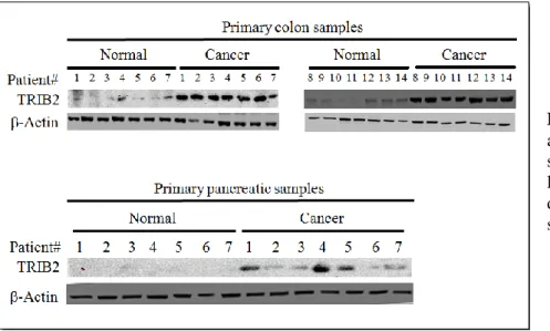
4.9. TRIB2 protein expression in primary clinical samples 40
CHAPTER 5: DISCUSSION AND CONCLUSION 43
5.1. Future Directions 47
CHAPTER 6: REFERENCES 48
Page 8 of 72 FIGURES LIST
Pages
Fig. 2.2.1.1. Process and development of Melanoma 13
Fig. 2.3.1.1. PTEN/PI3K/AKT pathway 16
Fig. 2.3.4.1. PTEN/PI3K/AKT pathway inhibition 18
Fig. 4.1.1. Expression level of TRIB2 and actin in isogenic cell lines 28 Fig. 4.1.2. Cell viability post exposure to chemotherapeutics 29 Fig. 4.2.1. Cell viability following BEZ235 treatment of U2OS cell line 30 Fig. 4.2.2. Cell viability following BEZ235 treatment of 293T cell line 30 Fig. 4.2.3. Cell viability following BEZ235 treatment of G361 cell line 30 Fig. 4.3.1. Immunoblots for caspase-3, cleaved caspase-3, TRIB2 and actin following BEZ235 or
DTIC treatment 30
Fig. 4.3.2. Immunoblots for caspase-3, cleaved caspase-3, TRIB2 and actin following BEZ235
treatment 32
Fig. 4.4.1. Immunoblots for total p53 and actin following BEZ235 or DTIC exposure 33 Fig. 4.5.1. Representative FACS profiles for p53+/+ and p53-/- 34 Fig. 4.5.2. Immunoblots for MDM2, total p53 and actin following BEZ235 exposure 35 Fig. 4.6.1. SDS-PAGE gel following IgG, MDM2, AKT or FOXO3a co-IP 36 Fig. 4.7.1. Immunoblots for diverse proteins after BEZ235 or DTIC exposure 36 Fig. 4.8.1. Immunoblots for diverse proteins after BEZ235 exposure 36 Fig. 4.8.2. Immunoblots for FOXO3a and TRIB2 after transfection of FOXO3a shRNA constructs 39 Fig. 4.8.3. Cell viability following BEZ235 treatment of U2OS-empty-FOXO3aKD or
U2OS-TRIB2-FOXO3aKD cancer cells 39
Fig. 4.9.1. Immunoblots for TRIB2 and actin in primary ex-vivo clinical samples 40 Fig. 4.9.2. Immunoblots for TRIB2 and actin in our primary ex-vivo in Melanoma clinical samples 41 Fig. 4.9.3. Immunoblots for a number of proteins and actin in our primary ex vivo Melanoma clinical
samples 42
Fig. 5.1. Proposed mechanism for the PTEN/PI3K/AKT pathway, without TRIB2 over expression 46 Fig. 5.2. Proposed mechanism for the PTEN/PI3K/AKT pathway, with TRIB2 over expression 46
Page 9 of 72 TABLES LIST
Pages Table 2.2.3.1. American Joint Commission on Cancer TNM system 14
Table 3.1.1. Cell lines 22
Table 3.1.2. Chemotherapeutics drugs 22
Table 3.1.3. Melanoma and normal tissue samples 23
Table 3.3.1.Primary Antibodies 24
Table 3.3.2.Secondary Antibodies 25
APPENDIX LIST
Pages
Fig. A. 1. Datasheet of Total Akt antibody 55
Fig. A. 2. Datasheet of phospho-Akt antibody 56
Fig. A. 3. Datasheet of Total FOXO antibody 57
Fig. A. 4. Datasheet of phospho-FOXO antibody 58
Fig. A. 5. Datasheet of Faz-L antibody 59
Fig. A. 6. Datasheet of MDM2 antibody 60
Fig. A. 7. Datasheet of PTEN antibody 61
Fig. A. 8. Datasheet of P53 antibody 62
Fig. A. 9. Datasheet of BIM antibody 63
Fig. A. 10. Datasheet of Total PRAS40 antibody 64
Fig. A. 11. Datasheet of phospho-PRAS40 antibody 65
Fig. A. 12. Datasheet of Total p70S6K antibody 66
Fig. A. 13. Datasheet of Actin antibody 67
Fig. A. 14. Datasheet of Caspase-3 antibody 68
Fig. A. 15. Datasheet of Cleaved Caspase-3 antibody 69
Fig. A. 16. Datasheet of Total PDK1 antibody 70
Fig. A. 17. Datasheet of phospho-PDK1 antibody 71
Page 10 of 72 ABBREVIATIONS LIST
AJCC American Joint Committee on Cancer AKT Protein kinase B
ATF4 Activating transcription factor 4 BRAF B-type Raf kinase
ChIP Chromatin Imunnoprecipitation Co-IP Co-Imunnoprecipitation
DNA Deoxyribonucleic acid DTIC Dacarbazine
FACS Fluorescent Activated Cell Scanning FasL Fas ligand
FDA Food and Drug Administration FOXO Forkhead transcription factor IGF-1 Insulin-like growth factor 1 IgG Immunoglobulin G
Il-2 Interleukin-2
IP Immunoprecipitation KD Kinase Domain
MAPK Mitogen-activated protein kinases MAPKK Mitogen-activated protein kinase kinase MDM2 Mouse double minute 2 homolog mTOR Mechanistic target of rapamycin
p70S6K Phosphorylation of 40S ribosomal protein S6 PBS Phosphate buffered saline
PDK1 Pyruvate dehydrogenase lipoamide kinase isozyme 1 PI3K Phosphatidylinositol 3 kinase
PIP3 Phosphatidylinositol (3,4,5)-trisphosphate PRAS40 proline-rich AKT substrate of 40 kDa PTEN Phosphatase with tensin homology RTK Receptor tyrosine kinases
USA United States of America UV Ultraviolet
Page 11 of 72
CHAPTER 2
Page 12 of 72 2. INTRODUCTION
2.1. Cancer.
Cancer is caused by a series of somatic alterations in DNA and the consequence of these events is an uncontrolled cellular proliferation. The majority of these alterations are caused by replication errors, defective DNA repair processes or exposure to carcinogens1,2. However, occasionally cancers can be caused by an alteration in a dominant gene that drives uncontrolled cell proliferation. The genes that can promote cell growth when altered are often called oncogenes3.
Most and perhaps all types of human cancers are characterized by six essential alterations in cell physiology: self-sufficiency in growth signals, insensitivity to growth-inhibitory signals, evasion of programmed cell death, limitless replicative potential, sustained angiogenesis, and tissue invasion and metastasis. These properties are exclusive from the cancer cell and are not found in the normal adult cell from which the tumor is derived4.
2.2. Melanoma.
2.2.1. General Issues.
Pigmented lesions are among the most common findings on skin examination. The challenge is to distinguish Melanomas, which account for the overwhelming majority of deaths resulting from skin cancer, from the remainder, which with rare exceptions are benign5. Cutaneous Melanoma, is a form of aggressive cancer that develops from melanocytes (Figure 1.2.1.1). It is most common in people between 30 and 60 years of age. This type of cancer is a heterogeneous disease that presents different genetic alterations and diversity of histological subtypes6,7. Having regarded what was referred before, the scientific investigation in this area has been vast. Recent studies have revealed that PI3K signaling is deregulated in a high proportion of Melanomas8,9. Despite all the research that has been done, Melanoma patients have only experienced a minor increase in life expectancy in stark contrast to other types of cancer. The rising number of cases per year has resulted in Melanoma mortality rising sharply over the along the last five decades10.
Page 13 of 72 Figure 2.2.1.1: Schematic illustration of the process and development of Melanoma and its metastases (from A to D).
2.2.2. Epidemiology.
Melanomas can occur in adults of all ages (the median age at diagnosis is the late fifties), in people of all colors and men are affected slightly more than women (1.3/1). It is located on the skin and originates following the transformation of melanocytes, the pigment-producing cells in the neural crest that migrate mainly to skin, mucous membranes, upper esophagus and eyes6,7. The highest incidence rates occurs in white-skinned peoples living at low latitudes. Accordingly, the association between sun exposure and Melanoma have been explored. An important risk factor for Melanoma is UV irradiation upon sun exposure10.
Melanoma primarily grows horizontally within the epidermis (Melanoma in situ) but in an advance stage it can grow in depth and penetrates into the dermis (invasive Melanoma)8. At this point, the patient prognosis is good, with surgical resection of the tumor conferring a 53-97% survival. However, if a distant metastasis is present, the patients exhibit a less than 5% survival independently of the therapeutic intervention. It is estimated that there were more than 1 million Melanoma survivors living in the USA as of January 1, 2014, and an additional 76 100 people will be diagnosed in 2014. Melanoma incidence rates have
Melanoma A B C D Epidermis Dermis Subcutaneous tissue Melanoma
Page 14 of 72 been increasing for at least 30 years. About 84% of Melanomas are diagnosed at a localized stage, when they are highly curable7,11.
2.2.3. Treatment of Melanoma by Stage.
The stage of a Melanoma is a description of how widespread it is. This includes its thickness in the skin, whether it has spread to nearby lymph nodes or any other organs, and certain other factors. A staging system is a standard way to sum up how far a cancer has spread. The system most often used to stage Melanoma is the American Joint Commission on Cancer (AJCC) system12,13 (Table 1.2.2.1).
Stage Description of Melanoma's Characteristics
0 It is in the epidermis but has not spread to the dermis
I It is smaller than 1.0 mm in thickness and has not been found in lymph nodes or distant organs II It is thicker than 4.0 mm and is ulcerated. It has not been found in lymph nodes or distant organs III It can be of any thickness, but it is not ulcerated. It has spread to 1 - 3 lymph nodes near the affected
skin area (no distant spread)
IV It has spread beyond the original area of skin and nearby lymph nodes to other organs such as the lung, liver, or brain, or to distant areas of the skin, subcutaneous tissue, or distant lymph nodes Table 2.2.3.1: American Joint Commission on Cancer (AJCC) system, which is used to stage Melanoma.
Surgery to remove the tumor and surrounding tissue is the primary treatment for most Melanomas. Less than 3% of all patients with Melanoma undergo radiation therapy. However, almost one-half (45%) of patients with metastatic disease who receive either chemotherapy or immunotherapy also receive radiation therapy14,15. Patients with stage III Melanoma are often offered adjuvant immunotherapy with interferon for about a year. However, this treatment has side effects that make it very difficult to tolerate. Treatment for patients with stage IV Melanoma has changed in recent years and typically includes immunotherapy or targeted therapy drugs. Patients with localized or regional metastatic disease are identified for surgical resection and could benefit from interferon-α adjuvant therapy, despite the significant toxicity associated with this treatment. In patients with distant metastasis, surgery is unlikely to be offered and the only therapeutic option available is systemic drug administration12,15,16.
2.2.4. Conventional Chemotherapy .
Conventional chemotherapy is based on the use of alkylating agents such as Fotemustine (Muphoran), Dacarbazine (DTIC), and Temozolomide (Temodal) which trigger cytotoxic effects by blocking cell replication. However, these chemotherapy drugs promote only 10% of objective response with
Page 15 of 72 no improvement of overall survival15,17. Since the major advance realized in 2011 with the FDA approval of Vemurafenib, for mutated BRAF Melanomas, these drugs are limited to patients harboring non-BRAF mutated Melanomas or for patients who developed resistance to previous treatments18,19.
It has been well documented that Melanoma is an immunogenic tumor but metastatic Melanoma cells have developed mechanisms to escape from immune surveillance in order to survive. The immune system involvement in protection against Melanoma is supported by the increased of Melanoma incidence under immunosuppression conditions20. In 1998, the first immunotherapy to be approved by the Food and Drug Administration (FDA) for treatment of advanced Melanoma was Interleukin-2 (IL-2) but, like Dacarbazine, response rates were low even at high-doses of treatment. Its use in clinical practice is limited by the severe toxic side-effects21,22. However, substantial advances in systemic cancer therapies have been reported since 2009 and a new immunotherapeutic drug (anti-CTL4-4 antibody) Ipilimumab23.
2.2.5. Novel Therapy.
The recent characterization of the molecular alterations in Melanoma leads to the development of targeted therapies in order to finish with the resistance to therapeutic agents, both chemical or biological, which remains the main problem in the management of the therapy in Melanoma. These treatments are designed to target tumors according to their molecular diversity and activated intracellular signaling pathways24–27. Advanced studies led to the development of inhibitors of PI3K which selectively target only the catalytic sites. The PI3K inhibitors, GSK2126458 and BEZ235, were evaluated in vitro showing an enhanced cell growth inhibition28–30.
The use of novel therapeutics should ideally be made following an immunological and genetic mutation screen of the patients tumor, in order to the treatment be specifically adapted based on mutations of a patient who have cancer. However, there are no currently available immunological biomarkers and those targeted agents for which mutations can be tested, frequently develop secondary resistance. New biomarkers could be useful for many different things like screening, early diagnosis, disease staging as well as for the identification of those patients who are in high risk of disease recurrence31,32.
2.3. The AKT Pathway.
2.3.1. General Issues
The AKT is a serine/threonine kinase downstream of PTEN/ PI3K, that exists as three isoforms in mammals (AKT1, AKT2 and AKT3) , which are encoded by three different genes. They are ubiquitously
Page 16 of 72 expressed, but their levels are variable, depending upon the tissue type33,34. In Melanoma cells, AKT3 is the form preferentially expressed. The AKT3 activation is found in about 60% of sporadic Melanomas35. The AKT kinase regulates multiple biological processes including cell survival, proliferation, growth, and glycogen metabolism through phosphorylation of many physiological substrates33. A variety of growth factors (e.g. IGF-1), hormones (e.g. Thyroid Hormone T3), cytokines (e.g. IL-2) and certain oncogenes (e.g.
Ras) activate AKT, by binding their cognate receptor tyrosine kinase (RTK) and triggering activation of the lipid kinase PI3K36–39. For instance, Ras activation of the AKT pathway confers protection from apoptosis in fibroblasts in response to DNA damage or oncogenic Myc. Although several AKT targets have been reported, it is not fully understood how AKT promotes survival9,40,41.
Figure 2.3.1.1: General scheme of the PTEN/PI3K/AKT pathway.
2.3.2. The Activation of AKT.
The PTEN is a dual lipid and a protein phosphatase. Its primary target is the lipid phosphatidylinositol-3,4,5-triphosphate (PIP3), the product of the phosphatidylinositol-3-kinase (PI3K)42,43. The loss of function of the PTEN (which has been implicated in many human cancers), as well as the activation of the PI3K, results in accumulation of PIP3 triggering for the activation of its downstream effectors, PDK1, AKT and Rac144,45. The activation of PI3K is induced by growth factors and insulin targeting by the catalytic subunit to the membrane where it is in close proximity with its substrate, mainly
Page 17 of 72 PIP2. PDK1 contains a C-terminal pleckstrin homology (PH) domain, which binds the membrane bound PIP3 triggering PDK1 activation. Activated PDK1 phosphorylates AKT at Thr308 activating its ser/thr kinase activity and further activation occurs by PDK2 by phosphorylation at Ser47346,47. Activation of AKT results in the suppression of apoptosis induced by a number of stimuli including growth factor withdrawal, detachment of extracellular matrix, UV irradiation and cell cycle discordance9. Furthermore, the abnormal expression and activity of the PI3K/AKT pathway proteins has been shown to promote Melanomagenesis by inducing cell survival signaling in Melanoma cells. Therefore, members of this signaling cascade are attractive targets for inhibiting Melanoma9.
2.3.3. The Importance of p53.
The PI3K-AKT pathway has recently been reported to inhibit the transcriptional activity of p53 and reduce the proapoptotic functions of it. The p53 is a tumor suppressor which plays a key role in the induction of apoptosis and cell cycle arrest in response to a variety of genotoxic stresses and to the activation of some oncogenes such as Myc, thereby preventing the propagation of damaged cells48,49. Its function is controlled by several mechanisms, including the regulation of p53 protein stability. Central to this process is MDM2, a ubiquitin ligase that targets p53 for ubiquitination and allows export of p53 from the nucleus to the cytoplasm, where p53 degradation by proteasomes takes place50.
Under normal circumstances, p53 is maintained at very low levels by continuous ubiquitination and degradation. Activation of p53 in response to cellular stresses is mediated partly by inhibition of MDM2 and rapid stabilization of p53 protein. The deregulated activation of mitogenic signals, caused by the oncogenic activation of Ras or Myc for example, leads to the activation of p53, which provides a mechanism to prevent the abnormal proliferation associated with tumor development51,52. However, this activation of p53 by mitogenic signals must be suppressed during normal cell proliferation to prevent p53 from inducing cell cycle arrest or apoptosis. Therefore, it appears reasonable to assume that mitogenic signals elicit both p53-activating and -inp53-activating signals53,54. Recent studies have indeed shown that Ras can inhibit or activate p53, depending on the cellular contexts and the duration of Ras activation. The Raf/MEK/MAPK pathway has been shown to mediate Ras activation of p53. Therefore, it is possible that the PI3K/AKT pathway opposes the MAPK pathway in activation of p53. However, it has yet to be determined how AKT suppresses
Page 18 of 72
2.3.4. The role of FOXO proteins.
As mentioned above, the AKT promotes cell survival directly by its ability to phosphorylate and inactivate several pro-apoptotic targets, like Bim, and the forkhead transcription factors (FOXO)58. The FOXO proteins (including FOXO3a) play an important role in longevity and tumor suppression by regulating a wide range of genes involved in stress resistance, metabolism, cell cycle arrest and apoptosis. Previous studies have shown that BEZ235 treatment of malignant Melanoma cells induces FOXO3a-dependent gene expression following the inhibition of PI3K1. Activation of this pathway can directly result in phosphorylation of FOXOs and their subsequent cytoplasmic sequestration and/or degradation via the ubiquitin-proteasome pathway. When FOXO is activated by the inhibition of the PI3K/AKT pathway, FOXO’s promotes a wide range of effects including cell cycle arrest, cell differentiation, autophagy and apoptosis via various mechanisms59,60.
Figure 2.3.4.1: General scheme of PTEN/PI3K/AKT pathway inhibition and FOXOs posphorilation, with FOXOs subsquent cytoplasmatic sequestration.
Page 19 of 72 2.4. The Tribbles Pseudokinases Family.
TheTribbles (TRIB) pseudokinases family members are the human homolog of Drosophila tribbles protein, which regulates the cell cycle during oogenesis and morphogenesis, and influences proliferation, motility, metabolism, and oncogenic transformation. All three TRIB (TRIB1, TRIB2, and TRIB3) pseudokinases are associated with a variety of human malignancies, acting as adaptors in important cellular signaling pathways extending from mitosis and cell activation to apoptosis and modulation of gene expression59. The members of Tribbles family have been reported to interact and modulate the activity of signal transduction pathways, including the PI3K/AKT and the MAPK systems, and with various signaling molecules and transcription factors, including ATF4, p65, CtIP, MAPKK, AKT and COP160,61.
The TRIB2 (and the others Tribbles proteins) are characterized by a central serine/threonine kinase-like domain (KD) and a C-terminal binding site for E3 ubiquitin ligases. However, these proteins are considered to be catalytically inactive because they lack conserved residues from the characteristic adenosine triphosphate binding site and catalytic core motif within the KD61. Therefore, Tribbles probably function as adapter or scaffold proteins. Although the three members of Tribbles family proteins are highly homologous in the KD and in the C-terminal E3 ubiquitin ligase binding site, they show restricted similarity in the N- and C-terminal domains62. The TRIB2 domains responsible for protein binding or functional/oncogenic activity are unknown. On the other hand, the integrity of the KD domains is required for both of these activities, and a mutation of critical residues within the KD interferes with these activities. Additionally, the binding of COP1 to the TRIB2 C-terminus is essential for TRIB2-induced AML. In the nonexistence of COP1 binding, Leukemia does not occur59,63,64.
Furthermore, TRIB2 has been implicated in the negative regulation of FOXO3a. The restoration of FOXO proteins have been suggested as a promising strategy to treat various types of cancer and accordingly the forced expression of nuclear FOXO has been shown to induce apoptosis in a wide range of in vitro cancer cell line models59,63. Additionally, the TRIB2, which is highly expressed in metastatic Melanoma cells, has been implicated in the resistance of various cancers to a range of chemotherapeutics, including PI3K inhibitors that are under clinical trial. It is hypothesized that this resistance is due to the repression of FOXO family members59,61,65.
2.5. Hypothesis.
Our group discovered that the kinase-like protein TRIB2 was highly expressed in metastatic Melanoma samples (and recently colon and pancreatic malignancies). Recent data within our laboratory revealed that the over expression of TRIB2 conferred drug resistance to a range of chemotherapeutic agents
Page 20 of 72 and importantly conferred resistance to a number of phosphatidylinositol 3-kinases (PI3Ks) inhibitors that are being tested in Melanoma clinical trials.
These results indicate that TRIB2 confers chemotherapeutic resistance. Based on our laboratory research, the present work intends to elucidate some of the mechanism(s) of action about how TRIB2 mediates PI3K inhibitor resistance and the role(s) of FOXO3a in this response.
In order to test our hypothesis, we need to confirm the inhibition effectiveness of our PI3K inhibitor and the cellular phenotype following TRIB2 over expression and to examine the recruitment of FOXO3a and the promoters of cell cycle arrest.
Page 21 of 72
CHAPTER 3
Page 22 of 72 3. MATERIALS AND METHODS
3.1. Cell Culture and Tissue Samples.
The cell lines used for this work and their respective information are represented in Table 3.1.1. The cells were cultivated in DMEM (Sigma) with 10% heat inactivated FCS (Sigma) supplemented with Pen/Strep (Gibco) and within 35 mm plates. The U2OS and the 293T cell lines was previously transfected with a plasmid containing the TRIB2. The G361 and the SK-Mel28 cell lines was stably transfected with a TRIB2 shRNA expressing plasmid. We treated the cells with several chemotherapeutic drugs. The chemotherapeutic drugs and their respective concentrations are represented in Table 3.1.2. We did several drug time courses with the different chemotherapeutic drugs.
Table 3.1.1: Cell lines used in this work and their origin.
Table 3.1.2: Chemotherapeutics drugs used in this work and their respective concentrations.
All used Melanoma and Normal tissue samples are represented in Table 3.1.3, and were provided by Dr Selma Ugurel (Julius-Maximilians-Universität Würzburg, Germany). They were sectioned for Immunohistochemistry (Faro Hospital) and the remaining tissue used for protein analysis.
Cell Line Origin
U2OS Human Osteosarcoma 293T Human Renal Cancer MCF-7 Human Breast Cancer MDA-489 Human Breast Cancer A375 Human Melanoma G361 Human Melanoma M14 Human Melanoma SK-Mel28 Human Melanoma UACC62 Human Melanoma
Chemotherapeutic Drugs Concentration
DTIC (Dizcarbacine) 100nm BEZ235 100nm BAYER COMPOUNDS (236; 439; 766; 931) 10nm Cyclohexamide 10ug Actinimycin D 10ug MG132 10ug/ml
Page 23 of 72 Table 3.1.3: Melanoma and Normal tissue samples used in this work.
3.2. Protein Extraction and Quantification.
The protein extraction is the total protein that was extracted from each cell line or our tissue samples. For cellular extraction, the cells were collected from the culture plates by first removing the growth medium and to then scrape the cells in 1 ml of PBS. This suspension was then transferred into a clean Eppendorf and spinned at 1100 rpm for 5 min at 4ºC. The PBS was removed from the pellet and RIPA buffer (Tris-HCL ph 7.4, NaCl, 10% Nonidet P-40, 10% sodium deoxycholate, 100 mM EDTA, PIC, 200 mM NA-F, 100mM Na3VO4 and protease inhibitors cocktail) was added to the pellet. The pellet was resuspended in this total lysis buffer and incubated on ice for 30 minutes. After 30 minutes the lysed cells were spun 15 minutes at 13000 rpm (maximum speed). The supernatant (containing our proteins) was collected and transferred to a fresh eppendorf prior to quantification.
For protein extraction from our tissue samples, tissue sections were placed inside a manual homogenizer (Sigma) with 500 μL of the RIPA buffer (described above) and vigorously homogeneized. After homogenization samples were incubated on ice for 30 minutes. Lysed samples were spun at 13000 rpm and the supernatant transferred to a fresh eppendorf prior to protein quantification. For both extraction protocols all extracted proteins were stored at -80ºC until required.
To determine the protein concentrations (protein quantification) in each sample we used the Quick StratTM Bradford Protein Assay (BioRad) and the NanoDrop 2000 UV-Vis Spectrophotometer (ThermoScientific) following the manufacturers guidelines.
Melanoma Samples Normal
Samples Stable Disease Complete response Progressive Disease CSM002 CSM066 CSM105 1274 CSM178 CSM209 CSM108 1440 CSM200 CSM203 CSM143 1454 CSM214 CSM068 CSM060 1425 CSM099 CSM006 CSM057 1408 CSM027 1474 CSM094 1412 CSM038 1489 CSM111 1428 CSM213 1508
Page 24 of 72 3.3. Western Blotting.
Our extracted protein samples were diluted in to 2x lammeli loading buffer (containing β-mercaptoethanol) and heatedat 95ºC for 5 minutes. Samples were loaded into our 10% SDS-PAGE gels. Separated proteins within each gel were transferred on to nitrocellulose membranes (Amersham) and were blocked with 5% BSA (in Tris-buffered-saline [TBS]) for 1 hour (preventing non-specific antibody binding). After blocking, membranes were immunoblotted with several primary antibodies (dilution of 1/1000 into BSA) overnight at 4ºC. The primary antibodies used in this work and their respective information are represented in Table 3.3.1.
After incubation, membranes were washed (x3) with TBS 0.1% tween20. After washing, corresponding secondary antibodies were added (dilution of 1/5000 into BSA) at room temperature for 1 hour. The secondary antibodies used in this work and their respective information are represented in Table 3.3.2. The membranes were washed (x3) times with TBS 0.1% tween20 and visualized using ECL+. Images were obtained using a Molecular Imager® ChemiDoc™ XRS System (BioRad).
Table 3.3.1: Primary antibodies used in this work and their respective information.
Primary Antibodies Supplier and Information
Total AKT C-20; sc-1618; Goat; Santa Cruz Biotechnology p-AKT Ser 473; sc-7985; Rabbit; Santa Cruz Biotechnology Total FOXO (FKHRL1) N-16; sc-9813; Goat; Santa Cruz Biotechnology
p-FOXO (p-FKHRL1) Ser253; sc-101683; Rabbit; Santa Cruz Biotechnology
TRIB2 Custom, Rabbit, Madrid
Fas-L C-178; sc-6237; Rabbit; Santa Cruz Biotechnology MDM2 C-18; sc-812; Rabbit;Santa Cruz Biotechnology
PTEN A2B1; sc-7974; Mouse; Santa Cruz Biotechnology P53 DO-1; sc-126; Mouse; Santa Cruz Biotechnology BIM H-191; sc-11425; Rabbit; Santa Cruz Biotechnology Total PRAS40 H-216; sc-67042; Rabbit; Santa Cruz Biotechnology p-PRAS40 Thr246; sc-32629; Rabbit; Santa Cruz Biotechnology Total p70S6K C-18; sc-230; Rabbit; Santa Cruz Biotechnology
Actin I-19; sc-1616; Goat; Santa Cruz Biotechnology Caspase-3 E-8; sc-7272; Mouse; Santa Cruz Biotechnology Cleaved Caspase-3 h176; sc-22171; Rabbit; Santa Cruz Biotechnology
Total PDK1 C-20; sc-7140; Goat; Santa Cruz Biotechnology p-PDK1 Ser241; #3061; Rabbit; Cell Signaling Technology 14-3-3σ N-14; sc-7681; Goat; Santa Cruz Biotechnology
Page 25 of 72 Table 3.3.2: Secondary antibodies used in this work and their respective information.
3.4. Co-Imunnoprecipitation (Co-IP).
The cells were washed with medium. Trypsin was added to the plate with the cultivated cells and then the cells were scraped. The solution was collected to new Eppendorf tubes and centrifuged, and the supernatant was collected to new tubes. The protein A/G-agarose beads (Sigma) were washed for 2 times with PBS and a 50% protein A/G agarose working solution (in PBS) was made. Each indicated antibody was added to the beads for 1 hour. After 1 hour the beads were washed (x2) with PBS. 500 μg of total protein lysate was added to each set of beads and incubated overnight at 4ºC. Samples were centrifuged (max speed), the pellet was kept, and washed with pre-chilled PBS (x3). SDS-loading buffer was added to beads and the samples heated to 95ºC for 5 minutes. Samples were extracted and run on an appropriate percentage SDS-gel.
3.5. JetPrime Transfection Protocol.
In order to transfect some of ours cell lines (see Table 3.1.1 from 3.1), we followed the JetPrime transfection Protocol. We started the protocol diluting 2 µg of our DNA into 200 µl jetPRIME® buffer and, after that, we mixed both by vortexing. Then, it was added 4 µl of jetPRIME® to the mix and then, the new mix, was vortexed for 10 seconds and, after that, spun down briefly. At that time, the mix was incubated for 10 minutes at room temperature. Following the incubation, it was added 200 µl transfection mix (drop wise), per plate and evenly, onto the cells in serum containing medium. Finally, the plates was gently rocked and, then, placed into the incubator for 24 hours.
Secondary
Antibodies Supplier and Information
Anti-rabbit IgG-HRP; sc-2004; Goat; Santa Cruz Biotechnology Anti-goat IgG-HRP; sc-2020; Donkey;
Santa Cruz Biotechnology Anti-mouse IgG-HRP; sc-2314; Donkey;
Page 26 of 72 3.6. Chromatin Imunnoprecipitation (ChIP).
The plates were washed with medium, and a 1% formaldehyde/PBS solution was added to cross-link proteins to DNA. The solution was removed, and the plates were washed with ice cold PBS (x3). Cells were scraped from the plates with 1M Tris-HCl with 10mM DTT, and transferred to Eppendorf tubes. After centrifugation, the pellets were washed with Buffer I and II. After centrifugation, the pellet was resuspended in lysis buffer (made fresh with PIC). The cells were sonicated (6 times for 10 sec each sample) on ice, to shear DNA to an average fragment size of 200-500 base pairs. After centrifugation (at max speed for 15 minutes) to pellet cell debris, the supernatant was transferred into a new Eppendorf. 300 μl of Buffer D and PIC were added to each sample. 100 μl input samples were removed at this stage. The input samples were heated overnight at 65°C.
To the remaining sample (after the inputs were removed), we added sheared salmon sperm DNA, the antibody of interest and protein G-fast flow beads (Sigma) and Buffer D. After incubation overnight at 4ºC, the beads were pulled down and washed with TSE I, II and III. Afterwards the beads were washed with ice cold TE. The DNA was extracted with three washes with a solution of NaCHO3 and SDS. Once extracted the samples were transferred to a fresh Eppendorf and heated overnight at 65ºC. Our input and the ChIP samples were loaded into Sigma-PCR clean-up columns and after washing our immunoprecipitated DNA eluted with 30 μl dH2O. Samples were stored at -20ºC
3.7. Fluorescent Activated Cell Scanning (FACS).
For cellular extraction (after treatment), we first collected the growth medium and then transferred this into a clean 15 ml Falcon Tube. After that, we added 1mL of Trypsin to the cells and then we placed them in an incubator for 15 minutes. After 15 minutes we detached the cells by mixing up and down. The trypsinized cells were then added to the corresponding Falcon Tube and spinned at 1100 rpm for 5 minutes. The medium/trypsin was removed from the pellet and 1 ml of cold PBS was added to the pellet. The pellet was resuspended in the PBS and spinned at 1100 rpm for 5 minutes. The PBS was removed from the pellet and 1 ml of 70% Ethanol was added to the pellet. The pellet was resuspended in the 70% Ethanol and then was stored at 4ºC until required for FACS. Samples were run on FACS after propidium iodide (2.5 mg mL-1) was added to the fixed, stained cells prior to analysis. 50,000 gated, total events were scored per study from triplicate studies. Data was analyzed using FACS-express 3 (De Novo software).
Page 27 of 72
CHAPTER 4
Page 28 of 72 4. RESULTS
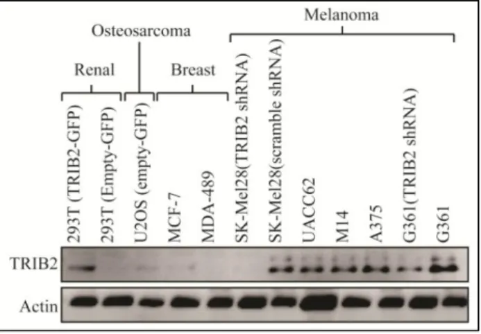
4.1. TRIB2 protein expression conferred resistance to classical chemotherapeutic modalities. Previous data from our laboratory demonstrated that the TRIB2 protein negatively regulated FOXO3a. However, the exact mechanism of this resistance remained elusive. Considering that forkhead transcription factors are important effector proteins following chemotherapeutic treatment, we hypothesized that elevated TRIB2 protein expression could confer resistance to a range of chemotherapeutic drugs. To test this hypothesis, we took matched cell lines (with either stable over expressed TRIB2-GFP or shRNA knocked down TRIB2). These cell lines are summarized in my materials and methods (Table 3.1.1, section 3.1, page 22). Prior to examining cell line sensitivity, we confirmed each cell line TRIB2 protein expression level (Figure 4.1.1)
Figure 4.1.1: A representative western blot showing the expression level of TRIB2 and actin in our isogenic cell lines. 50 μg of total protein lysate was loaded per lane and separated on a 10% SDS-PAGE gel.
Having confirmed that our cell lines had matched TRIB2 protein expression levels, we questioned if these isogenic lines show any significant viability difference when exposed to 20 μM dazcarbizine (DTIC), 10 μM gemcitabine (Gem) or 50 μg/ml 5-fluorouracil (5-FU). We note that 72 hours post treatment, cells with increased TRIB2 protein expression were significantly more resistant to each chemotherapy modality that they were exposed to (Figure 4.1.2).
Page 29 of 72 Figure 4.1.2: Cell viability was measured 72 hours post exposure to each indicated chemotherapeutic agent. The percentage of subG1 cells were
measured by FACS analysis after 50,000 total events (N=3). P values are indicated for each isogenic cell line comparison and error bars are indicative of standard deviation.
Recently, a wide range of PI3K inhibitors have been developed to treat Melanoma and in particular metastatic Melanoma that typically presents an extremely poor clinical prognosis. Considering the highly significant resistance to conventional chemotherapeutic modalities (including the standard Melanoma treatment agent DTIC), we next questioned if TRIB2 status conferred resistance to PI3K inhibitors. One of the most extensively tested agents is the PI3K inhibitor BEZ235 (Novartis) that, in combination with DTIC is the standard treatment regime.
4.2. TRIB2 expression conferred resistance to the PI3K inhibitor BEZ235.
We exposed our isogenic cell lines to 100 nM BEZ235 for 24, 48 and 72 hours. Prior to BEZ235 exposure, there was no significant difference in terms of cell cycle distribution or proliferation rate between each isogenic cell line (data not shown). However, 48 hours post 100 nM BEZ235 treatment we note a number of highly significant differences. In cells that do not over express the TRIB2 protein there is an almost complete loss of the G2 (4N) population (white bars in Figure 4.2.1, 4.2.2 and 4.2.3) and a
significantly increased sub-G1 population (the black bars shown in Figure 4.2.1, 4.2.2 and 4.2.3).
In contrast, cell lines with increased TRIB2 protein expression display a more distinct G1 population
(dark grey bars in Figure 4.2.1, 4.2.2 and 4.2.3). Furthermore the sub-G1 population is significantly lower,
indicative of viable cells. Overall these results indicate that the over expression of TRIB2 conferred significant resistance to the PI3K inhibitor BEZ235
Page 30 of 72 Figure 4.2.1, 4.2.2, 4.2.3: Cell viability following 100 nM BEZ235 treatment of U2OS, 293T or G361 isogenic cancer cell lines. Cells were measured after 24, 48 or 72 hours BEZ235 treatment. Using propidium iodide staining, cell cycle stage was determined (subG1, G1, S and G2 phase).
50,000 total events were scored, N=3 and error bars are indicative of standard deviation averaged from triplicate samples over three independent studies.
4.3. Increased TRIB2 expression significantly attenuates BEZ235 induced apoptosis.
Our data indicated that high TRIB2 protein significantly increased cell line viability (hence a lower subG1 cell population) to both conventional as well as novel chemotherapeutics. However, we do not know if this resistance was due to a reduction in apoptosis (programmed cell death) or more generalized (such as necrosis). To address this question, we treated our U2OS-empty-GFP and U2OS-TRIB2-GFP isogenic cells with 100 nM BEZ or 20 µM DTIC, and evaluated the expression of caspase-3 and cleaved (active) Caspase-3 up to 48 hours post-treatment (Figure 4.Caspase-3.1).
Figure 4.3.1: Representative immunoblots for caspase-3/cleaved Caspase-3, TRIB2 and actin following 100nM BEZ235 or 20 μM DTIC. For each SDS-PAGE gel, 100 μg of total protein was loaded. Antibodies were used as described in the materials and methods section.
Page 31 of 72 In each cell line prior to treatment we observe little to no Caspase-3 cleavage consistent with healthy, non-apoptotic cells. In addition up to 48 hours post treatment (BEZ235 or DTIC) that there was no significant change in the total protein level of Caspase-3 (top band in Figure 4.3.1). However, strikingly 48 hours post BEZ235 or DTIC treatment there was a significant accumulation of cleaved Caspase-3 (indicative of apoptosis induction). Strikingly, in cell lines with increased TRIB2 protein expression, there was reduced Caspase-3 cleavage and therefore less apoptosis (lane 2 and 4 compared to lane 6 and lane 8 in Figure 4.3.1). Taking into account that Caspase-3 cleavage is a critical process in both the classical and non-classical apoptotic cascade, we can conclude, consistent with cell morphology and detachment (observed under the microscope) that TRIB2 over expressing cells are significantly less apoptotic than isogenic cells with low/endogenous TRIB2 protein expression. Strikingly cells that over express TRIB2 show significantly reduced caspase-3 cleavage and that are consistent with the resistance to BEZ treatment (as well as other PI3K inhibitors).
When we examined TRIB2 protein expression, we note a very interesting result. While the total level of TRIB2 protein did not change following DTIC treatment (Figure 4.3.1 middle panel, lane 3-4 and lane 7-8), when cell lines were treated with the PI3K inhibitor BEZ235, the level of total TRIB2 protein significantly increased (Figure 4.3.1, lane 1-2 and lane 5-6). It is tempting to suggest that this increase in independent of TRIB2 transcription for a number of reasons. In the U2OS-TRIB2-GFP cells, TRIB2 expression in under the control of a cytomegalovirus (CMV) promoter. Consequently, the transcription rate from this promoter is unlikely to change as it is already a high-expression promoter. We cannot rule out an increase in endogenous TRIB2 transcription in each cell line although considering that expression is already extremely high in U2OS-TRIB2-GFP cells, it is tempting to hypothesize that this increase in TRIB2 protein expression is independent of transcription and could be the result of post-translational modification(s). This is unexpected result is under intense investigation in our group.
Our results so far have only examined one time point (48 hours) post chemotherapeutic treatment and while we note a striking difference in the amount of Caspase-3 cleavage between U2OS-empty versus U2OS-TRIB2 cells, we wanted to examine Caspase-3 cleavage over more time points (Figure 4.3.2).
Page 32 of 72 Figure 4.3.2: Representative immunoblots for caspase-3/cleaved Caspase-3, TRIB2 and actin following 100nM BEZ235. For each SDS-PAGE gel, 50 μg of total protein was loaded. Antibodies were used as described in the materials and methods section.
Consistent with our previous Caspase-3 immunoblots, we note no significant difference between the levels of non-cleavage Caspase-3 over time in each cell line. In contrast however (and again consistent with Figure 4.3.1), we note that compared to matched isogenic cells, U2OS-TRIB2 cells show significantly lower Caspase-3 cleavage after BEZ235 exposure. Strikingly, by halving the amount of total protein per lane (50 μg compared to 100 μg) that this difference is even more striking. Furthermore, it is now extremely difficult to detect endogenous TRIB2 protein expression in our U2OS-empty cell line (Figure 4.3.2 lane 2, 3 and 4). We note that in our U2OS-TRIB2 cells, that following BEZ235 exposure that as early as 12 hours post treatment that there was a noticeable increase in the total level of TRIB2 protein (compare Figure 4.3.2 lane 5 versus lane 6). Considering that this increase can be seen as early as 12 hours post-BEZ235 treatment, it supports the hypothesis that the increase is independent of transcription as this is a short time point to consider a transcriptional response. In contrast, a post-translational response increasing the stability of TRIB2 is highly plausible. However, this aspect is still only a hypothesis and significantly more investigation is required at this point. Interestingly, cells treated with the chemotherapeutic DTIC show little to no TRIB2 stabilization after DTIC treatment (Figure 4.3.1). This suggests that the stabilization of TRIB2 (whether a transcritional or post-translational) is PI3K-dependent. These results clearly shows that cell lines with significantly elevated TRIB2 have considerably lower Caspase-3 cleavage and that following PI3K inhibition that TRIB2 protein levels accumulate. Our actin immunoblot confirms an equivalent protein load per lane in our immunoblots.
Page 33 of 72 4.4. In contrast to BEZ235 exposure, DTIC treatment stabilizes p53.
We note that following DTIC or BEZ235 exposure cells that do not over express TRIB2 show significant apoptotic cell death compared to isogenic cells that have high levels of the TRIB2 protein. This is a striking observation as these chemotherapeutic agents have highly divergent mechanisms of action (as described in my introduction). As an alkylating agent, we questioned if the tumour suppressor p53 (a protein that is extremely similar to FOXO3a) was affected following the exposure to DTIC. We also included BEZ235 treatment hypothesizing that p53 would not be affected by PI3K inhibition (Figure 4.4.1).
Figure 4.4.1: Representative immunoblots for total p53 (DO1) and actin following 100nM BEZ235 or 20 μM DTIC exposure. For each SDS-PAGE gel, 50 μg of total protein was loaded. Antibodies were used as described in the materials and methods section.
We note that U2OS-empty cells show a small increase in total p53 up to 48 hours post BEZ235 (100nM) treatment that was not unexpected. U2OS-empty cells show robust induction of apoptosis at this time point (see Figure 4.1.2). Consequently, within this cellular environment, with approximately 30-40% of the cells inducing apoptosis, we would expect a slight accumulation of p53. However strikingly, we note that following DTIC treatment that there was a significant accumulation of p53 (Figure 4.4.1 lane 3-4 and lane 7-8). Furthermore, in cell lines with high TRIB2 protein expression that p53 accumulation was lower compared to matched isogenic cell lines (see Figure 4.4.1 lane 4 versus lane 8). From this result we can conclude that DTIC exposure leads to the robust accumulation of p53, that BEZ235 treatment does not trigger p53 accumulation and that importantly, cell lines with high TRIB2 expression show attenuated p53 accumulation compared to control cells.
Page 34 of 72 4.5. A role for p53?
Based on our results described in 4.4, we hypothesized that DTIC would trigger the subG1 accumulation and apoptosis in a p53-dependent manner. To address this question, we used the Vogelstein isogenic HCT-116 colon cancer cell line with matched p53 status. These cell lines were grown to 70% confluence and treated with 20 μM DTIC or 100 nM BEZ235. At 48 hours post exposure to either agent, cells were analyzed via FACS (Figure 4.5.1).
Figure 4.5.1: Representative FACS profiles for p53+/+ and p53-/- HCT116 cell viability 48 hours following 20 μM or 100 nM BEZ235 treatment. Using propidium iodide staining, cell cycle stage was determined (subG1,
G1 phase, S and G2 phase). 50,000 total events were
scored, N=3
As we would have predicted based on our previous immunoblot analysis, we note that p53+/+ cells are highly sensitive to DTIC treatment (Figure 4.5.1 top left FACS plot). In contrast, p53-/- HCT-116 cells are extremely resistant to DTIC (Figure 4.5.1 top right FACS plot). In contrast, BEZ235 treatment induced significant cell death/subG1 accumulation in a p53-independent manner (Figure 4.5.1 bottom left and bottom
right FACS plots). The most striking aspect of the data we have accumulated suggests that increased TRIB2 protein expression confers resistance to both DTIC (a p53-dependent chemotherapeutic) and BEZ235 (a PI3K inhibitor and likely FOXO3a-dependent chemotherapeutic).
As a negative regulator of FOXO3a, our data unexpectedly raised the hypothesis that TRIB2 could confer resistance to p53-dependent chemotherapeutics. To investigate this further we carried out immunoblotting for total p53 and the E3-ubiquitin ligase (and negative regulator of p53) mouse double minute 2 homolog (MDM2) in our isogenic U2OS cell lines (Figure 4.5.2).
Page 35 of 72 Figure 4.5.2: Representative immunoblots for MDM2, total p53 (DO1) and actin following 100nM BEZ235 exposure. For each SDS-PAGE gel, 100 μg of total protein was loaded. Antibodies were used as described in the materials and methods section.
First we note that incontrast to U2OS-empty cells that U2OS-TRIB2 cells have a slightly higher level of MDM2 under non-treatment conditions (Figure 4.5.2. lane 1 compared to lane 6). Second, following BEZ exposure, U2OS-empty cells show a steady reduction in MDM2 protein levels (Figure 4.5.2 lane 2-5). Unexpectedly, U2OS-TRIB2 cells show a rapid decrease in MDM2 protein expression and then a rapid rise in MDM2 protein level, oscillating up and down. As we would predict, U2OS-empty cells show a steady increase in total p53 level consistent with MDM2 levels decreasing (Figure 4.5.2, middle panel, lanes 2-5). Strikingly, while U2OS-TRIB2 cells have p53, the accumulation of total p53 is significantly attenuated (Figure 4.5.2 lane 5 versus lane 10). We note that U2OS-empty cells show a gradual increase in total p53 up to 12 hours post BEZ treatment. In contrast, U2OS-TRIB2 cells show a significantly lower level of p53 protein under non-treated conditions, an initial increase 60 minutes post BEZ exposure and then a noticeable decrease up to 12 hours post BEZ incubation. Strikingly, the exact opposite trend was observed for MDM2 raising the hypothesis that in cells which over express TRIB2, the p53/MDM2 regulatory balance is disrupted.
4.6. TRIB2 and MDM2 interaction.
The next question we asked after observing the deregulated p53/mDM2 axis in TRIB2 over expressing cells was if TRIB2 and MDM2 could interact. This question is particularly important as very little is known regarding TRIB2 protein regulation although it has been reported that TRIB2 can be regulated by ubiquitination66,67. We carried out co-immunoprecipitation (co-IP) assays 12 hours post 100 nM BEZ235 treatment or after mock-drug treatment in our isogenic U2OS cell line model, asking if TRIB2 would interact with MDM2, AKT or FOXO3a (Figure 4.6.1).
Page 36 of 72 Figure 4.6.1: SDS-PAGE gel following IgG, MDM2, AKT or FOXO3a co-IP. Captured lysates were probed for TRIB2. 500 μg of total protein was captured per sample and run in each lane. 25 μg of total protein lysate was run on a separate SDS-PAGE gel and probed for actin to ensure equivalent protein loading N=3.
Our co-IP studies indicate that TRIB2 and MDM2 interact, raising the hypothesis that MDM2 could, in principle regulate TRIB2. Overall, this result indicates that the TRIB2 and MDM2 interact and that the overexpression of TRIB2 perturbs the p53/MDM2 regulatory balance. This supports our previous data where unexpectedly, TRIB2 over expression confers cell line resistance to chemotherapeutics that induce p53-dependent apoptosis (including gemcitabine and 5-FU).
4.7. TRIB2 Expression and Stress Signaling.
So far, our data indicates that DTIC induces a potent p53-dependent apoptotic response in contrast to BEZ235 that causes cell death independently of p53. This raised two questions, specifically “how does
BEZ235 induce cell death?” and “would these drugs in combination significantly increase cell death?” To
address these questions we used our isogenic U2OS and 293T cell lines. Each cell line was treated with either 100 nM BEZ235, 20 μM DTIC or both chemotherapeutics (representative of the combinational therapy used to treat malignant Melanoma) for 48 hours. Following this drug treatment, total cell lysates were collected and immunoblot analysis conducted for a number of key signaling proteins and downstream effector proteins from these signaling networks (Figure 4.7.1).
Figure 4.7.1: Representative immunoblots for each indicated protein after 100nM BEZ235 or 20 μM DTIC exposure. 50 μg of total protein was loaded per lane. Antibodies were used as described in the materials and methods section.
Page 37 of 72 We initially investigated the protein expression levels for TRIB2, total-PDK1, phospho-PDK1, PDK1, total p53, FasL, 14-3-3σ, PTEN and β-Actin in our matched U2OS and 293T cell lines. These proteins are key components of the PI3K and PI3KK signaling networks. In particular PDK1 and PTEN being key members of the AKT signaling cascade, p53 (as the “guardian of the genome”), FasL (as a critical effector protein of the pro-apoptotic FOXO cascade) and 14-3-3σ (a protein regulated by p53, directing cell cycle arrest and regulating FOXO localization).
We note that in cell lines with low TRIB2 protein expression that BEZ235 exposure triggered the robust accumulation of FasL, a FOXO3a regulated gene. In contrast, DTIC did not induce any significant amount of FasL. Furthermore, BEZ235 exposure induced the de-phosphorylaion of PDK1, indicative of inhibiting PI3K signaling. As we would predict, DTIC treatment had no effect on PDK-1 phosphorylation indicating that the PI3K signaling pathway remains active in the presence of DTIC. PTEN protein expression remained constant regardless of chemotherapeutic exposure while 14-3-3σ protein expression also did not significantly change. When we look at these pathways in cells with increased TRIB2 expression, we see a striking observation; BEZ235 and dual DTIC/BEZ treated cells do not induce significant amounts of FasL, consistent with a lower overall sensitivity to these chemotherapies. This observation is very apparent in dual treated cells and while the lower protein accumulation is not as clear-cut in BEZ235 only TRIB2 over expressing cells, the accumulation of FasL is lower in both U2OS-TRIB2 and 293T-TRIB2 cells compared to their matched controls. This result implied that the high level of TRIB2 was affecting at least FasL protein expression, consistent with our previously published work showing TRIB2-dependent inhibition of FOXO3a59. Interestingly, our immunoblots for p53 also indicate that in high TRIB2 expressing cells that there is a slightly lower accumulation of p53 following DTIC treatment.
4.8. Implicating AKT and FOXO.
Based on our initial screen in Figure 4.7.1, we noticed that following BEZ235 and dual BEZ235/DTIC treatment that there was reduced (or ablated) FasL expression. As FasL is regulated by FOXO3a and is a critical effector protein of apoptosis, we questioned if other components of the PI3K network were affected by TRIB2 status and was contributing to resistance to BEZ235. To address these questions, we treated our matched U2OS cells with 100 nM BEZ235 and via immunoblotting, examined key components of the AKT signaling cascade (Figure 4.8.1).