A multistep study to
unveil the
involvement of
peptide signalling in
STK-dependent
pathways in
Arabidopsis thaliana
Maria João Nogueira Ferreira
Biologia Funcional e Biotecnologia de Plantas
Departamento Biologia 2018
Orientador
Sílvia Coimbra, Professor auxiliar, FCUP
Coorientador
Maria Isabel Amorim, Professor auxiliar, FCUP
Coorientador
Acknowledgements
À Professora Sílvia Coimbra por me ter recebido no seu grupo e ter-me dado uma oportunidade incrível que indubitavelmente me fez crescer pessoalmente e profissionalmente. Obrigada pelo apoio durante este ano de tese. À Professora Isabel Amorim, agradeço-lhe por ter acreditado em mim e desafiado a entrar no mundo das plantas quando ainda não imaginava que iria seguir esse rumo. Obrigada pela ajuda durante estes anos.
Aos meus companheiros SPReDianos, obrigada pela ajuda prestada, pelos conselhos e ensinamentos!
À minha família por sempre me apoiarem em todas as decisões que tomo e serem um fator determinante nessas escolhas. Pais e tia, é verdade que tudo se torna mais fácil quando sabemos que temos uma rede pronta a amparar-nos de uma queda. Mesmo que seja no Japão!
Aos meus amigos, agradeço pela vossa paciência para comigo. Às vezes o stress fala mais alto, mas é uma mais valia ter pessoas com quem podemos partilhar as nossas frustrações. Susana, Márcio, Débora, Mariana, Patrício, Antero e Inês, obrigada pela companhia que me fizeram, mesmo com as 8/9 horas de diferença que existiam entre nós.
To professor Higashiyama, for accepting me in his group and contribute for a pleasant stay in Nagoya. Thank you for giving me the tools I needed for my work and for the advices regarding it. I feel very privileged for having met you, Professor.
To Hide, I know you don’t like the word “supervisor”, so thank you for your guidance. It really helped me to become more confident and independent in working at the lab. It was very helpful for my work to talk and discuss some topics with you. I can say it also contributed to my critical thinking skills. To Kazu, you were a great lab host! Thank you for introducing me to the lab, for the advices about my work, and for always answer to my questions (I know I could be annoying sometimes). To Bob, for sharing this 6 months experience with me. You were an amazing Japanese culture teacher and an even greater friend and lab partner (thank you for cheering me up with ice cream). You showed me that the scientist life is not easy, but it can be very rewarding. I also need to thank
Takuya for sharing his teachings with me and to Kimata for his advices about my transcriptome experience.
To Kathy, Tamiris and Paolo, for their support and friendship. Even though you were from another group, I felt we were from the same. Kathy you were a great deskmate and our “juicy” talks were a good escape from work. Tamiris and Paolo we must have another karaoke night!
To my share house friends, you really were an important part of my experience in Japan. Mifuyu, Matt and Sean, you taught me a lot about Japan and its society. I enjoyed spending time with you. Thank you for hearing my complaints! Tingting, Pim, Haruna, Waka and Sasuke, I am very happy for having met you. Thank you for the good talks, the walks and the tasty meals.
Por fim, mas não menos importante, um agradecimento especial à Diana por ter partilhado esta experiência comigo. Obrigada pela tua companhia e ajuda durante o tempo infinito em que eu estava na sala das plantas, por seres uma boa companheira de viagem e por não te importares de ouvir tantas canções!
Resumo
Nos óvulos das plantas com flor, os tecidos esporofíticos envolvem e protegem o saco embrionário. O estudo de óvulos com alterações nos tecidos esporofíticos demonstrou que existe uma comunicação entre o esporófito e o megagametófito, sendo esta importante para o desenvolvimento de sementes viáveis. Todavia, os processos subjacentes à coordenação célula-célula continuam pouco claros. A sinalização por péptidos é uma hipótese plausível visto ser um processo biológico já reportado na reprodução em plantas. Resultados recentes mostraram que os tecidos esporofíticos do óvulo, nomeadamente o funículo e os tegumentos, podem estar envolvidos na sinalização por péptidos, apesar de ainda não ter sido descrito nenhum par de péptido-recetor relacionado com os processos reprodutivos. SEEDSTICK (STK) é um gene que codifica um fator de transcrição, regulador mestre no desenvolvimento do óvulo e cuja expressão é maioritariamente nos tecidos esporofíticos do óvulo. O trabalho aqui apresentado pertence ao projeto SEXSEED e pretende desvendar o envolvimento da sinalização por péptidos nas vias controladas pelo STK.
No decorrer deste estudo, dados de transcriptómica obtidos de inflorescências stk foram cruzados com listas disponíveis de péptidos ricos em cisteínas (PRCs), que são um grupo de péptidos de plantas que assumem a função de ligantes, mediando aspetos relacionados com a reprodução em plantas. A análise confirmou que STK controla a expressão de genes que darão origem a PRCs. Várias metodologias foram testadas utilizando uma linha repórter para o STK, a fim de se obter dados transcriptómicos de tegumentos e funículo. Contudo, as experiências não foram bem-sucedidas. Um novo procedimento para o isolamento de células de funículo foi estabelecido e os dados de transcriptómica de funículos stk e da variedade selvagem (VS), após a fecundação ter ocorrido, estão a ser analisados. Para complementar o estudo de transcriptómica, foi também realizada uma análise fenotípica do stk. A orientação do crescimento do tubo polínico em pistilos stk foi examinada por azul de anilina e o comprimento do funículo
stk e VS foi quantificado utilizando projeções de intensidade máxima de funículos. Esta
análise fenotípica irá facilitar a interpretação dos dados de transcriptómica, permitindo relacionar o fenótipo com os genes desregulados identificados no mutante.
O presente trabalho foi importante, uma vez que surgiram novas informações quanto à rede de genes regulada pelo STK. A análise do sequenciamento de RNA de funículos
stk e VS será extremamente vantajosa para decifrar as funções do funículo controladas
pelo STK e que são importantes para a correta formação da semente. É nosso intento que, num futuro próximo, esta análise culmine na transferência desta informação para espécies agrícolas de interesse económico.
Palavras-chave: Arabidopsis thaliana; bioinformática; comunicação célula-célula; funículo; sinalização por péptidos; SEEDSTICK; tegumentos; transcriptoma.
Abstract
In seed plant ovules, the sporophytic tissues surround and protect the embryo sac. The analysis of ovules with defects in the sporophytic tissues has shown that a communication between the sporophyte and megagametophyte exists and is important for the development of viable seeds. Nevertheless, the processes behind cell-to-cell coordination are still unclear. One plausible way could be by peptide signalling, a biological process already reported in plant’s reproduction. Recent results showed that the ovule’s sporophytic tissues, namely the funiculus and integuments, may be involved with peptide signalling, even though no pair of peptide-receptor related to the reproductive process has been described yet. SEEDSTICK (STK) encloses for a transcription factor, master regulator of ovule development and highly expressed in the ovule’s sporophytic tissues. The work presented here by is part of the SEXSEED project and intends to disclose the involvement of peptide signalling in the pathways controlled by STK.
In the course of this study, transcriptomic data obtained from stk inflorescences was crossed with available lists of cysteine-rich peptides (CRPs), a group of plant peptide ligands which mediates aspects of plant’s reproduction. The analysis confirmed that STK was controlling the expression of CRPs genes. Several methodologies were performed using a STK reporter line to obtain integuments and funiculus transcriptomic data, however the experiments were not succeeded. A new procedure for funiculus cells isolation was established and the transcriptomic data from stk and wild type (WT) funiculi, after fertilization took place and is now under analysis. To complement the transcriptomic study, a phenotypic analysis on stk was also performed. Pollen tube guidance in stk pistils was examined by aniline blue and both stk and WT funiculus length was quantified using maximum intensity projections of funiculi. This phenotypic analysis will facilitate the transcriptomic data interpretation, allowing to relate phenotype with the deregulated genes identified in the mutant.
This survey was of value since new information was discovered regarding the STK regulatory network. The high-throughput RNA-sequencing analysis of funiculi from stk and WT plants will be extremely helpful in deciphering the funiculus functions under the control of STK, important for a correct seed formation. Hopefully, in the near future, that will culminate in the transference of this information to agricultural crop species.
Keywords: Arabidopsis thaliana; bioinformatics; cell-to-cell communication; funiculus; integuments; peptide signalling; SEEDSTICK; transcriptome.
Index
Resumo ... iii
Abstract ... v
List of figures and tables ... ix
List of abbreviations ... xi
1. Introduction ... 1
1.1. Reproductive process in Arabidopsis thaliana ... 1
1.1.1. Ovule and megagametophyte: a synchronized development ... 2
1.1.2. Seed development in A. thaliana ... 3
1.1.3. SEEDSTICK: a regulator of ovule development ... 5
1.1.4. Funiculus: an umbilical-cord-like structure ... 7
1.1.5. Sporophyte and Megagametophyte: a cross talk during ovule development .... 9
1.1.6. Peptide Signalling: a way of communication in reproduction ... 10
1.2. Objectives ... 14
2. Materials and Methods ... 15
2.1. Bioinformatics analysis ... 15
2.2. Plant material and growth conditions ... 15
2.2.1. Preparation of plant material for microscopy ... 16
2.3. Genotyping ... 16
2.4. Protoplast isolation ... 18
2.5. Nuclei isolation ... 18
2.6. Transcriptome from funiculus cells ... 19
2.6.1. Collection of funiculi and ovules from A. thaliana ... 19
2.6.2. mRNA extraction and cDNA synthesis ... 20
2.6.3. Validation of mRNA and cDNA quality ... 20
2.6.4. RNA-sequencing library preparation ... 21
2.7. Aniline blue staining of pollen tubes ... 21
2.8. SR2200 staining ... 21
2.9. Propidium iodide staining ... 22
2.10. CalcoFluor White M2R staining ... 22
2.11. SyBr Green I staining... 22
2.12. Quantification of funiculus length ... 23
2.13. Imaging Processing ... 23
3.1. Bioinformatics analysis ... 24
3.2. Methods for the isolation of different ovular cells/nuclei ... 30
3.2.1. Isolation of protoplasts from ovule’s sporophytic tissues ... 30
3.2.2. Nuclei isolation from ovule’s sporophytic tissues ... 32
3.2.3. Obtaining a funiculus transcriptome ... 35
3.2.3.1. New procedure to isolate living funiculus cells ... 35
3.2.3.2. Assessment of funiculus cell mRNA quality ... 36
3.2.3.3. RNA-sequencing library preparation ... 40
3.3. Phenotypic analysis of stk mutant during ovule development ... 42
3.3.1. Analysis of STK involvement in PT guidance - aniline blue staining ... 42
3.3.2. Analysis of STK involvement on funiculus growth ... 43
3.3.2.1. Finding the ideal method for funiculus stained ... 44
3.3.2.2. Quantification of funiculus length ... 46
4. Discussion ... 49
4.1. Bioinformatics analysis ... 49
4.2. Methods for integuments and funiculus cells/nuclei isolation ... 51
4.2.1. Protoplasts isolation from ovule’s sporophytic tissues ... 51
4.2.2. Nuclei isolation from ovule’s sporophytic tissues ... 53
4.2.3. Transcriptome from funiculus: procedure and mRNA quality evaluation ... 54
4.2.3.1. RNA-sequencing library: from preparation to preliminary results ... 56
4.3. Phenotypic analysis of stk mutant during ovule development ... 58
4.3.1. Analysis of STK involvement in PT guidance ... 58
4.3.2. Analysis of STK involvement on funiculus growth ... 59
4.3.2.1. Optimizing the method to stain the funiculus ... 59
4.3.2.2. Measuring the funiculus growth ... 60
5. Conclusion ... 62
6. References ... 64
List of figures and tables
Figure 1. Schematic representation of the pollen tube growth through the pistil until it
reaches the embryo sac in Arabidopsis thaliana ... 2
Figure 2. Ovule and female gametophyte development are synchronized in A. thaliana ... 3
Figure 3. Schematic representation of A. thaliana seed development ... 5
Figure 4. Confocal laser-scanning images of pSTK:STK-GFP expression patterns during ovule and seed development ... 6
Figure 5. Schematic representation of the funiculus morphology... 8
Figure 6. Schematic representation of secreted peptides and their receptors involved in pre-zygotic communication of reproductive cells ... 13
Figure 7. Venn diagram representing the crossing of Differentially Expressed Genes of stk RNA sequencing (stk) with CRPs under-predicted in plants (CRPs_Silverstein) and CRPs found in Arabidopsis WT ovules (CRPs_Huang) ... 24
Figure 8. Isolation of protoplasts from WT ovules of A. thaliana ... 31
Figure 9. Nuclei isolation inspection from A. thaliana WT leaves ... 32
Figure 10. Examination of nuclei isolation quality from pSTK:STK-GFP A. thaliana ovules ... 34
Figure 11. Procedure for funiculus cells isolation, using stage 12 flowers (according to Smyth et al., 1990) ... 36
Figure 12. Electrophoretic analysis of semiquantitative RT-PCR from funiculus of WT flowers at stage 12 (according to Smyth et al., 1990) ... 39
Figure 13. Electrophoretic analysis of semiquantitative RT-PCR from funiculus of WT flowers at stage 17 (according to Smyth et al., 1990) ... 40
Figure 14. Experimental designed for RNA-seq of funiculi samples ... 41
Figure 15. Aniline blue staining of WT pollen tubes ... 43
Figure 17. Images of stained WT pistils... 46
Figure 18. Maximum-intensity projections of confocal z-stacks of A. thaliana ovules .. 47
Figure 19. Comparison of funiculus length between WT and stk, from flowers at stages 12 and 17 (according to Smyth et al., 1990) ... 48
Supplemental Figure 1. Nuclei isolation of A. thaliana fixed ovules from pSTK:STK-GFP line ... 77
Supplemental Figure 2. Electropherogram of mRNA samples ... 78
Supplemental Figure 3. Electrophoretic analysis of gDNA fragments corresponding to stk genotyping... 79
Table 1. Reaction mixture used for PCR... 17
Table 2. PCR conditions used for genotyping ... 17
Table 3. Reaction mixture used for RNA retro-transcription ... 20
Table 4. List of genes encoding for CRPs present in stk RNA sequencing (stk-WT) ... 26
Table 5. List of genes deregulated in stk RNA sequencing (stk-WT) ... 29
Table 6. Assessment of funiculi cells mRNA quality. mRNA concentration (pg/µL) was obtained using an Alignment 2100 Bioanalyzer... 37
Supplemental Table 1. List of primers used for genotyping and cDNA validation by RT-PCR ... 76
List of abbreviations
ABS - Arabidopsis B SisterACT7 - ACTIN 7
AGP - ARABINOGALACTAN PROTEIN
BEL1 – BELL 1
BSA – Bovine serum albumin
CCG - CENTRAL CELL GUIDANCE
Col-0 - Columbia 0 variety CRPs – Cysteine-rich peptides
CTR1 - CONSTITUTIVE TRIPLE RESPONSE 1
DAB – Decolorized aniline blue DAPI - 4′,6-diamidino-2-phenylindole DEFL – Defensin- like peptides DEG – Differentially expressed genes ECA1 - Early Culture Abundant 1 peptides
FACS – Fluorescence activated cell sorting
FANS - Fluorescence activated nuclei sorting
FDR – False discovery rate FG – Female gametophyte
FIS2 - FERTILIZATION INDEPENDENT SEED 2
gDNA - genomic Deoxyribonucleic acid GFP – Green fluorescent protein
INO – INNER NO OUTER
LTP - Lipid-transfer protein family MES - 4-morpholineethanesulfonic acid MMC - Megaspore mother cell
MPK - MITOGEN-ACTIVATED
PROTEIN KINASE
mRNA - messenger Ribonucleic acid MS - Murashige and Skoog media NIB – Nuclei isolation buffer NPB – Nuclei purification buffer PCR - Polymerase chain reaction PI - Propidium iodide
PT - Pollen tube
RALFs - Rapid alkalinization factors RLK - Receptor-like kinase
RNA-seq – Ribonucleic acid sequencing rRNA – ribossomal Ribonucleic acid RT-PCR – Reverse transcriptase PCR SAZ - Seed abscission zone
SCPL41 - SERINE CARBOXYPEPTIDASE- LIKE 41 SHP – SHATERPROOF SR2200 – SCRI Renaissance 2200 staining STK - SEEDSTICK
SUS4 - SUCROSE SYNTHASE 4
TAA1 - TRYPTOPHAN
AMINOTRANSFERASE OF
ARABIDOPSIS 1
TF – Transcription factor WT - Wild type
1. Introduction
1.1. Reproductive process in Arabidopsis thaliana
All land plants possess a life cycle with alternation of generations between a gametophyte, a haploid organism, and a sporophyte, a diploid organism (Lora et al., 2016). The sporophyte produces two types of spores, microspore and macrospore, which will give rise to microgametophyte and megagametophyte, respectively (Yadegari and Drews, 2004). The male gametophyte, also referred as pollen grain, develops inside the locus of the anther, which is part of the stamen (male reproductive organ). When mature, the pollen grain will be released, containing a vegetative cell that will form the pollen tube (PT) and a generative cell, which by mitosis will originate two male gametes (McCormick, 2004; Palanivelu and Tsukamoto, 2012). On the other hand, the female gametophyte, also known as embryo sac, is completely enveloped by the maternal sporophytic tissues, being found within the ovule, which in turn is localized inside the ovary. When completely formed, the embryo sac encages, besides other type of cells, two female gametes, the egg cell and central cell (Drews et al., 1998; Yadegari and Drews, 2004).
Angiosperms share an interesting and important process called double fertilization, where two fertilizations occur simultaneously (Raghavan, 2003). This unique feature is a key process for seed development and consequently, formation of the next plant generation (Bleckmann et al., 2014). The process (Figure 1) begins when the pollen grain contacts with the stigmatic cells, germinates and produces the PT. At that point, the PT grows between the walls of the stigmatic cells, into the style and across the extracellular matrix of the transmitting tract (Figure 1A) (Yadegari and Drews, 2004). Once near an ovule, the PT elongates along the surface of the funiculus (the stalk that supports the ovule), changing its direction abruptly to reach the micropyle - the ovule opening – accurately (Figure 1B) (Hülskamp et al., 1995; Shimizu and Okada, 2000). After growing through the micropyle, the PT enters in the female gametophyte, penetrating one of the two synergid cells. There, the tip of the PT bursts, releasing the two male gametes, which will fuse one with the egg cell, originating the diploid zygote, while the other will fuse with the central cell giving rise to the triploid endosperm, that will nurture the embryo during its development (Dresselhaus, 2006; Hamamura et al., 2011). The penetrated synergid degenerates and the remaining one stays intact until a process of programme cell death is triggered (Higashiyama et al., 2001; Dresselhaus and
Franklin-Tong, 2013).
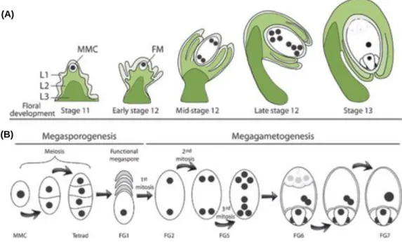
1.1.1. Ovule and megagametophyte: a synchronized development
A correct development of the ovule is required since inside of it, crucial events for sexual plant reproduction take place, such as the formation of the megagametophyte, followed by double fertilization, embryogenesis and finally, seed formation. The wild type (WT) ovule arises as a small fingertip-like structure from regions localized on the internal surface of the carpels. At this stage, the ovule primordium can be distinguished into three zones: the nucellus [where a megaspore mother cell will give rise by meiosis to the haploid megaspores], the chalaza [a central region where integuments formation occurs] and the funiculus [a supporting stalk which connects the ovule to the carpel’s placenta]. The nucellus provides a source for megasporocyte differentiation, where the megaspore mother cell (MMC) develops, without any cell division. Still during this stage, the two integuments (inner and outer) initiate their development. These sporophytic tissues will result in thick layers of cells, which will enclose the nucellus and protect the haploid generation during ovule development (Figure 2A). After MMC meiosis and spore tetrad formation, megasporogenesis ends and only one of the four megaspores, the functional megaspore, enters megagametogenesis (the other three spores will degenerate) (Figure 2B). After three rounds of mitosis and differentiation, the embryo sac is completed. InArabidopsis, the female gametophyte exhibits a Polygonum-type pattern (reviewed by
(A) (B)
Figure 1. Schematic representation of the pollen tube growth through the pistil until it reaches the embryo sac in Arabidopsis thaliana. (A) The figure illustrates a pollen grain landing on the stigma and germinating into a pollen tube.
The pollen tube grows along the style and the transmitting tract. (B) Once near an ovule, the direction of the pollen tube growth turns to the ovule entrance, where it will penetrate a synergid and release the sperm cells. (Adapted from Pereira
Maheshwari, 1950) composed by two synergid cells (cells responsible for pollen tube attraction, where pollen tube enters, burst and releases its gametes), the central cell (with two polar nucleus) and egg cell (the female gametes) and three antipodals (accessory cells that eventually will degenerate) (Schneitz et al., 1995).
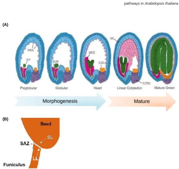
1.1.2. Seed development in A. thaliana
After fertilization of the egg and central cell, the development of a seed is initiated, resulting in a seed that can be divided into three different regions: embryo, endosperm and seed coat (SC) (Figure 3A) (for review see Nowack et al., 2010). The embryo goes through stereotypic cell-division patterns, which culminate in the embryo proper (the body of the vegetative plant) and the suspensor (an ephemeral structure that allows
Figure 2. Ovule and female gametophyte development are synchronized in A. thaliana. (A) Integument initiation occurs
in parallel with megasporogenesis (stage 11). As megagametogenesis continues, the integuments elongate (Early Stage 12). The integuments grow upward around the nucellus (mid-stage 12); and as the embryo sac is maturing (late stage 12), the outer integument begins to cover both the inner integument and the nucellus (floral development stages are according to Smyth et al., 1990). At maturity, the outer integument delimits the micropyle, which is close to the funiculus. FM, functional megaspore. (B) Megasporogenesis and megagametogenesis in Arabidopsis. A megaspore mother cell (MMC) undergoes two successive meiosis to form a tetrad, of which three cells degenerate. The remaining FM undergoes three successive rounds of mitosis within a syncytium, followed by nucleus positioning and cell differentiation. The mature embryo sac has two synergids, one egg cell and one central cell. FG1-7, female gametophyte stage 1–7, according to Christensen et al. (1997). (Adapted from Chevalier et al., 2011).
(B) (A)
communication between the embryo proper and the SC) (Belmonte et al., 2013; Lafon-Placette and Köhler, 2014). The endosperm, a nourishing tissue that supports embryo growth and germination, undergoes nuclei divisions and the nuclei migrate to form three subregions: micropylar, near the young embryo; peripheral, in the centre of the endosperm region; and chalazal, at the opposite pole of the embryo (Brown et al., 1999; Lafon-Placette and Köhler, 2014). The SC derives from the maternal integuments, which show a rapid growth upon fertilization (Moïse et al., 2005; Ezquer et al., 2016). The distinct cell types of the SC surround the embryo and endosperm, protecting them from external factors such as UV radiation or pathogens (Haughn and Chaudhury, 2005). Furthermore, the SC is involved in transferring nutrients from the maternal plant to the embryo and endosperm, and functions as a barrier to precocious germination (Becker et
al, 2014). These morphological alterations represent the morphogenesis phase of seed
development. The maturation phase is characterized by an accumulation of macromolecules that help protecting the embryo and preparing it for desiccation. Once the seed is mature, it must separate from the plant in a process called seed abscission (Lewis et al., 2006; Balanzà et al., 2016). It requires a seed abscission zone (SAZ) composed of: a separation layer, with thin cell walls near the seed body, that will degenerate at the end of the process and an adjacent layer with lignified cells (lignified layer), which will produce the tension necessary for the separation of the seed from the funiculus (Figure 3B).
1.1.3. SEEDSTICK: a regulator of ovule development
SEEDSTICK (STK) is a transcription factor (TF) belonging to the MADS-box family and essential for a viable seed formation. Generally, the MADS TFs regulate the transcription process by forming tetramers: one TF dimerizes with another to form a homo or heterodimer and then, the two dimers form a heterotetramer. The MADS-domain TFs that composed the quartet will bind to two nearby CArG box, which are the DNA-binding site of the MADS proteins, in the target promoter. This will cause a loop in the DNA (reviewed by Yan et al., 2016).
(A)
Morphogenesis Mature
(B)
Funiculus
Figure 3. Schematic representation of A. thaliana seed development. (A) Representation of seed subregions in Arabidopsis from the preglobular to mature green stages of development. Green, embryo proper (EP); dark pink,
micropylar endosperm (MCE); light pink, peripheral endosperm (PEN); orange, chalazal endosperm (CZE); purple, chalazal seed coat (CZSC); blue, seed coat (SC). (Adapted from Becker et al., 2014). (B) Representation of the seed abscission zone in A. thaliana seed. LL - lignification layer; SL - separation layer; SAZ - seed abscission zone. (Adapted from Balanzà et al., 2016).
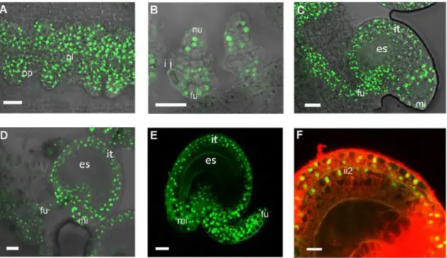
By using a SEEDSTICK-green fluorescent protein (GFP) reporter line, Mizzotti et al. (2014) were able to demonstrate the expression pattern of STK. At the beginning of ovule development, STK was detected in the placental tissues and along the ovule primordia (Figure 4A). Later, as the inner and outer integuments start to develop, the GFP signal appears only in the nucellus and funiculus (Figure 4B). At the subsequent stages until a mature seed is formed, STK continues to be expressed in the growing funiculus and its signal detection extends to the outer integument and outer layer of the inner integument (Figure 4C-E).
This expression pattern together with many genetic-based studies performed over the years supports the involvement of STK in ovule development until seed formation. STK is a powerful ovule-identity TF capable of converting WT sepals into carpeloid organs containing ovule-like structures, when overexpressed in A. thaliana plants (Favaro et al.,
Figure 4. Confocal laser-scanning images of pSTK:STK-GFP expression patterns during ovule and seed development. (A) STK-GFP protein is expressed in the placenta and in the ovule primordia. (B) When integuments arise, STK-GFP
signal is localized in the nucellus and in the funiculus. (C, D) when the ovule is mature, GFP signal can be detected throughout the integuments, funiculus and the adjacent placental region. (E) After fertilization, the STK-GFP signal is present in the outer integuments and funiculus of developing seeds. (F) Magnification of figure E with an overlay image of propidium iodide staining (specific staining of the cell wall). STK-GFP can be detected in the two layers of the outer integument and in the outer layer of the inner integument. fu - funiculus; it - integuments; ii2 - outer layer of inner integument; mi - micropyle; nu - nucellus; op - ovule primordia; pl – placenta. Scale bars = 50 µm (A and B), 40 µm (C, D and E) and 20 µm (F). (Adapted from Mizzotti et al., 2014).
2003). It redundantly controls ovule identity together with SHATTERPROOF1 (SHP1) and SHP2. In the stk shp1 shp2 triple mutant, ovules are converted into carpel-like or leaf-like structures, while single or double mutant combinations of these genes do not affect ovule identity (Pinyopich et al., 2003). The phenotype analysis of bell1 (bel1) stk
shp1 shp2 quadruple mutant, plus the results from yeast-three-hybrid and pull-down
assays, revealed that an ovule identity complex, which STK is part of, regulates the outer integument development by stabilizing the complex BEL1-AGAMOUS-SEPALLATA3. Then, this complex will activate the expression of INNER NO OUTER (INO) (Baker et
al., 1997), a gene needed for the outer integument outgrowth (Brambilla et al., 2007,
2008; Battaglia et al., 2008). As mentioned before, STK is also involved in seed formation. stk seeds were smaller compared to the WT ones and the funiculus was thicker and longer, at floral stage 17 according to Smyth et al. (1990). Furthermore, the mutant displayed defects in the SAZ that resulted in a lack of seed dispersion (Pinyopich
et al., 2003). Recently, a model was proposed in which STK would act as a repressor of
lignin deposition in the SAZ, enabling seed detachment (Balanzà et al., 2016). Together with Arabidopsis B Sister (ABS), STK regulates endothelium formation, the innermost layer of the inner integument. The abs mutant develops a deformed endothelium with flatter cells (Nesi et al., 2002). However, the abs stk double mutant has no endothelium, which led to a severely reduced seed set (Mizzotti et al., 2012).
1.1.4. Funiculus: an umbilical-cord-like structure
The funiculus is a structure that stays intact throughout ovule development until seed dehiscence, allowing the connection between the maternal plant and the ovule/seed, which highlights the importance of its biological function. Curiously, not many studies regarding morphological and histological analysis of A. thaliana funiculi are available, even though these subjects are extensively studied in other plants like Brassica napus (Chan and Belmonte, 2013) or Phaseolus vulgaris (Mawson et al., 1994). Light micrographs of the funiculus showed that this tissue is composed of an outer epidermis, a parenchymatous cortex, and an internal vascular core with xylem and phloem (Figure 5). Cells of epidermis and cortex are similar in size, while the ones of the vasculature are smaller. Additionally, by using an electron microscope, Khan et al. (2015) presented evidence of an endomembrane system and observed numerous plasmodesmata between the funiculus cell layers. Finally, it was demonstrated that the funiculus
vasculature proliferates and consequently, the epidermis and cortex cells also proliferate through anticlinal divisions.
Until now, only two genes were identified as being involved on funiculus development.
STK participates on funiculus cell division and proliferation and defines a SAZ on mature
funiculus (Pinyopich et al., 2003; Balanzà et al., 2016). On the other hand,
SPOROCYTLESS/NOZZLE indirectly controls funiculus growth by defining the chalaza
and integuments identity (Balasubramanian and Schneitz, 2000). The analysis of these mutants shows that alterations on the funiculus culminate on seed deformations. However, the network involved in these modifications has not yet been revealed. Recently, a transcriptomic analysis of funiculi was performed using a microdissection laser and the genetic profiles were compared to the ones of zygotic regions and subregions, during seed development (Khan et al., 2015). The results displayed the funiculus as a direct way for transportation of nutrients, minerals, sugars and maternal signals for the developing embryo and seed. For example, K+ ATPase 1 and REQUIRES
HIGH BORON 1 messenger RNAs (mRNAs) were found to be funiculus specific. The
accumulation of these transcripts related to transport increased along funiculus development. Moreover, the involvement of the funiculus in biological processes such as the glucosinolate metabolism and auxin response was predicted. Later, Larsson et al. (2017) confirmed the involvement of the funiculus in auxin response since it was found
Figure 5. Schematic representation of the funiculus morphology. Cross-section of a funiculus showing the epidermis (E),
that auxin biosynthesis genes and auxin transport proteins define a pre-patterning of vascular cell identity in the pre-anthesis funiculus.
1.1.5. Sporophyte and Megagametophyte: a cross talk during ovule
development
As explained before, in seed plant ovules, the maternal sporophytic tissues embed and support the haploid generation, therefore two different generations coexist in the same organ. An interaction between these two has been proposed based on the analysis of sporophytic ovule mutants and it seems to be necessary for a correct seed development. When there is a defect in the embryo’s sac surrounding tissues, megagametogenesis is severely impaired (for review see Bencivenga et al., 2011). For example, in stk shp1
shp2 ovules, integuments developed into carpel-like structures and the embryo sac
maturation was arrested after megasporogenesis (Brambilla et al., 2007; Battaglia et al., 2008). In bel1 ovules, an aberrant structure with carpel identity appears, instead of the integuments (Robinson-Beers et al., 1992) and the embryo sac is unable to develop since megagametogenesis stops at female gametophyte stage 1 (FG1). According to Christensen et al. (1997) (Figure2B) ino mutants have a normal inner integument, but the absence of the outer integument resulted in megagametogenesis disorders (Villanueva et al., 1999). The complete lack of endothelium in the abs stk ovules caused an extreme reduction in seed set (Mizzotti et al., 2012). It is worth noting that none of the genes stated above are expressed in the haploid lineage (Reiser et al., 1995; Villanueva
et al., 1999; Mizzotti et al., 2014; Ehlers et al., 2016). Hence, two important questions
arise: how is cell-to-cell signalling coordinated in ovule development and what is the nature of the players?
The correlation between hormones and ovule formation has been investigated and the analysis performed support the importance of hormones for the correct development of the ovule. CTR1 (CONSTITUTIVE TRIPLE RESPONSE 1) gene is related to ethylene signal transduction and encodes a Raf-like Ser/Thr protein kinase (Kieber et al., 1993).
ctr1 mutants show embryo sac defects that affect segregation ratios (Kieber and Ecker
1994; Drews et al., 1998). Regarding auxin, TRYPTOPHAN AMINOTRANSFERASE OF
ARABIDOPSIS 1 (TAA1) expression is highly reduced in aintegumenta ovules, where
integuments are severally affected, and the embryo sac is blocked at FG1 stage (Baker
the synthesis of auxin (Stepanova et al., 2008; Tao et al., 2008), thereby demonstrating a sporophytic-megagametophytic dialogue via auxin biosynthesis.
Nevertheless, these hormone studies require a deep analysis and it is still possible that another type of messengers, such as peptides, ligands or small RNAs, might also be involved in the cross talk between the two ovule generations.
1.1.6. Peptide Signalling: a way of communication in reproduction
Emerging evidence clarified that peptide signalling is essential for plant development (Bedinger et al., 2010; Kumpf et al., 2013; Breiden and Simon, 2016), and reproduction is not an exception. Commonly, peptides that act as signalling molecules are released through the classical secretory pathway and diffuse to the neighbour cells over short distances to bind to its receptor. Over the years, different peptides and receptors have been identified as important molecules involved in pollen-pistil interactions, including gametophyte interactions (Figure 6) (for review see Higashiyama and Yang, 2017). These include determinants of self-incompatibility, factors for pollen germination and tube growth, and pollen tube attractants. Many of the identified peptides are considered cysteine-rich peptides (CRPs). This family of peptides includes gene classes encoding for defensin-like (DEFL) peptides, lipid-transfer proteins (LTP), early culture abundant 1 (ECA1) gametogenesis-associated peptides, rapid alkalinization factors (RALFs), among others. LURE peptides (Okuda et al., 2009; Kanaoka et al., 2011; Takeuchi and Higashiyama, 2012) belong to the DEFL class and were first discovered in Toreniafournieri (a fascinating plant species with a protruding embryo sac that has been used
as a model plant for PT guidance studies). LURE1 peptides from Arabidopsis are secreted by the synergid cells and diffuse along the pathway of the PT until the surface of the septum, via the surface of the funiculus (Takeuchi and Higashiyama, 2012). The interaction with the PT is only accomplished due to the existence of tip-localized receptor-like kinases (RLK) (MDIS1-MIK [Wang et al., 2016] and PRK3-PRK6 [Takeuchi and Higashiyama, 2016]), which sense LURE1 attractant peptides. Only recently it was demonstrated the involvement of RALFs with reproductive processes. RALFs are ubiquitous in the plant kingdom and known to be involved in physiological and developmental processes in Arabidopsis (Murphy and Smet, 2014). They are ligands for receptors of the Catharanthus roseus RLK1-like family, which are associated not only with cell expansion regulation (Bai et al., 2014; Li et al., 2016) but also with functions in
pollen tube reception at the point of contact with the female gametophyte (Escobar-Restrepo et al., 2007; Miyazaki et al., 2009). Indeed, it was discovered that RALF4 and RALF19 (two PT expressed peptide ligands) (Mecchia et al., 2017) bind to the set of receptors ANXUR/BUPS, which are also localized in the PT. This interaction is in competition with RALF34 (a female derived ligand) at the interface of PT–female gametophyte contact. RALF34, then, replaces RALF4 and RALF19, allowing the rupture of PT and consequently, sperm release (Ge et al., 2017).
Despite the fact that no peptide-receptor pairs have been reported for ovule’s sporophytic tissues during ovule and/or seed development, recent studies suggest that the maternal sporophytic tissues might release small molecules as signals during important processes for reproduction. For example, the funiculus is a plausible tissue that emits a repellent (maybe a CRP), which blocks polytubey (Shimizu and Okada, 2000). The paths on the funiculus of WT pistils for PT orientation are divided into two phases: funicular guidance and micropyle guidance (Higashiyama et al., 2003). The first one would guide a PT from the transmitting tissue onto the surface of a funiculus. Then, another guidance signal, the micropyle guidance signal, would lead the PT to the micropyle. mitogen-activated
protein kinase 3 (mpk3) mpk6 mutant pollen tubes were defective in the funicular
guidance phase, while no alterations were observed during the micropylar phase (Guan
et al., 2014). This suggests the existence of ligands in the funiculus tissue that will bind
to receptors such as MPK3 and MPK6 present in the PT, allowing its orientation. Funicular guidance cues were reported to be governed by the female gametophyte (Shimizu and Okada, 2000). Recently, data revealed on the 25th International Congress
on Sexual Plant Reproduction corroborates this idea. In a poster titled “Analysis molecular mechanism of FG1 in regulating pollen tube guidance and funiculus development”, presented by Hui Zhou (Institute of Genetics and Developmental Biology, Chinese Academy of Sciences), a TF (FG1), specifically expressed in the funiculus, was shown to be down regulated on the central cell guidance mutant (ccg). CCG is a gene expressed in the central cell, which encodes for a TF (Chen et al., 2007). The analysis of fg1 mutant displayed defects in funicular guidance with addition of a high rate of unfertilized ovules. The fact that CCG might be controlling the expression of FG1 supports the idea of funicular guidance cues coming from the megagametophyte. However, it can also be indirectly derived from sporophytic tissues such as the integuments (Higashiyama, 2010). ino mutants display a disruption in PT guidance
(Baker et al., 1997), indicating that the outer integument might be involved in this process too.
Regarding post-fertilization events, a SAZ is formed in the funiculus, allowing seed abscission. STK is a key factor for the establishment of the lignin pattern in the SAZ. This TF interacts with SEUSS co-repressor, and together they function as repressors of
HECATE3, enabling a correct deposition of lignin in the vasculature of funiculus cells
(Balanzà et al., 2016). Remarkably, this study only reported transcription factors acting downstream of STK network. Floral abscission in Arabidopsis is a similar process to seed abscission. It was reported that floral abscission is regulated by peptide signalling (Aalen
et al., 2013; Meng et al., 2016), consisting of a peptide ligand, INFLORESCENCE
DEFICIENT IN ABSCISSION, its RLKs, HAESA and HAESA-LIKE2, and a downstream MPK cascade. Therefore, it is reasonable to hypothesize that a ligand-receptor pair may also play a role in the correct formation of the SAZ.
Figure 6. Schematic representation of secreted peptides and their receptors involved in pre-zygotic communication of
reproductive cells. (A) Pollen-pistil interactions in self-incompatibility responses, pollen tube growth and guidance. Ovular guidance is mediated by signals derived from sporophytic ovule tissues while micropylar guidance is mediated by signals from female gametophytic cells. (B) Enlarged image of an ovule right before sperm cell release is shown. The role of peptides and receptors is detailed in the text. Please note that peptide targets including ion channels such as KZM1 and enzymes such as PME (indicated by one asterisk) are not receptors in the ‘classical’ sense. (Adapted from Qu et al., 2015).
1.2. Objectives
By 2050, the world’s population will reach 10 billion people, which means that a solution to feed this growing population has to be found (FAO, 2017). It will be necessary to increase agricultural yields without using more arable land and water. The core purpose of agriculture is to produce viable seeds since they account for the majority of calories consumed by humans, are significant components of animal feed and are a major source for the production of high-value-added products. Hence, understanding the factors involved in seed production will help in obtaining plants with higher yields. The work presented hereby is included in the SEXSEED project, created to improve our knowledge on seed formation in model plants species and then transfer this data horizontally into agricultural crop species with economic value. Furthermore, this consortium, which is funded by the EU Horizon 2020 Research and Innovation Staff Exchange programme, aims to provide new insights into the role and the network of genes controlled by SEEDSTICK (STK), a transcription factor critical for plant reproduction and for a successful seed formation.
The analysis of ovule’s sporophytic mutant tissues clearly point to a cross talk between the sporophyte and megagametophyte. However, how this communication occur is still unknown. It might be accomplished by peptide signalling, where a peptide ligand binds to a specific receptor, originating a cascade of events. Currently, there is a gap regarding the peptide and receptors pairs present in the sporophytic ovule tissues, namely the funiculus and integuments. Since STK is highly expressed in these tissues, a bioinformatic analysis on the stk RNA-seq data was done with the purpose of inquire if peptide signalling related genes expression was being controlled by STK. The second aim was to identified ligand-receptor pairs present in the integuments and/or in the funiculus. Therefore, STK reporter line and stk mutant line were used to obtain a transcriptome from these sporophytic tissues during two different stages: before and after fertilization. To finalize, it was important to address STK involvement during PT guidance and regarding the development of the funiculus because they are processes which occur during the stages and involving the tissues selected for the transcriptome. Furthermore, the phenotypes in the stk mutant are not completely elucidative. Hence, accurate techniques were performed. This multifaceted work purposed here will shed lights on STK regulatory network.
2. Materials and Methods
2.1. Bioinformatics analysis
The Differentially Expressed Genes (DEG) of stk inflorescences transcriptomic data (Mizzotti et al., 2014) was crossed with a list of CRPs under-predicted in plants (Silverstein et al., 2007), which was named as CRPs_Silverstein, and a list of CRPs found in A. thaliana WT ovules (Huang et al., 2015), represented as CRPs_Huang. A threshold of a False Discovery Rate (FDR) < 0.05 was defined and the selected genes were briefly described using TAIR (https://www.arabidopsis.org/). The predicted expression pattern was based on e-FP Browser tool (http://bar.utoronto.ca/efp/cgi-bin/efpWeb.cgi), Klepikova Arabidopsis Atlas e-FP Browser (Klepikova et al., 2016) and the expression pattern defined by Huang et al. (2015).
2.2. Plant material and growth conditions
For all experiments, accession Columbia (Col-0) was used as a wild type A. thaliana. WT seeds were obtained from the Nottingham Arabidopsis Stock Center (NASC), United Kingdom. stk-2 (Pinyopich et al., 2003) mutant line and pSTK:STK-GFP (Mizzotti et al., 2014) reporter line were kindly provide by Lucia Colombo Lab, Universitá degli Studi di Milano. The stk-2 allele contains a 74 nucleotide insertion near the splice site of the third intron. In all the experiments, flowers utilized were from either stage 12 or 17 (according to Smyth et al., 1990). Stage 12 flowers used had the sepals opened, allowing to see a small portion of the petals. Stage 17 siliques used in the studies were defined as the first three siliques counting from the top to the base of the stem, starting on the younger silique, whose floral parts fell.
Seeds were surface sterilized in the laminar flow hood, using a bleach solution. Less than 100 µL of each seeds line were placed in 1.5 mL eppendorf tubes, where it was added 1 mL of 1% NaClO/ 0.02% Tween-20 solution. The tubes were shaken for 3 minutes to soak the seeds and then, a washing step with ddH2O was performed and
repeated 3 times. 1 mL of 0.1% Plant preservative mixture/0.02% Tween-20 solution was added to each tube and they were placed in the dark for 1-2 days at 4⁰C, in order to break the seeds dormancy. Afterwards, the supernatant was removed, 0.1% agar was added to the tubes and, using a tip, the seeds were transferred to 1/2 MS (Murashige
and Skoog) media plates, containing 1% (w/v) sucrose (pH 5.8 with KOH) solidified with 0.8% (w/v) agar. The plated seeds were placed in a growth chamber at 22⁰C and continuous light. When seedlings were 7 to 10 days old, they were moved to soil pots and were grown in a growth chamber under long-day conditions: 16 hours light at 21⁰C and 8 hours dark at 18⁰C.
2.2.1. Preparation of plant material for microscopy
To check the fluorescence of pSTK:STK-GFP line, under a SZ61 stereo microscope (Olympus) and using hypodermic needles, pistils were removed from the flowers, dissected and ovules were mounted in a microscope slide with H2O. Then, observation
of the ovules occurred using an inverted microscope (IX71, Olympus) equipped with GFP channel. Images were captured with a 3CCD camera (C7780-20, Hamamatsu Photonics), using the cellSens software.
Pistils and siliques from WT and stk plants from both stages 12 and 17 (Figure 14) were collected and placed on a microscope slide, under the SZX16 stereo microscope (Olympus). Images were captured using the U-CMAD3 camera (Olympus).
Pictures from Figure 11 were captured while the procedure was being done, using a SZX16 stereo microscope (Olympus) equipped with a U-CMAD3 camera (Olympus).
2.3. Genotyping
stk mutant line was confirmed by Polymerase Chain Reaction (PCR). A small leaf disk
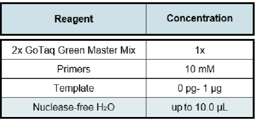
was enough to extract the amount of genomic DNA (gDNA) required for PCR amplification. The gDNA was extracted according to Edwards et al. (1991). The PCR was carried out using GoTaq® Green Master Mix (Promega). Specific primer sequences and expected band sizes are listed in Supplemental Table 1 and PCR mixture is represented on Table 1.
A PCR Thermal Cycler Dice® Gradient (TaKaRa) was used and the program was set according to Table 2.
PCR products were separated in a 2% (w/v) agarose gel in 1X TAE (40 mM Tris-acetate and 1 mM EDTA, pH 8.3); 0.5 mg/mL ethidium bromide was added prior to polymerisation. Using the Gene Ladder 100 (Nippon Gene) as a molecular weight marker and 1X TAE as running buffer, the electrophoretic separation was conducted at 100 V and non-limiting amperage for 30 minutes. The DNA was visualized in a UV transilluminator (302-365 nm).
Table 1. Reaction mixture used for PCR.
2.4. Protoplast isolation
Emasculated pistils from stage 12 were dissected under a SZ61 stereo microscope (Olympus), using hypodermic needles. Ovules from one pistil were released to a plastic disk containing 100 µL of enzymatic solution, and the disk was placed at 28⁰C with different incubation times. Three enzymatic solutions were used. Solution 1 was as described by Kawano et al. (2011) with some modifications: 1% cellulase (CEL, Worthington), 0.3% maceroenzyme (R-10, Yakult), 0.05% pectolyase (Y-23, Yakult) and 0.45 M mannitol. Solution 2 had the same composition as solution 1 with the only difference being the addition of two enzymatic enhancers: 1 mM CaCl2 and 0.2% BSA
(Bovine Serum Albumin). Solution 3 was as described by Park et al. (2016) and consisted of 1% cellulose (CEL, Worthington), 0.5% maceroenzyme (R-10, Yakult), 0.1% pectolyase (Y-23, Yakult), 0.4 M mannitol, 10 mM CaCl2, 0.1% BSA, 20 mM KCl and 20
mM MES (4-morpholineethanesulfonic acid), pH 5.7. Before the addition of enzymes and CaCl2, the MES solution containing mannitol and KCl was preheated at 70⁰C for 5
minutes to resolubilize any crystals. The solution was then allowed to cool to room temperature, and enzymes were added, followed by incubation of the solution at 55⁰C for 10 minutes, enhancing enzyme solubility. 10 mM CaCl2 was added after the solution
reached room temperature. The final enzyme solution was filter sterilized by syringing through a 0.45 μm membrane. Protoplasts were observed in bright field under an inverted microscope (IX71, Olympus) and pictures were taken with a 3CCD camera (C7780-20, Hamamatsu Photonics), using the cellSens software.
2.5. Nuclei isolation
Nuclei isolation procedure from WT and ATML1::NLS-VENUS leaves was adapted from Deal and Henikoff (2011) and performed as follows: 0.1 g of leaves were grinded in liquid N2 and the powder was resuspended in 5 mL of cold nuclei purification buffer (NPB) [20
mM MOPS, pH 7; 40 mM NaCl; 90 mM KCl; 2 mM EDTA; 0.5 mM EGTA, pH 8; 0.5 mM spermidine]. Afterwards, the extracts were filtered to a 50 mL falcon tube through a 70 µm nylon mesh and they were centrifuged at 1000 rpm for 3 minutes. The nuclei were gently resuspended in 500 mL of cold NPB and moved to a 1.5 mL eppendorf tube. Cell sorting of ATML1::NLS-VENUS leaves nuclei solution was performed using the SH800 cell sorter (Sony, Tokyo, Japan).
The isolation of nuclei from pSTK:STK-GFP line was performed according to DW-K et
al. (2012) with some modifications. Ten pistils per sample were emasculated, and 24
hours later, they were dissected under a SZ61 stereo microscope (Olympus), using hypodermic needles. Ovules were placed inside of an eppendorf tube, containing 20 µL of nuclei isolation buffer (NIB) [0.25 M sucrose; 15 mM Piperazine-N, N′-bis (2-ethane-sulfonic acid), pH 6.8; 5 mM MgCl2; 60 mM KCl; 15 mM NaCl; 1 mM CaCl2; 0.9% Triton
X-100; 1 mM PMSF (phenylmethanesulfonyl fluoride)]. With a pre-cooled pestle, the ovules were macerated and 300 µL of NIB was added. The samples were incubated on ice for 15–30 minutes, subjected to centrifugation at 1000 g for 4 minutes at 4⁰C, ressuspended and filtered through a 0.45 μm membrane. All the steps were performed on ice whenever possible. For the isolation of nuclei from pre-fixed ovules, ovules were obtained in the same way as described above, but they were first placed in 50 µL of RNAlater™ Stabilization Solution (Ambion) and 4% paraformaldehyde solution was added. After 5 minutes incubation at room temperature, the samples were macerated with a pestle and centrifuged at 1500 g at 4⁰C for 1 minute. Supernatant was removed for the solution become 2x concentrated, 100 µL of NIB was added and the tubes were placed on ice for 15 minutes. The samples were centrifuged, and supernatant was removed. Finally, samples were filtered through a 0.45 μm membrane.
To each sample analysed under the microscope, 5 μM of 4′,6-diamidino-2-phenylindole (DAPI) was added. The nuclei samples were analysed with an Axio Imager A2 microscope (Zeiss) equipped with a DAPI and a GFP channel. Images were captured with an Axiocam 506 color (Zeiss), using Zen 2 software (Zeiss).
2.6. Transcriptome from funiculus cells
2.6.1. Collection of funiculi and ovules from A. thaliana
Under a SZ61 stereo microscope (Olympus), WT and stk pistils from both stages 12 and 17 flowers were first dissected, and ovules were placed into a 50 µL droplet of medium (whose composition had been described in Gooh et al., 2015) in a plastic dish (Φ 3.5 cm). Each funiculus was separated, using a surgical knife and, one at a time, they were transferred with a tungsten needle to another plastic dish, containing 10 μL of medium. Then, each funiculus was transferred to an eppendorf, which already had 40 μL of RNAlater™ Stabilization Solution (Ambion). For samples containing only ovules, 5 pistils
were dissected in the same conditions as described before and all the ovules were transferred to the inside of an eppendorf tube with 40 μL of RNAlater™ Stabilization Solution (Ambion).
2.6.2. mRNA extraction and cDNA synthesis
mRNA was extracted from both WT and stk funiculi and ovules, from stages 12 and 17 flowers, using the DynabeadsTM mRNA DIRECTTM Micro Kit (Invitrogen) according to
the manufacturer’s instructions. RNA was retro-transcribed using the High-Capacity RNA-to-cDNA™ Kit (ThermoFisher). Reaction mixture was as described in Table 3.
The reaction was incubated at 37⁰C for 60 minutes. Then, it was stopped by heating to 95⁰C for 5 minutes. cDNA samples were kept at -30⁰C.
2.6.3. Validation of mRNA and cDNA quality
mRNA quality from funiculi and ovules samples were validated using the Agilent RNA 6000 Pico Kit (Agilent Technologies), followed by a running of the chip in an Agilent 2100 Bioanalyzer (Agilent). cDNA was tested by a semiquantitative reverse transcriptase (RT)-PCR using AGP9 (AT2G14890), AGP4 (AT5G10430), SUS4 (AT3G43190), FIS2 (AT2G35670) and ACT7 (AT5G09810) primers. Specific primer sequences and
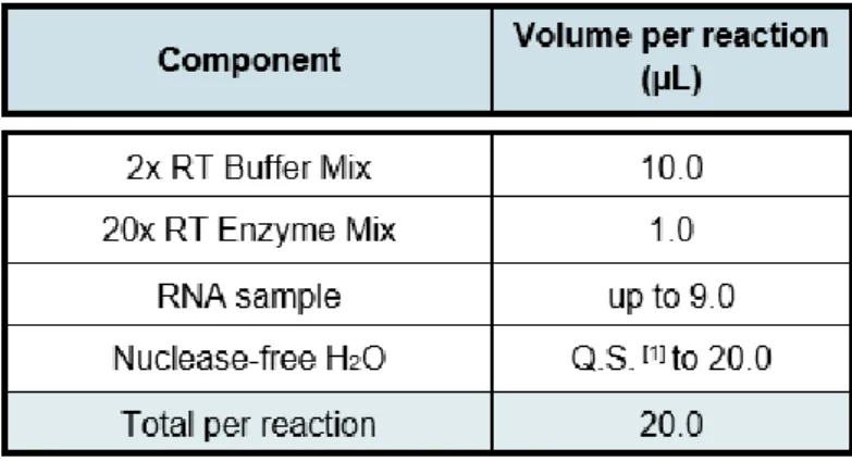
Table 3. Reaction mixture used for RNA retro-transcription. RT – Reverse
expected band sizes are listed in Supplemental Table 1. PCR and electrophoretic separation were performed as described earlier.
2.6.4. RNA-sequencing library preparation
mRNA was extracted from one sample containing 100 funiculi from stage 12 of both WT and stk, from three biological replicates of 100 funiculi from stage 17 of both WT and stk and from another three biological replicates of ovules from 5 pistils at both stage 12 and 17 of WT and stk. Sequencing libraries were prepared according to the manufacturer’s instructions, using the TruSeq® RNA Sample Preparation v2 (Illumina Inc.) and
sequenced with a NextSeq 500 sequencer (Illumina). Sequencing and data analysis were performed as reported in Kadokura et al. (2018).
2.7. Aniline blue staining of pollen tubes
WT and stk pistils from stage 12 flowers were emasculated and 24 hours later, they were hand pollinated with WT pollen. 12 hours after, pistils were collected and fixed in absolute ethanol and glacial acetic acid in a 3:1 ratio and left for more than 2 hours at 4⁰C. The material was washed in 70% ethanol, 50% ethanol, 30% ethanol, 10% ethanol and finally in ddH2O, for 20 minutes each. After overnight incubation 1 M NaOH at 4⁰C, the material
was washed two times with ddH2O and stained with 0.1% (w/v) decolorized aniline blue
solution (DAB) (in 0.1 M K3PO4). The material was observed under an ultraviolet
illumination using an upright Axio Imager A2 microscope (Zeiss). Images were captured with an Axiocam 506 color (Zeiss), using Zen 2 software (Zeiss).
2.8. SR2200 staining
SCRI Renaissance 2200 (SR2200) staining was performed as reported in Musielak et
al. (2016). Briefly, WT ovules were manually dissected out of the pistil and collected in a
drop of staining solution on a microscope slide. Soft vacuum was applied for 5 minutes at room temperature. Afterward, the staining solution was replaced by ddH2O and again
incubated under soft vacuum for 5 minutes. After replacing the water with 10% glycerol, the sample was mounted under a coverslip. Images were obtained with a LSM780NLO
confocal microscope (Zeiss) and processed with ZEN software (Zeiss). SR2200 was excited with a 405 nm laser line and emission recorded between 415 and 476 nm (405/415–476). Two pistils from different plants were used.
2.9. Propidium iodide staining
Propidium iodide (PI) staining was used to stain funiculi. For that, WT ovules from one pistil were collected, mounted in 10 µg/mL of propidium iodide in water, and analysed. A LSM780NLO confocal microscope (Zeiss) was used and images were processed with ZEN software (Zeiss). PI was excited with a 543 nm laser line and emissions were detected between 600 and 640 nm (543/600-640). For observations, ovules from 2 pistils were analysed.
2.10. CalcoFluor White M2R staining
Pistils previous emasculated and siliques were harvested and fixed in absolute ethanol and glacial acetic acid in a 3:1 ratio and left for more than 2 hours at 4⁰C. The material was washed in 70% ethanol, 50% ethanol, 30% ethanol, 10% ethanol and finally in ddH2O, for 20 minutes each. After incubation on 1 M NaOH at 4⁰C for more than two
days, the material was washed in ddH2O for 30 minutes, stained with 1% Calcofluor
White M2R, freshly made, for 1 hour and washed again in ddH2O for another 30 minutes.
After mounting the samples in water in a slide microscope, they were immediately observed. A LSM780NLO confocal microscope (Zeiss) equipped with a chameleon laser was used and images were processed with ZEN software (Zeiss). For confocal imaging, Calcofluor White M2R was excited with a. 405 nm laser line and emissions were detected between 425 and 475 nm (405/425-475), while for two-photon imaging, the excitation was in 900 nm and emissions were around 780 to 810 nm (900/780-810). The observations were done in two pistils and two siliques.
2.11. SyBr Green I staining
Pistils previous emasculated and siliques were collected and cleared in ClearSee solution following the protocol of Kurihara et al. (2015). After one-month incubation in ClearSee, the samples were stained in 0.1% Calcofluor White M2R (prepared in
ClearSee) for 1 hour and washed in ClearSee for 30 minutes. The samples were stained with SYBR™ Green I Nucleic Acid Gel Stain (Invitrogen) diluted 10 times in ClearSee for 30 minutes, washed 3 times for at least 30 minutes in ClearSee and mounted on slides with ClearSee solution for imaging. A LSM780NLO confocal microscope (Zeiss) was used and images were processed with ZEN software (Zeiss). Calcofluor White M2R was excited with a 405 nm laser line and emissions were detected between 425 and 475 nm (405/425-475), while SyBr Green I was excited with a 494 nm laser and emission was detected at 519 nm (494/519). Observations were performed in two siliques and two pistils.
2.12. Quantification of funiculus length
Pistils previous emasculated and siliques were collected, the valves were removed and the remain biological material was cleared in 1 M NaOH and stained with 1% Calcofluor White M2R, as described before. The samples were placed on a microscope slide and mounted in water, so that the funiculus could be as straight as possible. Maximum intensity projections taken with LSM780NLO confocal microscope (Zeiss) and processed with ZEN software (Zeiss) were analysed with Image J software (Schneider et al., 2012). Representative pictures were from 18 WT and 15 stk funiculi (among two plants) from stage 12 flowers and 17 WT and 19 stk funiculi (among five plants) from stage 17 flowers. Straight lines were designed along the funiculus to measure its length. The statistical significance of the differences was assessed using a Student’s t-test (α = 0.05).
2.13. Imaging Processing
Regarding scale bars and small image adjustments, all images were processed using the software Image J (Schneider et al., 2012).
3. Results
3.1. Bioinformatics analysis
With the aim of identify putative CRPs targets of STK, the transcriptomic data of stk inflorescences (Mizzotti et al., 2014) was crossed with available lists of CRPs under-predicted in plants (Silverstein et al., 2007) and CRPs found in A. thaliana WT ovules (Huang et al., 2015).
stk RNA sequencing (RNA-seq) data contained 17741 genes from which only 1822
genes had a significant p-value (p < 0.05). These DEGs were crossed with CRPs lists and the results showed that 3.8% of the genes present in the stk transcriptome are CRPs. Only 10 genes were common to the three sets of data, 57 genes were found in the stk and CRPs from ovules data, 3 genes were exclusively in the stk inflorescences and in the list of predicted CRPs in plants, and 134 genes were expressed in both CRPs lists (Figure 7).
From the lists obtained by overlapping stk transcriptome with CRPs data, the genes that were presented in the 3 sets of data and the ones only present in stk and in one of the CRPs lists were selected. After filtering the genes by choosing a False Discovery Rate (FDR) below 0.05, each gene was briefly described, using TAIR (http://www.arabidopsis.org/). Additionally, the putative expression pattern of the genes
stk
Figure 7. Venn diagram representing the crossing of Differentially Expressed Genes of stk RNA sequencing (stk) with
CRPs under-predicted in plants (CRPs_Silverstein) and CRPs found in A. thaliana WT ovules (CRPs_Huang).
CRPs_Huang CRPs_Silverstein
was also investigated based on the e-FP Browser tool (http://bar.utoronto.ca/efp/cgi-bin/efpWeb.cgi), Klepikova Arabidopsis Atlas e-FP Browser (Klepikova et al., 2016) and on Huang et al. (2015), which grouped the CRPs into six different clades: CRPs enriched in immature ovules (A), in mature ovules (B), in pollinated ovules (C), in fertilized ovules at two successive stages (D and E) and during early seed development (F). This step was performed in order to find genes preferentially expressed in reproductive tissues. With the information collected, some interesting genes were selected to further be subject of study.
Overall, the stk transcriptome presented genes, which belong to different CRPs classes: DEFL, RALF, LTP, ECA1, etc. Among the CRP genes existing in stk inflorescences and with a FDR < 0.05 (Table 4), the gene with the highest fold change (6.1x) was
AT1G56415. This gene has not yet been described and so, the only information available
was the expression pattern according to Huang et al. (2015), which included this gene in pattern D clade (mainly expression on fertilized ovules). LCR73 and AT1G13605 were the next two genes with the highest fold change (4.9x). Their expression was mainly on seeds. AT2G43530 and RALFL24 were two genes that, even though with a lower fold change in stk data (3x and 2.1x, respectively), their expression pattern was quite interesting since it was similar to STK’s (highly expressed in ovary tissues). In this list, most of the genes were mainly expressed in reproductive tissues, after fertilization stages. It is worth noting the fact of some CRPs from Table 4 have a sequence similarity to the pollen coat protein gene family (LCR73, LCR38, LCR48, LCR68) or to the male component of the self-incompatibility response (SCRL10) gene family.
Table 4. List of genes encoding for CRPs present in stk RNA sequencing (stk-WT). Gene locus, Log2 of fold change (FC), a brief description of the gene’s function,
the gene expression pattern according to Huang et al. (2015) and the expression profile according to eFP Browser and Klepikova Arabidopsis Atlas eFP Browser are detailed for each gene. Huang et al. (2015) defined six distinct expression patterns: CRPs enriched in immature ovules (A), in mature ovules (B), in pollinated ovules (C), in fertilized ovules at two successive stages (D and E) and during early seed development (F). It is also presented the false discovery rate (FDR) for stk RNA-seq (FDR < 0.05). The list is organized in a descendant order according to the fold changes in the stk RNA-seq.
At the same time, a manual analysis on stk transcriptome was performed. The purpose was not only to look for CRPs that did not appear when the transcriptomes were crossed, but also receptors with a kinase domain and other genes deregulated in stk inflorescences and possibly thrilling to study in the future. Once again, a description of each gene along with the expression pattern was performed when possible. Analysing Table 5, the gene with the highest absolute fold change (4.3x) was AT5G63087, a CRP that was not identified on the previous analysis and whose expression is specific on seeds. SERINE CARBOXYPEPTIDASE- LIKE 41 (SCPL41) was downregulated 1.5x in the absence of STK, with a correspondent FDR < 0.05. Although SCPL41 is not a CRP or receptor, it is a gene to take in account as a possible player of STK network, since both share a very similar expression pattern. From this list, the genes with a FDR above 0.05 were NET3C, RALFL8, RALFL9 and LCR69. Again, a link between STK and the
male reproductive tissues was found, since CRPs specifically expressed on pollen grain and PT were upregulated on the stk transcriptome.
One of the objectives of the present work was to obtain a transcriptome from ovule’s sporophytic tissues, where STK is localized. Therefore, the bioinformatics analysis performed was a starting point on understanding if STK could possibly be regulating genes related to peptide signalling such as CRPs. The information collected will be very helpful for the analysis of specific transcriptomic data from integuments and/or funiculus cells.