UNIVERSIDADE DA BEIRA INTERIOR
Ciências
New signalling pathways in reproductive tissues
associated with mitochondrial dysfunction
induced by metabolic diseases
Antónia Teixeira Diniz
Dissertação para obtenção do Grau de Mestre em
Bioquímica
(2º ciclo de estudos)
Orientador: Doutor Luís Pedro Rato
Co-orientador: Prof. Doutor Pedro Fontes Oliveira
Co-orientador: Profª. Doutora Branca Silva
ii O conteúdo do presente trabalho é da exclusiva responsabilidade do autor:
iii
Acknowledgments
First of all, to my advisor Doctor Luís Pedro Rato for all the patience and demonstrated availability, for all the teachings, support and advises throughout the work, as well the criticisms, corrections and relevant suggestions made during this orientation.
To my co-advisers Professor Branca Silva and Professor Pedro Fontes Oliveira for all the availability, for the scientific competence, the suggestions, criticisms, and opinions that contributed to the development of this work.
A special thanks to Doctor Marco G. Alves for all the advices, corrections and suggestions that contributed to the elaboration of this thesis.
To my laboratory colleagues, especially to my co-worker Ana Silveira for companionship and for all help and support along this year.
To all my friends who accompanied me on this journey, for all the good advices and for being there at less good times.
And as it could not be, a special big thank you to my parents for always being there when needed, motivation and unconditional support always. This work is also yours.
iv
Resumo
A diabetes mellitus é uma doença metabólica cuja incidência está a aumentar na população mundial. Classicamente pode ser divida em dois tipos: tipo 1 que se carateriza por um estado de insulinodependência, e o tipo 2 em que se verifica uma resistência à ação da hormona. Fatores externos associados ao estilo de vida, como os maus hábitos alimentares, em particular o consumo frequente de dietas altamente calóricas, em combinação com outros fatores como o sedentarismo, são as principais causas para o incremento de patologias como a diabetes mellitus tipo 2 (DMT2). A desregulação metabólica associada à DMT2 leva ao aparecimento de outras comorbidades, nomeadamente uma diminuição da fertilidade masculina. O eixo hipotálamo-hipófise-testículo, conhecido como eixo reprodutivo, é sensível às alterações metabólicas induzidas pela DMT2. Estudos recentes têm mostrado que alterações endócrinas e metabólicas associadas à DMT2 afetam a fisiologia dos órgãos reprodutivos, nomeadamente a bioenergética mitocondrial pelo que a manutenção da função mitocondrial nos órgãos reprodutivos é essencial. Assim, pretendemos estudar qual o impacto da DMT2 nas vias moleculares subjacentes à regulação da função mitocondrial testicular e epididimal. Para isso usou-se o modelo animal de DMT2 onde se avaliou a expressão de proteínas-chave envolvidas tanto regulação da biogénese mitocondrial, como na ativação do sistema de defesa antioxidante. Também avaliámos os efeitos da DMT2 no número de cópias de DNA mitocondrial (mtDNA) e na expressão dos níveis dos complexos da cadeia respiratória mitocondrial em ambos os tecidos. Avaliámos ainda os danos induzidos pelo stress oxidativo, como a carbonilação e nitração de proteínas, assim como a peroxidação lipídica. Os resultados demonstraram que a DMT2 diminui a expressão da sirtuína 1 (SIRT1) e sirtuína 3 (SIRT3) no tecido testicular. Verificou-se uma diminuição na expressão dos complexos III e V da cadeia respiratória mitocondrial. Não se verificaram alterações no conteúdo do mtDNA testicular e nas atividades das enzimas antioxidantes. No entanto, estes resultados foram acompanhados por um aumento da peroxidação lipídica e nitração de proteínas. Ao nível epididimal, observou-se uma diminuição na expressão do regulador-chave da biogénese mitocondrial, o coativador 1 α do receptor γ activado pelo proliferador de peroxissoma, assim como a expressão das SIRT1 e SIRT3. Embora não se tenham verificado alterações significativas no conteúdo do mtDNA, a DMT2 induziu uma diminuição significativa da expressão dos complexos II, III e V. Também se observou uma diminuição significativa na atividade das enzimas envolvidas no sistema de defesas antioxidantes, que culminou com o aumento da nitração de proteínas. Os resultados obtidos sugerem-nos que a DMT2 induz uma diminuição da expressão de reguladores-chave da biogénese mitocondrial dos órgãos reprodutivos, comprometendo assim as vias moleculares envolvidas na regulação da função mitocondrial e, consequentemente, na manutenção do sistema de defesas antioxidantes. Desta forma, torna-se essencial aprofundar os conhecimentos na bioenergética mitocondrial para que se possam desenvolver novas abordagens terapêuticas, de modo a atenuar o
v aumento da infertilidade masculina, principalmente nos países mais desenvolvidos onde a elevada prevalência das doenças metabólicas é considerado um problema de saúde pública.
Palavras-chave:
Diabetes mellitus; Fertilidade masculina; Testículos; Epidídimo; Biogénese mitocondrial; Stress oxidativo
vi
Resumo Alargado
Os testículos são os elementos centrais do sistema reprodutor masculino. As suas principais funções consistem na produção das células germinativas, processo designado de espermatogénese, bem como na síntese das hormonas esteroides. Nos testículos estão presentes diferentes tipos de células entre elas as células de Sertoli e as células de Leydig. As células de Sertoli desempenham um papel fundamental no desenvolvimento das células germinativas, uma vez que representam o suporte físico e nutricional para o desenvolvimento da espermatogénese. Após serem libertados nos túbulos seminíferos os espermatozoides são células altamente diferenciadas, mas incapazes de fertilizar. É no epidídimo que vão sofrer diversas modificações que vão permitir que esses espermatozoides se tornem maduros, adquirindo mobilidade e capacidade de fertilizar. Este ducto permite também o armazenamento dos espermatozoides férteis num estado viável, dentro da cauda do epidídimo, até serem ejaculados. Dado que tanto o tecido epididimal como o testicular requerem níveis adequados de energia é imprescindível assegurar o metabolismo celular para a manutenção da capacidade reprodutiva do indivíduo.
A diabetes mellitus (DM) é considerada como uma das doenças crónicas mais prevalentes no mundo, que causa sérias perturbações no metabolismo celular. Dado o aumento crescente de número de casos que se tem verificado de ano para ano, a DM é considerada como uma ameaça à saúde humana a nível global. Esta patologia é considerada como um distúrbio metabólico que pode ser dividido em dois tipos principais, tipo 1 e tipo 2. Estes dois tipos podem ser descritos sumariamente como desordens metabólicas caraterizadas por um estado de hiperglicemia (valores elevados de glucose no sangue), resultando de uma secreção defeituosa de insulina, resistência à ação da insulina, ou ambas.
A DM tipo 1 desenvolve-se geralmente em idade jovem e é caraterizada por uma destruição das células beta pancreáticas pelo sistema imune, resultando na dependência de um tratamento com insulina exógena. A DM tipo 2 ocorre quando a produção de insulina pelas células beta não é suficiente para manter os níveis de glucose no sangue dentro de valores fisiológicos normais (72-99 mg/dl), levando a exaustão funcional das células beta. Ao longo dos anos tem se vindo a verificar que a doença começa a ser detetada cada vez mais cedo em crianças e adolescentes.
Devido ao estado hiperglicémico, a DM está associada a uma exacerbada produção de espécies reativas de oxigénio (EROS), sobretudo na mitocôndria, onde ocorre a grande maioria dos processos metabólicos. Esta superprodução destes agentes oxidantes leva a danos celulares a nível do DNA (principalmente do DNA mitocondrial), dos lípidos e das proteínas, sendo um potencial alvo as células germinativas que se encontram desguarnecidas de defesas antioxidantes e dependentes do microambiente criado pelo tecido testicular e epididimal. Numa forma de tentar perceber quais os processos que levam à criação de defesas antioxidantes de forma a neutralizar as EROs, têm-se vindo a elucidar possíveis vias moleculares que ativam as defesas antioxidantes. Em várias delas podemos encontrar
vii proteínas chave na regulação da homeostase mitocondrial, como é o caso da sirtuína 1 (SIRT1), sirtuína (SIRT3) e o coativador 1 α do receptor γ activado pelo proliferador de peroxissoma (PGC-1α). O PGC-1α é considerado como o maior regulador da biogénese mitocondrial, sendo ativado em resposta a diversos estímulos ambientais, tais como a exposição ao calor ou em períodos de fome prolongada. Esta proteína tem a capacidade de ativar a biogénese mitocondrial através da ativação de diversos fatores de transcrição envolvidos no crescimento e divisão mitocondrial, e induz a expressão de proteínas que ativam enzimas antioxidantes, como é o caso da SIRT3. A SIRT3 pertence à família das desacetilases NAD+-dependentes, sendo também uma proteína chave como reguladora do
metabolismo. SIRT3 exerce a sua função removendo grupos acetilo de proteínas envolvidas em repostas celulares ao stress oxidativo, como é o caso PGC-1α. Além disso, a SIRT3 tem a capacidade de induzir a expressão de enzimas antioxidantes, como é o caso da superóxido dismutase e da catalase.
Assim o objetivo do trabalho foi avaliar os efeitos da DM tipo 2 na expressão das proteínas SIRT1, PGC-1α e SIRT3 e as implicações em termos oxidativos. Para isso utilizou-se o modelo animal da DM tipo 2, ratos Gotokakizaki (modelo de ratos não obesos que desenvolvem espontaneamente a DM tipo 2). Os animais foram divididos em dois grupos, o primeiro grupo constituído por sete ratos saudáveis e o segundo constituído por sete animais com DM tipo 2. Após oito meses, os animais foram sacrificados e procedeu-se à recolha do tecido testicular e epididimal e avaliou-se a expressão de proteínas-chave envolvidas na regulação da biogénese mitocondrial e a atividade das enzimas envolvidas nas defesas antioxidantes, em particular a glutationa peroxidase, glutationa redutase, superóxido dismutase e catalase, bem como os danos causados pelo stress oxidativo nas proteínas e nos lípidos. Para além disso, avaliamos, também o número de cópias mitocondriais e a expressão dos complexos mitocondriais em ambos os tecidos.
Os resultados demonstraram que em ambos os tecidos o eixo molecular SIRT1/PGC-1α/ SIRT3 encontra-se desregulado, uma vez que as expressões destas proteínas se encontram diminuídas. Consequentemente, essa desregulação leva a uma diminuição na atividade das enzimas do sistema antioxidante no tecido epididimal, contrariamente ao que acontece no tecido testicular, onde as atividades das defesas antioxidantes permaneceram inalteradas. Apesar destas observações, verificou-se em ambos os tecidos um aumento da nitração das proteínas e da peroxidação dos lípidos. Para além disso verificamos uma diminuição nos complexos III e V no tecido testicular e uma diminuição mais acentuada nos complexos II, III e V no tecido epididimal.
Podemos assim concluir que a DM tipo 2 causou uma desregulação das vias moleculares responsáveis pela manutenção da biogénese mitocondrial, como é o caso do eixo molecular SIRT1/PGC-1α/SIRT3. Por conseguinte o sistema antioxidante também é afetado, promovendo danos oxidativos. No entanto, devem ser feitos mais estudos para elucidar estes mecanismos moleculares tendo em vista o desenvolvimento de novas abordagens terapêuticas de modo a diminuir a incidência da infertilidade masculina.
viii
Abstract
Diabetes mellitus is a metabolic disease and its incidence is reaching epidemic proportions. Classically it can be divided into two types: type 1 diabetes mellitus, characterized by an insulin-dependent state, and type 2 diabetes mellitus (T2DM), where there is a resistance to the action of the hormone. External factors associated with lifestyle, such as eating behaviors, in particular, the excessive consumption of high caloric diets in combination with other factors, such as sedentary lifestyle, are the main causes for the increased incidence of T2DM. The metabolic deregulation associated with T2DM leads to the emergence of other comorbidities, notably a deregulation of male fertility. The hypothalamus-pituitary-testicle axis, also known as a reproductive axis, is sensitive to the metabolic changes induced by T2DM. Recent studies have shown that endocrine and metabolic alterations associated with T2DM affect the physiology of reproductive organs, mainly their mitochondrial bioenergetics. Maintaining mitochondrial function in reproductive organs is imperativefor the maintenance of the reproductive capacity of the individual. Thus, we aimed to study the impact of T2DM on the molecular pathways underlying the control of the testicular and epididymal mitochondrial function. For this, we used an animal model of T2DM, in which we evaluated the expression of key proteins involved in the regulation of mitochondrial biogenesis and in mitochondrial function. We measured the activity of the enzymes of the antioxidant defense system. We also evaluated the effects of T2DM on the number of mitochondrial DNA copies and on the expression of the levels of the mitochondrial respiratory chain complexes in both tissues. Finally, we evaluated the parameters of oxidative stress (OS), such as carbonylation and proteins nitration, as well as lipid peroxidation.
Our results showed that T2DM decreased the expression of sirtuin 1 (SIRT1) and sirtuin 3 (SIRT3) in the testicular tissue. There was also a decrease in the expression of complexes III and V of the mitochondrial respiratory chain, but the content of mitochondrial DNA (mtDNA) remained unchanged in the testicular tissue. There were no alterations on the activities of the antioxidant enzymes, however, these results were accompanied by an increase in lipid peroxidation and nitrate of proteins. In the epididymal tissue, a decrease was observed on the expression of the key regulator of mitochondrial biogenesis, the peroxisome proliferator activated receptor γ co-activator 1 α (PGC-1α), as well as on the SIRT1 and SIRT3 expression levels. Although there were no changes in the mtDNA content, DMT2 has induced a significant decrease in the expression of complexes II, III and V in the epididymis. There were also decreases in the activities of the enzymes involved in the system of antioxidant defenses, which were accompanied by an increase of protein nitration. The results suggested that T2DM disrupted the expression of key regulators of the mitochondrial biogenesis of the reproductive organs, thereby compromising the molecular pathways involved in the regulation of the mitochondrial function and, consequently in the maintenance of the antioxidant defense system. In this way, it is essential to deepen the knowledge in mitochondrial bioenergetics in order to develop possible therapeutic approaches to attenuate the increased decline of male
ix fertility, especially in developed countries where the prevalence of metabolic diseases is a major public health concern
Keywords:
Diabetes mellitus; Male fertility; Testes; Epididymis; Mitochondrial biogenesis; oxidative parameters.x
List of Abbreviations
3-NT - 3-nitrotyrosine
4-HNE - 4-hydroxy-2-nonenals
β2MG - nuclear-encoded beta-2-microglobulin gene
AMPK - 5’ adenosine monophosphate-activated protein kinase ATP - adenosine triphosphate
BMP4 - bone morphogenic protein 4 BSA - bovine serum albumin BTB – blood-testis barrier CAT - catalase
CREB - cyclic AMP response-element-binding protein
DHAP - fructose-1-phosphate aldolase yielding dihydroxyacetone phosphate DM - diabetes mellitus
DNA - deoxyribonucleic acid DNP - 2,4-dinitrophenyl hydrazone ETC - electron transport chain
FOXOs- forkhead box transcription factors FOXO3a- forkhead box O3
FSH - follicle-stimulating hormone GCs – germ cells
GnRH – gonadotropin releasing hormone GPx - glutathione peroxidase
GK rats- Goto-Kakizaki rats GR - glutathione reductase GSSG- oxidized glutathione GTT-glucose tolerance test
HOMA-IR- Homeostasis assessment model-insulin resistance HPT-axis - hypothalamus-pituitary-testicles axis
LCs - Leydig cells
LDH - lactate dehydrogenase LH - luteinizing hormone
mtDNA – mitochondrial deoxyribonucleic acid mtND1- mitochondrial ND1 gene
NAD+ - nicotinamide adenine dinucleotide
NRF1 -nuclear respiratory factor 1 NRF2-nuclear respiratory factor 2 O2 – oxygen
OAA - oxaloacetate OS – oxidative stress
xi PBS - phosphate buffer saline
PGC-1α - peroxisome proliferator activated receptor γ co-activator 1α PPARs - peroxisome proliferator activated receptors
PUFAs - polyunsaturated fatty acids
PVDF - transferred to a polyvinylidenedifluoride qPCR - quantitative polymerase chain reaction ROS - reactive oxygen species
RNS - reactive nitrogen species SCs - Sertoli cells
SCF- stem cell factor SIRT1 - sirtuin 1 SIRT3 - sirtuin 3
SOD - superoxide dismutase T - testosterone
T1DM - type 1 DM T2DM - type 2 DM
TFAM - mitochondrial transcription factor A TBS-T - Tris buffer solution with 0.05% Tween 20
xii
Table of contents
I. Introduction ... 1 1.Testicular Anatomy ... 2 1.1. Sertoli cells ... 3 1.2. Leydig cells ... 4 1.3. Spermatogenesis ... 41.4. Hormonal control of spermatogenesis... 6
2. The Epididymis ... 8
2.1. Functions of the epididymis ... 11
2.1.1. Epididymal sperm maturation ... 11
2.1.2. Epididymal sperm storage ... 12
3. Diabetes mellitus and male fertility ... 13
3.1. Diabetes mellitus and oxidative stress ... 14
4. Mitochondria an overview ... 16
4.1. Mitochondrial Biogenesis ... 17
4.2. The role of mitochondria bioenergetics in male fertility ... 20
II. Aims of the study ... 23
III. Materials and methods ... 25
1. Chemicals ... 26
2. Animal model and experimental design ... 26
3. Glucose tolerance test ... 27
4. Insulin levels... 27
5. Total protein extraction... 27
6. Analysis of nitration and lipid peroxidation ... 27
7. Analysis of carbonyl groups ... 28
8. Western Blot... 29
9. Mitochondrial DNA relative copy number ... 30
10. Glutathione peroxidase activity ... 31
11. Glutathione reductase activity ... 31
xiii
13. Catalase activity ... 32
Statistical Analysis ... 32
IV. Results ... 33
1. General characteristics of the T2DM animal model ... 34
2. T2DM decreased testicular levels of SIRT1 and SIRT3 ... 37
3. T2DM did not alter testicular mtDNA copy number ... 38
4. The expression of testicular mitochondrial respiratory complexes III and V was decreased by T2DM ... 39
5. T2DM did not alter the activity of the testicular antioxidant enzymes ... 40
6. T2DM increased testicular protein nitration and lipid peroxidation ... 41
7. T2DM compromised the expression of key regulators of the mitochondrial biogenesis in the epididymis... 43
8. T2DM significantly decrease the activity of antioxidant enzymes in the epididymis ... 44
9. T2DM did not alter epididymal mtDNA content ... 45
10. Expression of the epididymal mitochondrial complexes II, III and V was altered under T2DM ... 46
11. T2DM increased protein nitration in the epididymal tissue ... 47
V. Discussion ... 49
VI. Conclusion ... 56
xiv
List of figures
Figure 1: Representation of the human testis.. ... 2
Figure 2: Representation of spermatogenesis.. ... 6
Figure 3: Schematic representation of the Epididymis (A) and typical patterns of rat epididymal segmentation (B). ... 8
Figure 4: Different cells types and organization of the epididymis. ... 10
Figure 5: The reaction catalyzed by Sirtuins. ... 18
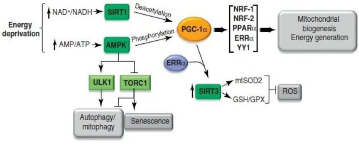
Figure 6: Integration of energy-generating and antioxidant pathways. ... 20
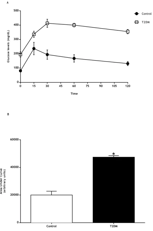
Figure 7: A) Blood glucose levels of control and T2DM groups measured during the intraperitoneal glucose tolerance test. B) AUCGTT (area under curve of glucose tolerance test) values in T2DM rats and in the control group. ... 36
Figure 8: A) Effect of type 2 diabetes mellitus in testicular proteins levels of Sirtuin 1 (SIRT1), peroxisome proliferator-activated receptor coactivator 1α (PGC-1α) and Sirtuin 3 (SIRT3). Panel B) Illustrative representation of Western Blot experiment. ... 37
Figure 9: Relative testicular mtDNA copy number from the control and T2DM groups. .... 38
Figure 10: Expression of the testicular mitochondrial complexes I, II, III, IV, V from the control and T2DM groups. ... 39
Figure 11: Activity of the testicular antioxidant enzymes Glutathione reductase, Glutathione peroxidase, Superoxide dismutase and Catalase from the control and T2DM groups………..40
Figure 12: A) Testicular lipid peroxidation, B) 3-NT (3-nitrotyrosine) content and C) proteins carbonylation in control rats and T2DM rats. ... 42
Figure 13: A) Effect of type 2 diabetes mellitus in epididymal proteins levels of Sirtuin 1 (SIRT1), peroxisome proliferator-activated receptor coactivator 1α (PGC-1α) and Sirtuin 3 (SIRT3). Panel B) Illustrative representation of Western Blot experiment. ... 43
Figure 14: Activity of the antioxidant enzymes Glutathione reductase, Glutathione peroxidase, Superoxide dismutase and Catalase in the epididymal tissue from the control and T2DM groups. ... 44
xv
Figure 15: Relative mtDNA copy number in epididymal tissue from control and T2DM groups. ... 45 Figure 16: Expression of the mitochondrial complexes I, II, III, IV, V in the epididymal tissue from control and T2DM groups. ... 46 Figure17: Epididymal lipid peroxidation, 3-NT (3-nitrotyrosine) content and proteins carbonylation in control rats and T2DM rats. ... 48
xvi
List of Tables
Table 1: List of primary and secondary antibodies used in western blot technique. ... 29 Table 2: Genes, oligonucleotide sequence and respective conditions for PCR amplification. ... 30 Table 3: Effect of T2DM on average weight, glycaemia, HbA1c, insulin and reproductive organs weight from the control group and T2DM group. ... 35
1
I. Introduction
2
1.Testicular Anatomy
The reproductive organs have an important role to ensure the survival of the species, once it is in the reproductive system that is created the fundamental units of life, the gametes. The sexual reproduction allows the transmission of the genetic code over generations through the variability induced by a combination of genes that ensures the evolution of the species. The reproductive system is the only body system that is not fully functional at the time of birth, it requires the actions of sex hormones released at puberty to become fully functional. In addition, there are clearly differences between the male and female reproductive system which does not occur in the other body systems (Alves and Oliveira 2017).
Testes are the central element of the male reproductive tract, are paired, whitish, ovoid-shaped organs, suspended outside the abdomen located within the scrotum, which serves as a protective envelope and keeps the testicular temperature approximately 35ºC (Alves and Oliveira 2017). The testicular capsule is composed by two layers of membranes: tunica vaginalis, tunica albuginea. The tunica vaginalis is the outer membrane that covers the surface of each testes, except where the testes attaches with epididymis and spermatic cord. The tunica albuginea is a tough fibrous membrane. Extensions of this membrane, in to the testes as fibrous septa result in the formation of approximately 250 pyramidal lobules each of which contains the seminiferous tubules (STs) (Alves and Oliveira 2017).
Figure 1: Representation of the human testis. The testicular capsule is composed by tunica
albuginea which results in the formation of pyramidal lobules containing the seminiferous tubules culminating in the rete testis which transports the spermatozoa from the seminiferous tubules into the efferent ducts. Adapted from (Alves and Oliveira 2017).
3 Testes present a bi-functional and a compartmental organization which is highly conserved throughout evolution (Schlatt et al. 1997). Testes show an interstitial space where it can be found the somatic Leydig cells (LCs), blood and lymphatic vessels and cells of the immune system, whereas the seminiferous tubules are composed by peritubular myoid cells (PMCs), Sertoli cells (SCs) and germ cells (Schlatt et al. 1997). These organs present two main functions: the production of sex steroids hormones, mainly testosterone, and the formation of germ cells. The process of production of sperm designated, spermatogenesis, occurs in seminiferous tubules and the steroidogenesis in LCs (Schlatt et al. 1997).
STs are the functional unities of the testes which represents about 80% of the testicular mass (Rato et al. 2010). SCs are specialized polarized epithelial cells that extend from the base of the STs to its lumen. SCs are the first somatic cells to differentiate in the testes and are pivotal in the testes development. These cells contribute to the formation of the BTB (blood-testis barrier) which protects the germ cells from the immune system. The establishment of BTB provides a specific intratubular environment that is dependent on the function of SCs. This creates the adequate condition for the development of spermatogenesis (T. Dias et al. 2016). This is a complex and coordinated process, that takes place in the STs (Komsky-Elbaz et al. 2018). It consists of cell division and differentiation of spermatogonia which culminates in the production of the mature male gametes.
1.1. Sertoli cells
The main function of these cells is nursing germ cells since, behind the physical support, they also provide nutritional support to germ cell line (Rato et al. 2012). SCs produce and secrete, several proteins, and factors that are essential for the germ cell’s development (Mruk and Cheng 2004). For instance, SCs produce transferrin, vitamin transporters, lactate, acetate, extracellular matrix components, glial cell-derived neurotrophic factor, fibroblast growth factor 2, growth regulatory factors (stem cell factor, transforming growth factors alpha and beta, bone morphogenic protein 4, and stem cell factor which initiate the differentiation of spermatogonia stem cells, and interleukins (Islam et al. 2017; Walker and Cheng 2005). On the other hand, these cells have an important role in order to maintain the balance in the number of germ cells, using phagocytosis to clean degenerating GCs or residual body from spermatids. This is one of the most critical function, because a considerable fraction of GCs is discarded during spermatogenesis, and the presence of these dead cells into the STs lumen could lead to the release of noxious contents that negatively impact sperm production. SCs can interact with other SCs but also with germ cells, this cell-to-cell interactions are crucial for the regulation of spermatogenesis (Mruk and Cheng 2004). This contact is possible through adherent, tight and gap junctions (Islam et al. 2017). The communication between germ cells and SCs is due to paracrine factors or ligand/receptor-mediated interactions (Rato et al. 2010). Besides that, studies have evidenced that SCs are capable to maintain a unique antiviral defense system through productions of some interferons (IFNs), ILs and Cytokines
4 (Dias et al. 2016). Metabolically SCs exert their action, producing various substrates, preferentially using glucose through the process glycolysis (Alves et al. 2013). The glucose enters in the cells mainly through GLUT 1 and GLUT 3 and is metabolized by glycolysis in pyruvate which can be converted into alanine, lactate or acetyl-CoA in mitochondria (Mruk and Cheng 2004; Rato et al. 2013). However, the preferred route of glucose is the production of lactate which is then transported to the germ cells mediated by active membrane monocarboxylate transporters (MCTs) via MCT4 (Dias et al. 2016). Lactate stimulates the synthesis of ribonucleic acid and proteins in developing germ cells and also helps to promote the activation of alternative metabolic pathways, such as gluconeogenesis and the pentose-phosphate pathway. Besides that, SCs also produce acetate, which is converted in acetyl-CoA in the cytoplasm or in the mitochondrial matrix by acetyl-CoA synthase. This metabolite is involved in the synthesis of cholesterol and other lipids and it has been proposed that acetate is required to the maintenance of the high rate of synthesis of the lipids and for the intense remodeling of the germ cells membranes during their development. Although, lipids are also used by SC as a source of energy by -oxidation of fatty acids pathway which is the preferential way to obtain energy. Furthermore, SCs are also capable to convert the branched-chain amino acids in the corresponding branched-chain acids to produce ATP (Alves et al. 2017).
1.2. Leydig cells
Leydig (or interstitial) cells arise from interstitial mesenchymal tissue between the tubules during the eighth week of human embryonic development. These cells are located in clusters in the interstitium between blood vessels and STs and the presence of peritubular myoid cells at the basement membrane of the STs prevent direct physical contact between SCs (Xu et al. 2007). LCs play vital roles in downstream masculinization events and in descent of the testes into the scrotum (Akingbemi, 2005) and, also, act as a support to spermatogenesis, producing the hormone testosterone, under the regulation of pituitary luteinizing hormone (LH), to keep SCs and germ cells functionally and to regulate germ cells maturation (Wong and Cheng, 2009). The secretion of testosterone is around 3-10 mg/day, accounting for more than 95% of total circulating T in post-pubertal men. The hormone acts on SCs stimulating the release of several chemical messengers that function as paracrine agents to stimulate the proliferation and differentiation of the germ cells (Neto et al. 2016; Sharpe et al. 2003).
1.3. Spermatogenesis
Spermatogenesis is a complex and coordinated process, that occurs in the seminiferous epithelium of the STs (Komsky-Elbaz et al. 2018). It consists of cell division and differentiation of spermatogonia which culminates in the production of the mature male
5 gamete. It can be divide in several steps: the proliferation of spermatogonia; spermatogonial differentiation into spermatocytes; meiotic division of spermatocytes producing spermatids; maturation of round spermatids; and the release of highly specialized mature spermatozoa into the testicular tubule lumen (Bell et al. 2014). Firstly, the spermatogonia (2n) suffers mitosis, followed by a cellular transformation from type B spermatogonia into spermatocytes, that suffers meiosis giving arise the spermatids (1n), that differentiate into spermatozoa (Wong and Cheng, 2009). Once formed these cells are released into the lumen and then proceed their journey through the epididymis where several molecular events will occur in order to become capable to fertilize (Bell et al. 2014). Even when spermatozoa reach the female reproductive tract it must ensure efficient energy production to successfully achieve acrosome reaction and capacitation, otherwise, fertilization will not occur. Thus, spermatozoa are able to maintain their energy levels through two manners: through glycolysis and oxidative phosphorylation (OXPHOS). Due to the highly specialized structure of spermatozoa, which allows the compartmentalization of energy production, the ATP generated through OXPHOS occurs in mitochondria, while the synthesis of ATP through glycolysis takes place in the head and the fibrous sheath, due to the absence of respiratory enzymes. Several glycolytic enzymes, such as hexokinase, PFK, LDH and glyceraldehyde-3-phosphate dehydrogenase are present in these segments of spermatozoa.
In fact, both processes may occur independently or in combination depending on the surrounding environment. The preferred substrates of spermatozoa are glucose and fructose. These hexoses are taken up from the seminal plasma fluid through several glucose transporters (GLUTs) present along the plasma membrane (Alves et al. 2013). Then, glucose is metabolized by germ cells originating two molecules of ATP, pyruvate, and nicotinamide adenine nucleotide (NADH). Pyruvate is further metabolized in lactate as an energetic substrate. Furthermore, fructose is internalized and phosphorylate by fructokinase to fructose-1-phosphate, which undergoes hydrolysis by fructose-1-phosphate aldolase yielding dihydroxyacetone phosphate (DHAP) and glyceraldehyde. DHAP is converted to glyceraldehyde-3-phosphate by trioseohosphate isomerase or glyceraldehyde kinase. Thus, the intermediaries for the glycolytic pathway are obtained and can finally be converted to pyruvate and lactate for energy production (Dias et al. 2014).
However, in conditions when the availability of glycolytic substrates is scarce, the energy production is ensured by the oxidative phosphorylation in mitochondria. It is known that the most efficient energy process occurs under aerobic conditions and in these conditions, pyruvate is converted in acetyl-CoA by pyruvate dehydrogenase, which then enters the Krebs cycle to combine with oxaloacetate (OAA). OAA is then further oxidized to reduce NAD+ and
the complex II prosthetic group flavin adenine dinucleotide generating carbon dioxide. The oxidation of these substrates is coupled with the phosphorylation of adenosine diphosphate via the mitochondrial electron transport chain, with the subsequent production of ATP. These are the two main metabolic pathways in sperm metabolism, however, the spermatogenesis
6 process is a high energy process with a high consumption of oxygen (O2), which consequently
leads to an exacerbated production of ROS (reactive oxygen species). This uncontrolled and excessive ROS production, accompanied with a deficient capture of this agents, leads to an oxidative environment. The spermatozoa are the cells more prone to this species, so they are completely dependent (Alves et al. 2017).
Figure 2: Representation of spermatogenesis. The seminiferous epithelium is composed of Sertoli
cells and developing germ cells at different stages. Leydig cells and blood vessels are located in the interstitium. Spermatogenesis is the cellular division and transformation that produces male haploid germ cells from diploid spermatogonial stem cells. Adapted from (Rato et al. 2012).
1.4. Hormonal control of spermatogenesis
The regulation of spermatogenesis is complex and includes the communication between the hypothalamus-hypophysis axis and the gonads. It begins with the release of the gonadotropin-releasing hormone (GnRH) a decapeptide produced by specialized neurons in the hypothalamus that reaches the anterior pituitary via the hypotalamo-pituitary vessels and triggers the release of both luteinizing hormone (LH) and follicle-stimulating hormone (FSH) (Walker and Cheng 2005). Each of these hormones acts in different sites in the testes but work in concert to control spermatogenesis. FSH acts on the SCs activating the secretion of paracrine agents that are essential for spermatogenesis (Ramaswamy and Weinbauer 2014). LH acts on the LCs stimulating the secretion of the hormone testosterone, that acts as a paracrine agent, moving from the interstitial spaces into the seminiferous tubules (Ramaswamy and Weinbauer 2014). This hormone is essential for the development of the
7 spermatogenesis and plays an important role in masculinization and the maintenance of male secondary sexual characteristics.
However, some negative feedbacks exerted by testicular hormones are important in order to regulate the process of spermatogenesis. The inhibin secreted mainly by SCs can also supress the release of FSH. Inhibin acts on the anterior pituitary decreasing the biosynthesis and secretion of FSH through a controlled mechanism that not affects the releasing of LH.
In the other hand, the testosterone produced by LC inhibits mainly LH secretion, through two different mechanisms: testosterone can act in the hypothalamic GnRH release, resulting in a decrease of FSH and LH hormones; or can acts directly into anterior pituitary leading to a decrease in the secretion of LH (Alves and Oliveira 2017). Thus, testosterone have an important role in the maintenance of males infertility once it plays a central role in the development of males reproductive tract and genitalia organs and also is required for the initiation and maintenance of spermatogenesis (Alves and Oliveira 2017). In the absence of testosterone, germ cells cannot undergo beyond meiosis, the formation of BTB is compromised and it prevents the release of mature spermatozoa. In sum, any disruption in this process can compromises the spermatogenesis process and infertility (Walker, 2010).
8
2. The Epididymis
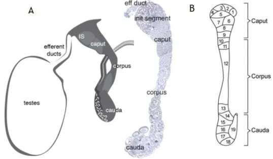
The epididymis is characterized has a long-convoluted organ of the male reproductive tract. After leaving the STs, spermatozoa follow through the rete testis and efferent ducts, reaching the epididymis (Žaja et al., 2016). The epididymis of mammals, such as mouse and rat, is structurally organized into four main regions: initial segment, the caput (head), corpus (body) and cauda (tail) (Figure 3A), while the human epididymis is poorly differentiated since no initial segment can be distinguished (Cheng et al. 2017). Each section of epididymis has distinct functions: the caput and the corpus are responsible for early and late sperm maturation, respectively, and the cauda storage of functionally spermatozoa (Cheng et al. 2017; Dias et al. 2016). Each region of the epididymis is organized into lobules separated by connective tissue septa, which serves as a support for the organ, but also as the separation between lobules which allows the expression of several genes and proteins within individual lobules and may play a role in creating specialized micro-environments. Each of the epididymal regions has a complex and different epithelium and can be further divided into 10 and 19 intraregional segments, that are separated by septae (Figure 3B) (Sullivan and Mieusset, 2016).
Figure 3: Schematic representation of the Epididymis (A) and typical patterns of rat epididymal segmentation (B). The epididymis can be divided into different parts: the initial segment,
the caput and the corpus which are responsible for the early and late maturation of the germ cells respectively; and the cauda which stores the spermatozoa Adapted from (A) Joseph et al. (2011) and (B) Jelinsky et al. (2007)
The epididymal epithelium it is composed of several types of cells: principal, apical, basal, narrow cells and halo cells, surrounded by multiple layers of peritubular myoid cells (Figure 4). Each cell type contributes to the establishment and regulation of a unique luminal
9 environment for the concentration, maturation, storage and viability of spermatozoa (Cornwall 2009; Shum et al. 2011).
The principal cells are the main cell type in epididymis corresponding a 65-80% of the epithelium. Morphologically, these cells present a columnar structure in the initial region which is converted into low cuboidal cells in the cauda of the epididymis. However, each segment presents differences in the structure and function, as the appearance and organization of the secretory apparatus, such as endoplasmic reticulum, secretory granules and Golgi apparatus, and endocytic apparatus (vesicular bodies, endosomes and lysosomes) (Abou-Haïla & Fain-Maurel, 1984). These cells play important functions in transport, synthesize a large number of proteins that are secreted to epididymal lumen and are directly involved in the control of luminal protein concentrations (Robaire and Viger, 1995).
Apical cells have many mitochondria in the apical cytoplasm by few microvilli at the luminal border and by a spherical nucleus that is located in the upper half of the cell cytoplasm. These cells are present in the epithelium of the initial segment and in the epithelium of the intermediate segment and they do not contact with the basement membrane (Sun and Flickinger, 1980). Apical cells differ from principal cells and narrow cells in terms of protein expression. In terms of functions, little is known, but it is suggested that these cells may have an important role in the regulation of pH in the lumen and a possible role in the maintenance of spermatozoa in the quiescent state, through the production of enzymes of the carbonic anhydrase family (Hermo et al. 2005).
Narrow (pencil) cells can be characterized as elongated cells that are present within the epithelium of the initial region and intermediated zone of the epididymis (Adamali and Hermo 1996; Sun and Flickinger 1980). A characteristic of these cells is the fact that present numerous apically located cup-shaped vesicles that are involved in endocytosis and in the secretion of H+ ions into the lumen (Hermo et al. 2000). These cells display an important role
in process of endocytosis as well in the intracellular transport between the epididymal lumen and the epithelial cells (Hermo et al. 2000). One of the most important features of narrow cells is the important role in protecting spermatozoa against the electrolytic imbalance and for the degradation of specific proteins and carbohydrates within their lysosomes (Adamali and Hermo, 1996).
Clear cells are localized in the caput, corpus and cauda regions of the epididymis, these cells are characterized for being large endocytic cells with an apical region containing numerous coated pits, vesicles, endosomes, multivesicular bodies, lysosomes and lipid droplets (Hermo et al. 1988) . Indeed, the endocytic activity by clear cells is more pronounced in the cauda of the epididymis. Furthermore, these cells are responsible for removing the contents of cytoplasmic droplets, that were created when spermatozoa are released and contain Golgi saccular components which are related to modifications of the structure of sperm membrane.
10 These cells are also in charge of the clearance of proteins from the epididymal lumen and participate in the regulation of luminal fluid acidification (Cornwall 2009; Hermo et al. 1988) Basal cells are triangular and flat cells which adhere to the basement membrane and do not have direct access to the lumen of the duct (Veri et al. 1993). These cells have cytoplasmatic extensions which suggest a close association with principal cells, and thus may regulate its functions. Indeed, their plasma membrane is constituted by coated pits which may be associated with the receptor-mediated endocytosis of factors from the blood or principal cells. Besides that, it also being demonstrated that basal cells regulate principal cell electrolyte transport by releasing paracrine factors (Cornwall 2009).
Halo cells are easily recognized for being small cells with a narrow rim of clear cytoplasm and are present in all epididymal segments localized in the base of the epithelium. These cells belong to the immune system and are characterized for containing a variable number of dense core granules. In young adult animals, halo cells can be described as helper T lymphocytes, cytotoxic T lymphocytes, and monocytes. With age, in some specific regions, it was verified an increase of these immune cells but also some eosinophils and B lymphocytes, occasionally (Robaire and Viger 1995).
Figure 4: Different cells types and organization of the epididymis. The three epididymal
compartments, as well as the relative position and distribution of each of the main cell types, are illustrated. The major function(s) associated with each cell type is also identified. Adapted from (Cornwall 2009).
11
2.1. Functions of the epididymis
The epididymis duct is a canal, which has the main functions to transport, mature spermatozoa in order to transform immature testicular sperm in competent cells to undergo fertilization, and their storage in a viable state in the cauda epididymis until they are ejaculated (Robaire and Viger, 1995). During the maturational process, spermatozoa include many changes in sperm physiological properties, such as the acquisition of forwarding motility, the ability to recognize and bind to the zona pellucida, and the capacity to fuse with the plasma membrane of an oocyte (Sullivan and Mieusset, 2016). Indeed, the epididymal lumen environment is extremely complex and it shows continuous and progressive changes in its composition from the caput to cauda regions.
Finally, the mature spermatozoa are stored for several days until ejaculation, in a quiescent state in the cauda epididymis. Thus, during the spermatozoa journey along the epididymal tract, it must ensure a controlled microenvironment in order to maintain the sperm viability. Mostly, protect the spermatozoa against the OS since they are highly vulnerable to ROS damages.
2.1.1. Epididymal sperm maturation
In the final stage of spermatogenesis, the spermatozoa are not completely mature, they leave the seminiferous tubules still immatures, without mobility and unable to fertilize the oocyte. Usually, sperm maturation takes place in the caput and corpus region of epididymis, where complex biochemical and physiological changes occur, to become movable and capable to fertilize (Cheng et al. 2017; Hu et al. 2017). In the epididymis lumen there are many components such as specific inorganic ions, small organic molecules, and proteins, that change continuously from the proximal to the distal epididymis to provide a specific environment for sperm maturation (Hu et al. 2017). The process of maturation it is accompanied by biochemical changes as formation of disulphide bonds within the sperm tail and nucleus, oxidation of sperm membrane sulfhydryl groups, increased capacity of glycolysis, modification of adenylate cyclase activity, and alteration in content in the lipid and protein composition of the membrane of spermatozoa (El-taieb et al. 2009). This includes the addition of new proteins, the removal or translocation of some specific proteins, or the modification of the structure of the proteins (Jankovičová et al. 2017). To help the whole process it secretes a specific fluid substance and it is essential for the transport, concentration, storage of spermatozoa and protection (Kamani et al. 2017).
Additionally, epididymis provides the adequate conditions to protect spermatozoa from oxidative injuries through antioxidant scavengers present in the luminal fluid that can be divided into enzymatic and enzymatic scavengers molecules (Hu et al. 2017). The
non-12 enzymatic antioxidants include taurine, glutathione, thioredoxin, and several studies also identified ascorbate as an important non-enzymatic scavenger antioxidant in caput epididymis. The antioxidant enzymes present in the epididymis are SOD (superoxide dismutase), CAT (catalase), GPx (glutathione peroxidase) and the idolamina dioxygenase.
2.1.2. Epididymal sperm storage
When spermatozoa enter in the cauda of the epididymis they already get mature. This compartment of the epididymis not only stores the spermatozoa but also contribute to protect spermatozoa against from oxidative damage and to keep the quiescence state through ionic and non-ionic changes (Ghosh et al. 2017).
It takes approximately 10 days until the spermatozoa reach out the cauda of epididymis where they will be stored before ejaculation (Robaire and Hermo 1988). The survival of spermatozoa in the cauda epididymis depends on the species and incubation temperature. In scrotal mammals, the combination of a unique luminal milieu and lower temperatures (30– 32ºC) are thought to be major contributors to sperm survival. However, if spermatozoa are removed from the cauda and incubated at 32ºC in vitro, their fertility and viability are measured in hours rather than days (Jones, 2014).
During this journey, spermatozoa are at risk due to the extreme susceptibility to oxidative damages because of membrane structure that is mainly composed of high quantities of PUFA. Interestingly, it is this membrane structure that allows the intrinsic fusogenic properties that will need to engage the membrane fusion events associated with fertilization (Vernet et al. 2004).However, the presence of high concentrations of PUFA in the membrane became spermatozoa a susceptible target to attack from ROS. Indeed, ROS are important in processes of signal transduction related with several physiological processes in sperm cells such as hyperactivation, capacitation, acrosome reaction, zona pellucida binding and oocyte penetration (Vernet et al. 2004). However, the uncontrolled production of ROS that exceeds the antioxidant capacity of the seminal plasma leads to oxidative stress (OS) which is harmful to spermatozoa.
All cellular components including lipids, proteins, nucleic acids are potential targets of ROS (Agarwal et al. 2008). The production of ROS by spermatozoa correlates with lipid peroxidation, DNA oxidation, poor sperm function and reduced fertility (Moustafa et al. 2004; Aitken and Koppers 2011). Thus, it is essential to the proper functionality of the spermatozoa to maintain a delicate balance in ROS production and recycling. It has been estimated that OS is a contributor in 30–80% of cases of male infertility thereby making it an important area of research (Tremellen 2008).
13
3. Diabetes mellitus and male fertility
Diabetes mellitus (DM) is one of the diseases of greatest global concern, with a progressive increase in the incidence of the disease over the years. At the beginning of the millennium, the World Health Organization (WHO) reported that 177 million people were affected by DM and around the year of 2030, it is estimated that more the 500 million people may suffer from DM (Agbaje et al. 2007). Many factors have contributed to the DM epidemics, such as the lifestyle of modern societies based on the erroneous eating habits, the lack of physical activity that all together predispose individuals to the development of DM.
DM is a metabolic disorder involving the derangement in carbohydrate, lipids and proteins metabolism. It can be divided into two types: type I and type II. Type I diabetes mellitus (T1DM) or insulin dependents diabetes mellitus which results in the destruction of the insulin-producing pancreatic beta cells by the autoimmune system, which generally develops at a young age but it may affect people of any age (Agbaje et al. 2007). The individuals with T1DM produce low quantities or no insulin, due to this condition is necessary exogeneous insulin to control their blood glucose levels (Canivell and Gomis 2014).
Type 2 diabetes mellitus (T2DM) or non-insulin dependent is the more common form of diabetes that results from defects in action or secretion of insulin. This type of diabetes can be described as the inability of cells to properly respond to insulin in a first stage, compensated by an increase in insulin levels to keep the normoglycemia, but in individuals predisposed to DM type II this mechanism is compromised resulting in a state of hyperglycemia (Asmat et al. 2016; Golay and Ybarra 2005). This is a progressive process because of the limited capacity of pancreatic cells to augment the secretion of insulin to counterbalance insulin resistance, maintaining glucose tolerance at normal levels. Normally the individuals in this condition do not need exogenous insulin action to survive (Golay and Ybarra 2005).
It has been observed an increase of T2DM in children and adolescents, which represents a threat to global health. The sustained hyperglycaemia can cause long term complications which include neuropathies, ophthalmopathies, kidney impairments, cardiovascular diseases and sexual dysfunctions that threaten the quality of life (Kyathanahalli et al. 2014; Aguirre-Arias et al. 2017). Indeed, the prevalence of DM has been increased in young people below reproductive age, moreover, data from animal models strongly suggest that DM impairs male fertility at multiple levels, such as endocrine control of spermatogenesis, spermatogenesis itself, or by impairing penile erection and ejaculation.
Male fertility is defined has the inability of males to produce or deliver fully functioning sperm. According to a medical point of view a couple is considered infertile to conceive if the pregnancy does not occur after one or two years of unprotected intercourse. It is estimated that over 10-15% of couples worldwide are infertile (Hosen et al. 2015). The causes behind
14 infertility, in some cases, are difficult to define, however, a large number of infertility cases are associated with the sperm abnormalities, such as morphology, motility, concentration and DNA fragmentation (Tunc et al. 2009).
Indeed, it was reported about 90% of chronic diabetic patients suffer from sexual dysfunctions including decreases in sexual libido, potency, erectile dysfunction and ejaculation difficulties (Al-Roujeaie et al. 2017). As well-known DM causes a hormonal and metabolic disruption which cause deleterious effects to male reproductive health. Several reporters had linked diabetes has a cause of infertility, the increase in glycaemic levels can lead to an oxidative environment which becomes toxic to the cells, causing several damages in DNA, proteins, and lipids (Rato et al. 2013). This is consistent with several studies in rats and humans that verified several deleterious effects of diabetes in male fertility. A study performed in type 2 diabetic men showed a decrease in semen parameters and an increased lipid peroxidation, so the authors concluded that OS may have detrimental effects on male fertility potential (Singh et al. 2014). Others have verified a significant reproductive dysfunction that resulted from a decrease in the reproductive organ weights and in sperm counts (Scarano et al. 2006). Also, a study reported in diabetic rats a deficient sperm quality, with a decreased in sperm concentration and motility and fertilization capacity as well as subsequent embryo development, they conclude that these abnormalities may be caused by alterations in steroidogenesis as a consequence of diabetes (Kim and Moley, 2008).
3.1. Diabetes mellitus and oxidative stress
In the testicular and epididymal tissue, spermatozoa possess metabolic processes to ensures the demands of their metabolism. One of the organelles that contribute the energetic of the germ cells are mitochondria which are known to produce significant amounts of ROS. It is due to hyperglycaemic state that increases the pyruvate production with a consequent production of the electron transfer donors, NADH and FADH, that subsequently enhances the electron flux through the mitochondrial electron transport chain. Consequently, there is an increase in the ATP/ADP ratio which leads to a hyperpolarization of the mitochondrial membrane potential. This creates a proton gradient, has a result of the high electrochemical potential difference, which leads to partial inhibition of the electron transport in complex III, resulting in the accumulation of electrons to coenzyme Q. Consequently, this leads to premature pass of electrons to O2 originating the superoxide anion (Ahmad et al. 2017). It is thought that is
this enhanced reduction of coenzyme Q and the generation of ROS that is the main cause for mitochondrial dysfunction, which is critical in diabetes-related metabolic disorders and tissue histopathology (Rolo and Palmeira 2006).
It is important to refer that in adequate concentrations this species plays an important role in some cellular signaling process such as the proliferation, differentiation, apoptosis and
15 growth regulation (Dobrakowski et al. 2017). For the germ cells, in normal concentrations, ROS are implicated in signal transduction mechanisms including the rate of hyperactivation, capacitation, the ability of the sperm to undergo acrosome reaction, spermatozoa-oocyte fusion and other processes implicated in fertility (Dobrakowski et al. 2017; Ferramosca et al. 2012). The problem is when there is an overproduction of this species and the rate of the removal, by the antioxidant defenses, is not sufficient to neutralize them, which makes them toxic to germ cells.
Sperm cells are vulnerable cells since the cytoplasm of germ cells contains a low concentration of scavenging enzymes and due to the chemistry of the cellular membrane which is mainly composed by large quantities of polyunsaturated fatty acids (PUFA). The main constituent of the lipid membrane is omega-3 polyunsaturated fatty acids which confer the fluidity of the plasma membrane and contribute to sperm structure formation, acrosome reaction, and sperm-oocyte fusion (Nichi et al. 2007; Tang et al. 2014). This species attacks the double bonds in molecules, which weaken the carbon–hydrogen bond on the adjacent hydrogen atom, making it susceptible to cleavage (Nichi et al. 2007).
Another important factor that can be responsible for the infertility is the fragmentation of DNA of the sperm nucleus, which is also a target for ROS (Naji et al. 2016). This species causes nucleotide modifications, such as, the 8-hydroxy-2´deoxyguanosine, the most oxidized base, when DNA it is damage in infertile men (Hosen et al. 2015). Furthermore, in spermatogenesis occurs the packaging of the sperm DNA, when nuclear histones are replaced by protamines. This process requires transient breaks in sperm DNA through topoisomerase II that breaks DNA strains and replace the histones by protamines. However, if the enzyme topoisomerase has a deficient activity may lead to defective repair and residual DNA fragmentation (Tunc et al. 2009). In fact, a study showed that the levels of DNA damages are more pronounced in the sperms of males with diabetes compared to those without diabetes (Agbaje et al. 2007).
Thus, the damages caused by OS affected the sperm quality and the fertilizing ability (Said et al. 2005). In fact, Rato and collaborators (Rato et al. 2014b) found that diabetes alters testicular metabolism which it is linked with deficient spermatozoa, a decrease in concentration of sperm, the abnormal morphology, and reduced motility. Another study by Kamani and collaborators found that in diabetic rats occurs a decrease of epithelium height, density of epithelium, an increase of fibromuscular layer thickness and lower density of interstitial cells because of the overproduction of ROS by the disease (Kamani et al. 2017).
16
4. Mitochondria an overview
Mitochondria are pivotal organelles present in almost all eukaryotes, being responsible for multiple important metabolic events, such as the citric acid cycle (also known as Krebs cycle), β-oxidation of fatty acids, and OXPHOS. Besides that, it also plays an important role as a mediator in the induction and execution of apoptosis, to determinate the life and death of the cells. These organelles are classically called as the “powerhouse” of the cell, due to their role in the production of ATP, mitochondria are the principal local where ROS was produced, specifically during OXPHOS.
Each mammalian cell contains several hundreds of mitochondria, with different sizes, shapes, and abundance depending on the cell type. The number of mitochondria in a cell is determined by the processes of biogenesis and division of the organelles in order to maintain functional all the process underlying ATP production, to ensures an adequate supplying of energy to cells. Furthermore, mitochondria may also change under different energy demand and different physiological or environmental conditions (Lee and Wei 2005). This organelle is central in the human physiology so dysfunctional mitochondria may give rise to several pathologies has been implicated in a large range of diseases, such as Alzheimer’s disease. According to the endosymbiotic theory, mitochondria are descendants of the ancient bacterial having their own genome and the capacity of auto replication. The mitochondrial DNA is constituted by a double strand molecule containing 37 genes encoding for 13 proteins, 2rRNAs, and 22 tRNAs. The 13 proteins are the essential subunits of oxidative phosphorylation system, the complexes I, III, IV, and V. The other mitochondrial products are 22 tRNAs (transfer RNAs) and 2 rRNAs (ribosomal RNAs) important for the translocation of the respiratory subunit mRNAs within mitochondrial matrix (Benkhalifa et al. 2014). However, some mitochondrial proteins are encoded by the nucleus, in the cytoplasm of the cell and imported to the mitochondria (Wenz 2013) These proteins are transported by molecular chaperones, unfolded, and imported into mitochondria via the translocase of the outer membrane complex. After transfer across the outer membrane, certain precursors are directed through the import machinery of the inner membrane complex into the mitochondrial matrix in a membrane potential-dependent manner (Ventura-Clapier et al. 2008).
Indeed, the oxidative phosphorylation pathway in mitochondria is the major source of ATP production in eukaryotic cells. During this process, the electrons flow from the reduced substrates, NADH and FADH, through the electron chain composed of respiratory H+ pumps
(complexes I-IV) until being delivered to O2. The complex I and II received the electrons from
NADH and FADH, respectively, and delivers those electrons to O2, at the complex IV. This
electron flow is coupled to proton ejection, forming an electrochemical gradient that is used to produce ATP at complex V (ATP synthase). Although, during the electron flow the, can electrons form complex I and III can directly react to O2 or other electron acceptors and origin
17 ROS. In fact, mitochondria are the major source of ROS production. Furthermore, it is estimated that during the oxidative phosphorylation 1–2% of the electrons are converted in the superoxide anion and hydrogen peroxide, through the incomplete reduction of O2 (Santos
et al. 2001).
Furthermore, mitochondria play an important role in the apoptosis process. This organelle has the important function to trigger cellular dead in order to make a rapid decision (if necessary) to initiate programmed cell death (Lee and Wei 2005).
4.1. Mitochondrial Biogenesis
Mitochondrial biogenesis can be characterized as the growth and the division of pre-existing mitochondria which is campaigned by several variations in mitochondrial size, number, and mass. This is a well-coordinated process involving several proteins that regulates all the process. One of the most important is PGC-1α (peroxisome proliferator activated receptor γ co-activator 1α) that belongs to the family of transcriptional co-activators, being the most studied member. This protein is expressed in tissues with high energetic demands like the brain, liver, cardiac tissue and skeletal muscle. It is not only an important regulator of mitochondrial biogenesis program but is also involved in several stress programs and might thereby integrate mitochondrial biogenesis into cellular stress adaption.
PGC-1α can be activated via PPAR (peroxisome proliferator activated receptors), AMPK (5’ adenosine monophosphate-activated protein kinase) or SIRT1(sirtuin1) pathway (Hofer et al. 2014) and it interacts with several transcription factors including nuclear hormone receptors, nuclear respiratory factors, and specific transcription factors to regulate glucose metabolism and response to environmental stimuli such as cold exposure and prolonged starvation (Fu et al. 2016). Thus, PGC-1α stimulate the mitochondrial biogenesis activating nuclear transcription factors leading to the transcription of nuclear respiratory factors 1 and 2 (NRF1 and NRF2). which up-regulates the transcription of several nuclear-encoded respiratory genes and induced the expression of mitochondrial transcription factor A (TFAM). Therefore, TFAM activates the mitochondrial transcription factors B1 and B2, essential components of the mtDNA (mitochondrial deoxyribonucleic acid) transcriptional machinery inducing the transcription and replication of mitochondrial genome. This is support by studies done in muscle cells that demonstrate the ability of PGC-1α to bind and co-activate the transcription factor NRF1 e NRF2.
In addition, PGC-1α interacts to with other transcription factors such as hormones, glucocorticoids, estrogen, an estrogen-related receptor α, and peroxisome proliferator-activated receptor α to enhance the transcription of mitochondrial fatty acid and -oxidation (Fu et al. 2016). In cases of energy deprivation PGC-1α has activated through NAD+ dependent
deacetylase, SIRT1 which deacetylates multiple residues in PGC-alfa promoting mitochondrial fatty acid oxidation in response to low levels of glucose (Zachary Gerhart-Hines).
18 Furthermore, it has been reported that PGC-1α in muscle cells can increase the expression of ROS scavenging enzymes, preventing mitochondrial OS, like GPx1 and SOD2 (Kong et al. 2010; St-Pierre et al. 2003). Besides that, PGC-1α regulates the expression of genes involved in gluconeogenesis, as is the case of the protein phosphoenolpyruvate carboxyl kinase and glucose-6-fosfato (Kong et al. 2010; Wenz 2013). SIRT1 belongs to a conserved family of proteins, the Sirtuins (SIRTs1-7) which are class III NAD+ dependent histone deacetylase
(Kolthur-Seetharam et al., 2009) distinct from class I and II histone deacetylases. SIRTs also possess ADP-ribosyltransferase activity (Kong et al. 2010). This group of proteins is activated in response to different factors, such as an increase in NAD+ levels associated with metabolic
and redox stresses, including several cellular processes like energy turnover, glucose metabolism, DNA repair and autophagy (Rogacka et al. 2016).
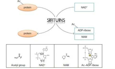
Figure 5: The reaction catalyzed by Sirtuins. Sirtuins deacetylates, i.e., removes the acetyl groups
from their substrates in the lysine residue, in a reaction that consumes NAD+
(nicotinamide adenine dinucleotide) releasing NAM (nicotinamide), Acetyl-ADP (adenosine diphosphate) ribose and the deacetylated protein. Adapted from (Tatone et al. 2018).
SIRT1 is the most conserved mammalian NAD+-dependent protein deacetylase, occurring in
various metabolic tissues and has been recognized as a key metabolic regulator. In the presence of NAD+, this protein catalyzes deacetylation of a variety of non-histone targets,
with O-acetyl-ADP-ribose and nicotinamide as side products. NAD+ is an essential coenzyme
found in all living cells, and its level regulates SIRT1 activity. The involvement of NAD+ in
deacetylation associates the SIRT1 activity with metabolism (Rogacka et al. 2016).
In situations of caloric restrictions more, carbons are oxidized producing NAD+ from NADH
what shifts the activations of sirtuins activity (Wang 2014). Indeed, when the ratio NAD+/NADH is high or the levels of ATP/AMP are reduced the AMPK becomes active (Cantó et