Ro le o f nitric o xide in hypo xia-induce d
hype rve ntilatio n and hypo the rm ia:
participatio n o f the lo cus co e rule us
1Departamento de Fisiologia, Faculdade de Medicina de Ribeirão Preto and 2Departamento de Morfologia, Estomatologia e Fisiologia,
Faculdade de O dontologia de Ribeirão Preto, Universidade de São Paulo, Ribeirão Preto, SP, Brasil
G. Fabris1,
J.A. Anselmo-Franci2
and L.G.S. Branco2
Abstract
Hypoxia elicits hyperventilation and hypothermia, but the mechan-isms involved are not well understood. The nitric oxide (NO) pathway is involved in hypoxia-induced hypothermia and hyperventilation, and works as a neuromodulator in the central nervous system, includ-ing the locus coeruleus (LC), which is a noradrenergic nucleus in the pons. The LC plays a role in a number of stress-induced responses, but its participation in the control of breathing and thermoregulation is unclear. Thus, in the present study, we tested the hypothesis that LC plays a role in the hypoxia-induced hypothermia and hyperventilation, and that NO is involved in these responses. Electrolytic lesions were performed bilaterally within the LC in awake unrestrained adult male Wistar rats weighing 250-350 g. Body temperature and pulmonary ventilation (VE) were measured. The rats were divided into 3 groups:
control (N = 16), sham operated (N = 7) and LC lesioned (N = 19), and each group received a saline or an NG
-nitro-L-arginine methyl ester (L-NAME, 250 µg/µl) intracerebroventricular (icv) injection. No signifi-cant difference was observed between control and sham-operated rats. Hypoxia (7% inspired O2) caused hyperventilation and hypothermia in
both control (from 541.62 ± 35.02 to 1816.18 ± 170.7 and 36.3 ± 0.12 to 34.4 ± 0.09, respectively) and LC-lesioned rats (LCLR) (from 694.65 ± 63.17 to 2670.29 ± 471.33 and 36 ± 0.12 to 35.3 ± 0.12, respectively), but the increase in VE was higher (P<0.05) and
hypo-thermia was reduced (P<0.05) in LCLR. L-NAME caused no signifi-cant change in VE or in body temperature under normoxia, but
abol-ished both the hypoxia-induced hyperventilation and hypothermia. Hypoxia-induced hyperventilation was reduced in LCLR treated with L-NAME. L-NAME also abolished the hypoxia-induced hypothermia in LCLR. The present data indicate that hypoxia-induced hyperventi-lation and hypothermia may be related to the LC, and that NO is involved in these responses.
Co rre spo nde nce
L.G.S. Branco
Departamento de Fisiologia Faculdade de O dontologia de Ribeirão Preto, USP 14040-904 Ribeirão Preto, SP Brasil
Fax: + 55-16-633-0999 E-mail: branco@ forp.usp.br Presented at the Meeting “NO Brazil, Basic and Clinical Aspects of Nitric O xide”, Foz do Iguaçu, PR, Brazil, March 10-13, 1999.
Research supported by FAPESP and CNPq. G. Fabris was the recipient of a postgraduate fellowship from FAPESP.
Received June 17, 1999 Accepted September 9, 1999
Ke y wo rds
·Nitric oxide
·Locus coeruleus
·Hypoxia
·Ventilation
·Body temperature
Intro ductio n
Hypoxic activation of arterial chemore-ceptors increases the excitatory synaptic drive of respiratory neurons in vivo (1). An in-crease in central respiratory activity also occurs in in vitro preparations in which chemoreceptor afferents are deleted (2,3). Hence, activation of medullary chemosen-sory neurons (4) or direct stimulation of respiratory neurons (2) contributes to the increased respiratory response to hypoxia.
Moreover, oxygen consumption and body temperature decrease during acute exposure to hypoxia in a variety of animal species (5). Although the precise nature for this hypoxic hypothermia is not fully understood, it is generally admitted that it results from a re-duction in the thermoregulatory set point, which has been referred to as anapyrexia (6), rather than from a reduction in body temper-ature due to an impairment of the thermoregu-latory effector mechanisms caused by a limi-tation in oxygen availability (7). The impor-tance of this response is emphasized by re-ports that show an increase in survival of the tested species if they are allowed to become hypothermic during hypoxia exposure (8).
Sympathetic activation is one of the ma-jor components of the adaptive responses to oxygen reduction, especially in the regula-tion of cardiovascular events that accom-pany hyperventilation (9-11). Sympathetic neurons receive both excitatory and inhibi-tory influences from noradrenergic cell groups located in the brainstem (12). The locus coeruleus (LC), a pair of nuclei located in the pons, is an assembly of densely packed noradrenergic neurons; its extensive projec-tions provide noradrenergic innervation to many brain areas and to the spinal cord (13). Hypoxia causes a significant increase in c-fos in the LC (14). Although little is known about the central pathways and neuromodu-lators involved in the neuro-physiological adjustments occurring during hypoxia, some candidates have been proposed, including
nitric oxide (15).
The diffusible lipophilic gas nitric oxide (NO) has been recognized as a physiological molecule because of its role in body temper-ature regulation (16,17). A previous study indicated that the hypoxia-induced hypo-thermia depends on the NO pathway (17). Also, NO may serve as a neurotransmitter in the central nervous system (CNS) and inhib-it inhibinhib-itory synaptic transmission that is triggered by CNS hypoxia (15).
The purpose of the present study was to test the hypothesis that the LC participates in the control of body temperature and pulmo-nary ventilation (VE) under normoxic and hypoxic conditions, and that NO is involved in this process in the CNS.
Mate rial and Me tho ds
Anim als
Experiments were performed on adult male Wistar rats weighing 250-350 g, housed at controlled temperature (25 ± 2o
C) and exposed to a daily 12:12-h light-dark cycle. The animals were allowed free access to water and food. Experiments were performed between 10:00 a.m. and 3:00 p.m.
Animals were divided into three groups: control (N = 16), sham-operated (N = 7) and LC-lesioned rats (LCLR) (N = 19). The ex-perimental group comprised rats submitted to bilateral lesion within the LC. Each group was divided into 2 other subgroups: 1) ani-mals which received an intracerebroventricu-lar (icv) injection of saline (1 µl) and 2) animals which received an icv injection of
NG
-nitro-L-arginine methyl ester (L-NAME, 250 µg/µl; Sigma Chemical Co., St. Louis, MO, USA).
Surge ry
mounted in a Kopf stereotaxic apparatus. Bilateral electrolytic lesions (2 mA, 10 s, positive electrode) were made using a con-stant current source and coated stainless steel electrodes in the LC (coordinates: A -3.4 mm from lambda, L ±1.2 mm, D 6.8 mm) (18). Sham-operated rats were similarly pre-pared, but the electrode was placed 2 mm above the dorsal coordinate and no current was passed through the electrodes. This dis-tinction was made to test the possibility of alteration in body temperature and VE in-duced by the surgical intervention. Experi-ments were initiated 24 h or seven days after lesion.
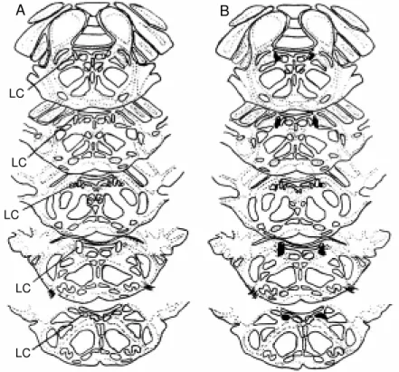
At the end of the experiment, the animal was killed with ether and the brain was removed and post-fixed in formol saline (10%) for 1 week. After fixation, the brain-stem was embedded in paraffin. Serial 13-µm frozen sections were cut and stained by the Nissl method for histological verifica-tion of the locaverifica-tion and extension of the lesions (or their absence in the case of sham-operated rats) and schematically presented in neuroanatomic maps (19). The LC lesions comprised a necrotic core surrounded by a zone of gliosis tissue. The sham-operated rats had an identifiable electrode tract, but no discernible lesion. Only the animals with 50 to 100% bilateral LC lesion were used for analysis of the results.
Rats used for icv injection of L-NAME were also anesthetized with 2,2,2-tribromo-ethanol and fixed in a Kopf stereotaxic appa-ratus. A stainless steel guide cannula (14 mm) was implanted into the right lateral ventricle of the brain (coordinates: A -1.0 mm from bregma, L ±1.6 mm, D -3.2 to -3.7 mm) (18). The displacement of the meniscus in a water manometer ensured correct posi-tioning of the cannula in the right lateral ventricle. The cannula was attached to the bone with stainless steel screws and acrylic cement. A tight-fitting stylet was kept inside the guide cannula to prevent occlusion. The surgical procedures were performed over a
period of 40 min. Six days after cannula placement, the animals were submitted to LC lesion or sham operation.
Expe rim e ntal pro to co l
Experiment 1. Determination of the com-bined effect of lesion and hypoxia on body temperature and VE. Experiments were per-formed using animals first exposed to hu-midified room air and then to a huhu-midified hypoxic mixture of 7% O2 (AGA, Sertãozi-nho, SP, Brazil). VE was determined by the plethysmograph method (20), and body tem-perature was determined by inserting a ther-mocouple probe into the colon. Before the experiment, the animals were habituated to temperature measurements in order to avoid stress-induced elevations in body tempera-ture.
Each animal was placed in a 5-liter plexiglass chamber and allowed to move about freely while the chamber was flushed with humidified air. After the animals re-mained calm (at least 60 min), control body temperature was measured. Control VE was measured and the test gas mixture (7% in-spired O2) was flushed through the chamber for 60 min. Ventilation was also measured after 5, 15, 45 and 60 min of exposure to hypoxia, and body temperature was meas-ured at the end of the hypoxic period. The chamber was then flushed with humidified air for 30 min and during this period VE was measured at 15-min intervals. At the end of the experiment, body temperature was meas-ured again.
re-Statistical analysis
Data are reported as means ± SEM. Changes in body temperature, VT, fR and VE were evaluated by ordinary analysis of vari-ance (ANOVA) or ANOVA for repeated measures to analyze the temporal effect of hypoxia on body temperature and VE. The differences between means were assessed by the Tukey-Kramer multiple comparisons test. Values of P<0.05 were considered to be significant.
Re sults
In all experimental protocols, the mean chamber temperature was 25.41 ± 0.52o
C (SEM), and the room temperature was 24.55 ± 0.47o
C. Figure 1 shows the lesion sites of individual rats (N = 7).
During normoxia and the recovery pe-riod after hypoxia, body temperature and VE did not differ between groups. Also, body temperature and VE during the return to air
Figure 1 - Representative draw ings show ing the lo-calization of the locus coe-ruleus (LC) (A) and lesions (dark areas, B) placed bi-laterally in the nucleus. Sequent ial sect ions of seven different animals.
corder (HP) allowed the calculation of respi-ratory frequency (fR) and tidal volume (VT) by appropriate correction factors (21).
Experiment 2. Determination of the com-bined effects of NOS blocker and hypoxia on body temperature and VE. Rats were treated with an icv injection of L-NAME or saline. A 705-LT, 50-µl Hamilton syringe and a Mizzy dental injection needle (200 µm OD) were used for icv injections. Sterile saline was the vehicle in which L-NAME was dis-solved, in a final volume of 1 µl. The injec-tion was performed over a period of 2 min, and 1 min was allowed to elapse before the injection needle was removed from the guide cannula, to avoid reflux. The same protocol described above was then used.
Experiment 3. Determination of the com-bined effects of LC lesion, NOS blocker and hypoxia on body temperature and VE.Six days after cannula placement, animals were sub-mitted to LC lesion or sham operation and the same protocol as described above, with an icv
injection of L-NAME or saline, was used.
A B
LC
LC
LC
LC
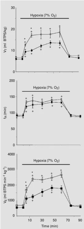
Figure 2 - Effects of hypoxia on tidal volume (VT),
respi-ratory frequency (fR) and pulmonary ventilation (VE) of
control rats (filled squares) (N = 16) and LC-lesioned rats (open squares) (N = 7). Values are reported as means ± SEM . The hypoxia-induced hyperventilation w as higher in LC-lesioned rats. +Significant increase
(P<0.05) in VT, fR and VE after hypoxia compared to
normoxia. * Significant increase in VT and VE of
LC-lesioned rats compared to control group. BTPS, Body temperature, pressure, saturated w ith w ater vapor.
VT
(
m
l
B
T
P
S
/k
g
)
30
20
10
0
Hypoxia (7% O2)
fR
(
m
in
)
200
150
100
0 50
VE
(
m
l
B
T
P
S
m
in
-1 k
g
-1) 4000
3000
2000
1000
0
Hypoxia (7% O2)
Hypoxia (7% O2)
10 30 50 70 90
Time (min)
after hypoxia did not differ from the baseline value in any group. The sham-operated group did not differ significantly from control ani-mals in any variable (data not shown). Data obtained from LCLR 1 week after lesion did not differ from control or sham-operated rats (data not shown).
Experiment 1. Combined effect of lesion and hypoxia on body temperature and VE. Figure 2 shows the effect of hypoxia on VT, fR and VE, in control and LCLR. When in-spired O2 was reduced from 21 to 7%, a significant (P<0.05) increase in ventilation was observed in both groups. The increase in VE was significantly higher (P<0.05) in LCLR than in control rats at 5 and 15 min of expo-sure to hypoxia. The hypoxia-induced hy-perventilation of LCLR was primarily the result of a significant elevation in VT, rather than a change in fR, which did not differ from control. The breathing pattern was recov-ered during return to air, when the experi-mental and control groups showed similar recordings.
Figure 3 shows the effect of hypoxia on body temperature of control and LCLR. Af-ter hypoxia, a significant decrease in body temperature was observed in both control (P<0.05) and LCLR (P<0.05), but the hy-poxia-induced hypothermia was reduced in LCLR. After hypoxia, the drop in body tem-perature was significantly lower (P<0.05) in LCLR compared to control.
Experiment 2. Combined effects of NOS
Figure 3 - Effects of hypoxia on body temperature of the control group (N = 16) and locus coeru-leus (LC)-lesioned rats (N = 7). Values are reported as means ± SEM . * Signif icant reduct ion (P<0.05) of mean body tempera-ture after hypoxia. +Significant
reduction (P<0.05) in the magni-tude of hypoxia-induced hypo-thermia.
37
B
o
d
y
t
e
m
p
e
ra
tu
re
(
oC
)
36
35
34
Control LC lesion
Air Hypoxia (7% O2)
* * *
+
*
+ + + +
+ + + + +
+ + +
+ + +
+ + +
*
+
*
+ + +
+
+ + +
+ +
+
Figure 5 - Combined effect of hypoxia and intracerebro-ventricular (icv) injection of NG-nitro-L-arginine methyl
ester (L-NAM E; 250 µg/µl) on body temperature of saline- and L-NAM E-treated rats (N = 7 for both groups). Values are reported as means ± SEM . * Significant drop (P<0.05) in body temperature after hypoxia of saline group compared to normoxia. +Significant
differ-ence (P<0.05) in the magnitude of hypoxia-induced hypothermia.
Figure 4 - Combined effect of hypoxia and int racerebroven-tricular injection of NG
-nitro-L-ar-ginine methyl ester (L-NAM E; 250 µg/µl) on tidal volume (VT),
respiratory frequency (fR) and
pulmonary ventilation (VE) in
L-NAM E group (open squares) (N = 7) and saline group (filled squares) (N = 7). Values are re-ported as means ± SEM . +
Sig-nificant increase (P<0.05) in VT,
fR and VE of the saline group
af-ter hypoxia compared to nor-moxia. * Significant difference (P<0.05), after hypoxia, betw een L-NAM E-treated rats and saline-treated rats. BTPS, Body tem-perature, pressure, saturated w ith w ater vapor.
VT
(
m
l
B
T
P
S
/k
g
)
30
20
10
0
fR
(
m
in
)
200
150
100
0 50
VE
(
m
l
B
T
P
S
m
in
-1 k
g
-1) 4000
3000
2000
1000
0
Hypoxia (7% O2)
Hypoxia (7% O2)
10 30 50 70 90
Time (min) Hypoxia (7% O2)
significant reduction in VT after hypoxia, rather than a change in fR.
Hypoxia failed to induce a reduction of body temperature when L-NAME was given intracerebroventricularly. These data are plot-ted in Figure 5.
Experiment 3. Combined effects of LC lesion, NOS blocker and hypoxia on body temperature and VE. Figure 6 shows a sig-nificant increase (P<0.05) in VE in LCLR + saline and LCLR + L-NAME after hypoxia, compared to normoxia, which was signifi-cantly higher (P<0.05) in LCLR + saline than in LCLR + L-NAME, at 5 and 15 min of exposure to hypoxia. The reduced hypoxia-induced hyperventilation of the LCLR + L-NAME group was primarily due to a signifi-cant reduction in VT after hypoxia, rather than a change in fR.
Figure 7 shows that the combination of LC lesion, saline and hypoxia caused a sig-nificant (P<0.05) drop in body temperature, similar to that obtained by LC lesion and hypoxia only. When L-NAME was injected
37
B
o
d
y
t
e
m
p
e
ra
tu
re
(
oC
)
36
35
34
Control + Saline Control + L-NAM E
Before injection
1 h after injection
Air
blocker and hypoxia on body temperature and VE. Figure 4 shows that the combination of saline injection and hypoxia caused a significant increase in VE, similar to that obtained by application of hypoxia only. When L-NAME was injected 30 min before hypoxia, hypoxia-induced hyperventilation was abolished, a fact primarily due to a
+ +
+
+ +
*
* * * *
*
* * * *
+
+ +
+ +
+
+ + +
+
+ +
+
+ +
+
*
30 min before hypoxia in LCLR, hypoxia-induced hypothermia was abolished, as ob-served by application of L-NAME in control rats. However, there was no significant dif-ference in body temperature between LCLR + saline and LCLR + L-NAME after hy-poxia.
D iscussio n
This study provides evidence that the LC participates in the control of pulmonary ven-tilation and body temperature during hy-poxia challenge, since bilateral electrolytic lesions of the nucleus caused an increased ventilatory response to hypoxia and a re-duced hypoxia-inre-duced hypothermia. A pos-sible link between the two responses cannot Figure 6 - Combined effect of hypoxia and
intracerebro-ventricular (icv) injection of NG-nitro-L-arginine methyl
ester (L-NAM E; 250 µg/µl) on tidal volume (VT),
respira-tory frequency (fR) and pulmonary ventilation (VE) of
LC-lesioned rats that received an icv injection of saline (open squares) (N = 5) or L-NAM E (filled squares) (N = 7). Values are reported as means ± SEM . +Significant
in-crease (P<0.05) in VT,fR and VE after hypoxia compared
to normoxia. * Significant difference (P<0.05) in VT and
VE of L-NAM E-treated rats compared to saline. BTPS,
Body temperature, pressure, saturated with water vapor.
Figure 7 - Combined effect of hypoxia and intracerebroven-tricular injection of NG
-nitro-L-arginine methyl ester (L-NAM E; 250 µg/µl) on body temperature of locus coeruleus (LC)-lesioned rats that received an icv injec-tion of saline (N = 5) or L-NAM E (N = 7). Values are reported as means ± SEM . * Significant drop (P<0.05) in body temperature after hypoxia.
VT
(
m
l
B
T
P
S
/k
g
)
30
20
10
0
fR
(
m
in
)
200
150
100
0 50
VE
(
m
l
B
T
P
S
m
in
-1 k
g
-1) 4000
3000
2000
1000
0
Hypoxia (7% O2)
Hypoxia (7% O2)
10 30 50 70 90 Time (min)
Hypoxia (7% O2)
37
B
o
d
y
t
e
m
p
e
ra
tu
re
(
oC
)
36
35
34
LC lesion + Saline LC lesion + L-NAM E
Before injection
1 h after injection
Air
Hypoxia (7% O2)
Hypoxia
L-NAM E
icv NOCNS LC
Tb VE
Figure 8 - Possible mechanisms through w hich the locus coeru-leus (LC) and nitric oxide (NO) could mediate hypoxia-induced hyperventilation and hypother-mia. Hypoxia leads to an in-crease in NO in the CNS and in the firing rate of LC neurons, w hich cause an increase in pul-monary ventilation (VE) and a
re-duction in body temperature (Tb). NG-nitro-L-arginine methyl
ester (L-NAM E) prevents hy-poxia-induced hyperventilation and hypothermia by inhibiting NO synthesis.
+
+ + +
+ +
+ + +
+
+ + + +
+
+
+ + +
+
+ +
+ + *
* *
*
+ *
+
*
+ *+ +
*
be excluded since a drop in body temperature leads to a reduced ventilatory drive. These data suggest that LC plays an inhibitory role in the hypoxia-induced hyperventilation and an excitatory role in the hypoxia-induced anapyrexia. Moreover, NO seems to play an important role in these responses to oxygen deprivation, since icv injection of L-NAME abolished both hypoxia-induced hyperventi-lation and hypothermia. Additionally, the inhibitory role of LC in pulmonary ventila-tion may depend on the NO pathway, since LCLR treated with L-NAME had a reduced ventilatory response to hypoxia.
In a wide variety of animal species, hy-poxia elicits a number of compensatory re-sponses, including increased ventilation and cardiac output (5). Decreases in PaO2 are monitored by peripheral arterial chemore-ceptors that evoke excitation of chemosen-sory fibers projecting in the brainstem within the nucleus tractus solitarius (NTS) (12). In the brainstem, these afferent inputs are pro-cessed and integrated together with other inputs to yield a final command to the respi-ratory motoneurons resulting in an increase in respiratory drive (22). However, the in-creased ventilation is O2 consuming, a fact that limits its beneficial effect. Hypoxia also elicits a decrease in body temperature and oxygen consumption (7), which is consid-ered to be an adaptive response because it decreases O2 demand according to the Q10 effect, promotes a leftward shift of the oxy-hemoglobin dissociation curve, and blunts the energetically costly response to hypoxia, e.g., increased cardiac output and ventilation (5). Previous studies have dem-onstrated that hypoxia per se causes hypo-thermia in a variety of organisms ranging from protozoans to mammals (23), but only recently did the mechanisms responsible for the hypoxia-induced anapyrexia start to be suggested.
The role of CNS in body temperature control is subjected to numerous modifiers, such as lactate, adenosine and histamine (for
review, see 23), but none of the possible candidates can trigger a complete hypother-mic response. Nitric oxide works as a physi-ological messenger molecule that may serve as a neurotransmitter in the CNS (24). NO in the CNS may have an important role in the hypoxia-induced hypothermia since during inhibition of the NO pathway hypoxia failed to reduce body temperature (17). Besides mediating hypoxia-induced hypothermia, oxygen deprivation was recently shown to lead to activation of the NO-cGMP pathway in the CNS, contributing to the induction and maintenance of the hypoxia-induced in-creased ventilation (15). The importance of NO can be demonstrated by inhibition of the effects of NO (25) using L-arginine analogs such as L-NAME. In the present study we have chosen L-NAME because it is a nonse-lective inhibitor of NOS and acts on both the constitutive and inducible isoforms of the enzymes.
re-ported that the LC is important in the modu-lation of sensory processing by the brain and is activated by a variety of stressful somatic and autonomic stimuli (26,27), but not dur-ing restdur-ing conditions (14). Activity in LC neurons is highest during wakefulness, par-ticularly under conditions requiring increased alertness (28). The major component of a-daptive responses to hypoxia is the stimula-tion of the sympathoadrenal system (9,11), and sympathetic neurons are under the con-trol of noradrenergic cell groups located in the brainstem (12), where the LC is the major noradrenergic nucleus. Recently, it has been reported that noradrenergic LC neu-rons send axons to spinal motoneuneu-rons, where they may participate in the control of respira-tory movements (29). Changes in the activity of LC neurons would then be expected to elicit widespread effects in the CNS, includ-ing those of a respiratory nature. A study demonstrating the distribution of neuronal pathways activated by hypoxia reported that the LC presents an increased c-fos staining during hypoxia (30). Also, Pérez et al. (31) observed LC-mediated inhibition of chemosensory responses in the rat NTS, which is the zone of termination of afferents from baroreceptors and chemoreceptors trav-elling in the carotid sinus (32) and aortic sinus (33). Furthermore, Moore et al. (34) observed that LC cooling blocks the fall in
respiratory output during hypoxia in anes-thetized neonatal sheep. They concluded that the fall in the biphasic respiratory response is mediated by the activation of neurons inhibitory to respiratory output and involves either axons of passage or cell bodies lying in the LC region. In agreement with these data, the present study suggests that if there is a CNS inhibitory mechanism of hypoxia-induced hyperventilation, it seems to depend on neuronal function within the LC.
As to the ventilatory response, LC seems to be involved in modulation of tidal vol-ume, since the hyperventilation response during hypoxia of LCLR differed from the control group due to a significant increase in tidal volume and not in respiratory frequency. In summary, the present results suggest that LC and NO pathway may participate in the control of body temperature and pulmo-nary ventilation under hypoxic conditions, and that the inhibitory role of the LC on hypoxia-induced hyperventilation and its ex-citatory role in hypoxia-induced hypother-mia may depend on the NO pathway.
Ackno wle dgm e nts
We thank Mauro Ferreira Silva, Nadir Martins Fernandes and Rute Aparecida de Freitas Marcon for excellent technical assis-tance.
Re fe re nce s
1. Law son EE, Richter DW, Ballantyne D & Lalley PM (1989). Peripheral chemorecep-tor inputs to medullary inspirachemorecep-tory and postinspiratory neurons of cat. Pflügers Archive, 414: 523-533.
2. Völker A, Ballanyi K & Richter DW (1995). Anoxic disturbance of the isolated respi-ratory netw ork of neonatal rats. Experi-mental Brain Research, 103: 9-19. 3. Ramirez JM , Quellmalz UJA, Wilken B &
Richt er DW (1998). The hypoxic re-sponses of neurons w ithin the in vitro mammalian respiratory netw ork. Journal of Physiology, 507: 571-582.
4. Kaw ai A, Ballantyne D, M ükenhoff K & Scheid P (1996). Chemosensitive medul-lary neurons in the brainstem-spinal cord preparation of the neonatal rat. Journal of Physiology, 492: 277-292.
5. Wood SC (1995). Oxygen as a modulator of body temperature. Brazilian Journal of M edical and Biological Research, 28: 1249-1256.
6. Branco LGS & M alvin GM (1996). Ther-moregulatory effects of cyanide and azide in the toad, Bufo marinus. American Jour-nal of Physiology, 270: R169-R173. 7. Gautier H & M urariu C (1998).
Neuro-modulators and hypoxic hypothermia in the rat. Respiration Physiology, 112: 315-324.
8. Hicks JW & Wood SC (1985). Tempera-ture regulation in lizards: effects of hy-poxia. American Journal of Physiology, 248: R595-R600.
Neurochemi-cal and Humoral M echanisms. Gordon and Breach, New York, 777-785. 10. M arshall JM (1987). Analysis of
cardio-vascular responses evoked f ollow ing changes in peripheral chemoreceptor ac-tivity in the rat. Journal of Physiology, 394: 385-403.
11. Richalet JP (1990). The heart and adrener-gic system in hypoxia. In: Sutton JR, Coatesand G & Remmers JE (Editors), Hypoxia: t he Adapt at ions. Dekker, Toronto, 231-240.
12. Soulier V, Cottet-Emard JM , Pequignot J, Hanshin F, Peyrin L & Pequignot JM (1992). Differential effects of long-term hypoxia on norepinephrine turnover in brain stem cell groups. Journal of Applied Physiology,73: 1810-1814.
13. Yang JJ, Chou YC, Lin M T & Chiu TH (1997). Hypoxia-induced differential elec-trophysiological changes in rat locus coe-ruleus neurons. Life Sciences, 61: 1763-1773.
14. Breen S, Rees S & Walker D (1996). Iden-tification of brainstem neurons respond-ing to hypoxia in fetal and new born sheep. Brain Research,748: 107-121.
15. Haxhiu M A, Chang CH, Dreshaj IA, Erokw u BNR & Prabhakar Cherniack NS (1995). Nitric oxide and ventilatory re-sponse to hypoxia. Respiration Physiolo-gy, 101: 257-266.
16. Scammell TE, Elmkist JK & Saper CB (1996). Inhibition of nitric oxide synthase produces hypothermia and depresses lip-opolysaccharide fever. American Journal of Physiology, 271: R333-R338. 17. Branco LGS, Cárnio EC & Barros RCH
(1997). Role of nitric oxide pathw ay in hypoxia-induced hypotherm ia of rats. American Journal of Physiology, 273: R967-R971.
18. Paxinos G & Watson C (1986). The Rat
Brain in Stereotaxic Coordinates. 2nd edn. Academic Press, San Diego.
19. Palkovits M & Jacobow itz DM (1974). To-pographic atlas of catecholamine and ace-tylcholinesterase-containing neurons in the rat brain. II. Hindbrain (mesencepha-lon, rhombencephalon). Journal of Com-parative Neurology, 157: 29-42.
20. Bartlett DJ & Tenney SM (1970). Control of breathing in experimental anemia. Res-piration Physiology, 10: 384-395. 21. M alan A (1973). Ventilation measured by
body plethysm ography in hibernating mammals and in poikilotherms. Respira-tion Physiology,17: 32-44.
22. Teppema LJ, Veening JG, Kranenburg A, Dahan A, Berkenbosch A & Olievier C (1997). Expression of c-fos in the rat brain-stem after exposure to hypoxia and to norm oxic and hyperoxic hypercapnia. Journal of Comparative Neurology, 388: 169-190.
23. Wood SC (1991). Interaction betw een hy-poxia and hypothermia. Annual Review of Physiology, 53: 71-85.
24. M oncada S, Palmer RM J & Higgs EA (1991). Nitric oxide: physiology, patho-physiology and pharmacology. Pharmaco-logical Review s, 43: 109-142.
25. Rees D, Palmer RM J, Schultz R, Hodson H & M oncada S (1990). Characterization of three inhibitors of endothelial nitric ox-ide synthase in vitro and in vivo. Brazilian Journal of Pharmacology, 101: 746-752. 26. Aston-Jones G, Shipley M T, Chouvet G,
Ennis M , Van Bockstaele E, Pieribone V, Shiekattar R, Akaoka H, Drolet G & Astier AB (1991). Afferent regulation of locus coeruleus neurons: Anatomy, physiology and pharmacology. Progress in Brain Re-search, 88: 47-75.
27. Van Bockstaele EJ, Akaoka H & Aston-Jones G (1995). Integration in the ventral
medulla and coordination of sympathetic, pain and arousal functions. Clinical and Experimental Hypertension, 17: 153-165. 28. Aston-Jones G, Foote SL & Bloom FE (1984). Anatomy and physiology of locus coeruleus neurons: Functional implica-tions. In: Ziegler M & Lake CR (Editors), Norepinephrine (Frontiers of Clinical Neu-roscience). Vol. 2. Williams & Wilkins, Bal-timore, 92-116.
29. Fung SJ, Chan JY, M anzoni D, White SR, Lai YY, Strahlendorf HK, Zhuo H, Liu RH, Reddy VK & Barnes CD (1994). Cotrans-mitter mediated locus coeruleus action on motoneurons. Brain Research Bulle-tin, 35: 423-432.
30. Nitsos I & Walker DW (1999). The distri-bution of FOS-immunoreactive neurons in the brainstem, midbrain and diencepha-lon of fetal sheep in response to acute hypoxia in mid and late gestation. Brain Research, Developm ent al Brain Re-search, 114: 9-26.
31. Pérez H, Ruiz S, Laurido C & Hernández A (1998). Locus coeruleus-mediated inhibi-tion of chemosensory responses in the rat nucleus tractus solitarius is mediated by alpha2-adrenoreceptors. Neuroscience Letters, 249: 37-40.
32. Erickson JT & M ilhorn DE (1991). Fos-like protein is induced in neurons of the me-dulla oblongata after stimulation of the carotid sinus nerve in aw ake and anesthe-tized rats. Brain Research, 567: 11-24. 33. Ciriello J (1983). Brainstem projections of
aortic baroreceptor afferent fibers in the rat. Neuroscience Letters, 36: 37-42. 34. M oore PJ, Ackland GL & Hanson M A