Published in final edited form as:
Biomarkers, March 2014, Vol. 19, No. 2, Pages 142-153. doi:10.3109/1354750X.2014.885084. http://informahealthcare.com/doi/abs/10.3109/1354750X.2014.885084
The decrease of NAD(P)H:quinone oxidoreductase 1 activity
and increase of ROS production by NADPH oxidases are early
biomarkers in doxorubicin cardiotoxicity
Ricardo Lagoaa,b,c, Carlos Gañánb, Carmen López-Sánchezb, Virginio García-Martínezb, Carlos Gutierrez-Merinoc,*
a ESTG- Polytechnic Institute of Leiria, 2401-951 Leiria, Portugal
b Dept. Human Anatomy and Embryology, Faculty of Medicine, University of Extremadura,
06006-Badajoz, Spain
c Dept. Biochemistry and Molecular Biology, Faculty of Sciences, University of Extremadura,
06006-Badajoz, Spain
(*) Correspondence should be addressed to Prof. Carlos Gutierrez-Merino. E-mail: [email protected]
Abstract
Context. Doxorubicin cardiotoxicity displays a complex and multifactorial progression.
Objective. Identify early biochemical mechanisms leading to a sustained imbalance of cellular bioenergetics. Methods. Measurements of the temporal evolution of selected biochemical markers after treatment of rats
with doxorubicin (20 mg/kg body weight).
Results. Doxorubicin treatment increased lipid oxidation, catalase activity and production of H2O2 by Nox-NADPH oxidases, and down-regulated NAD(P)H:quinone oxidoreductase-1 prior eliciting changes in reduced glutathione, protein carbonyls and protein nitrotyrosines. Alterations of mitochondrial and myofibrillar bioenergetics biomarkers were detected only after this oxidative imbalance was established.
Conclusions. NAD(P)H:quinone oxidoreductase-1 activity and increase of hydrogen peroxide production by
NADPH oxidases are early biomarkers in doxorubicin cardiotoxicity.
Keywords
1. Introduction
Doxorubicin is an antineoplastic anthracycline used for treatment of various hematological and solid tumor malignancies including breast cancer and leukemia. Although it is an effective chemotherapeutic drug, its clinical use is limited due to severe dose-dependent cardiotoxicity, which leads to dilated cardiomyopathy and congestive heart failure (Singal & Iliskovic 1998; Berthiaume & Wallace 2007; Takemura & Fujiwara 2007; Sterba et al. 2013).
Despite an extensive research on anthracycline pathophysiology, the molecular mechanism responsible for doxorubicin-induced cardiomyopathy is still controversial. Several mechanisms have been implicated in doxorubicin cardiotoxicity, such as apoptosis, disturbance of iron and calcium homeostasis and alterations of cardiomyocyte energetics, but many studies suggest that free radical-induced oxidative stress plays an important role (Tokarska-Schlattner et al. 2006;
Berthiaume & Wallace 2007; Takemura & Fujiwara 2007; Sterba et al. 2013).
It is well established that superoxide anion (O2·-) is generated during redox cycling of
doxorubicin or after oxido-reduction processes involving the anthracycline–iron complex, and that redox cycling of doxorubicin quinone ring can be catalyzed by several NAD(P)H dehydrogenases and oxidases (Doroshow & Davies 1986; Gille & Nohl 1997; Vásquez-Vivar et al. 1997; Deng et al. 2007; Guilleron et al., 2009). Cytochrome P450 reductase catalyzes the one-electron reduction of doxorubicin that can increase oxidative stress through the generation of superoxide anion (redox cycling) in vitro, but more recent in vivo studies have established this enzyme does not contribute to doxorubicin cardiotoxicity (Fang et al. 2008; Zhang et al. 2009; Dudka et al. 2012). Other
NAD(P)H dehydrogenases abundant in the heart tissue, such as xanthine oxidase, nitric oxide synthases and Nox (NADPH oxidases), are well described catalysts of doxorubicin redox cycling, hence they were targets examined in our work. Deng et al., (2007) pointed to Nox2 as the most probable NADPH oxidase isoform activated in the cardiotoxic process induced by doxorubicin.
and its activation requires the recruitment of several cytosolic subunits (p47phox, p67phox, p40phox and Rac1) (Santos et al. 2011). This hypothesis was later supported by the correlation found between inhibition of the regulatory subunit Rac 1 and the protective ability of statins against doxorubicin cardiotoxicity in vitro and in vivo (Huelsenbeck et al., 2011; Yoshida et al., 2009), as it was previously documented for cardiac hypertrophy (Santos et al., 2011; Takemoto et al., 2001). In addition, Nox2 can also be activated simply by exposure to H2O2 generated under oxidative stress
conditions (Li et al. 2001). Then, superoxide anion is converted to hydrogen peroxide (H2O2) or can
generate the hydroxyl radical in the presence of transition metals, and if nitric oxide is available, it reacts with superoxide anion producing peroxynitrite, another strong oxidative species that can cause cellular damage. The relevance of oxidative stress in doxorubicin cardiotoxic process has also been supported by the observation that antioxidant molecules like vitamin E or flavonoids (Milei et al. 1986; Sadzuka et al. 1997; Bast et al. 2007; Aluise et al. 2009; Kebieche et al. 2009; Mokni et al. 2012), as well as antioxidant enzymes such as superoxide dismutase (SOD) and glutaredoxin
(Xiong et al. 2006; Ichihara et al. 2007; Diotte et al. 2009), attenuate the cardio-pathological actions of the drug in animal models. However, no single therapeutic intervention has proven capable of efficiently reducing the adverse effects of doxorubicin in patients. Therefore, the elucidation of the pathological process and definition of the core of parameters indicative of biochemical changes behind doxorubicin-induced cardiomyopathy remain critical issues (Il'yasova et al. 2009; Panis et al. 2012).
The objective of the present work was to identify primary biomarkers inthe cardiotoxic process of doxorubicin. By examining the temporal relationships between doxorubicin-triggered events and the possible underlying molecular mechanisms, especially at the levels of cellular bioenergetics and redox balance, we aimed to clarify the relative importance of different pathways and targets proposed to explain doxorubicin toxicity.
2. Materials and Methods
2.1. Chemicals and drugs
Doxorubicin administered to animals was supplied by Pfizer (Madrid, Spain) and that used for in vitro studies was obtained from Sigma (St. Louis, MO, USA). Sodium chloride, potassium chloride, di-sodium hydrogen phosphate, potassium dihydrogen phosphate, malic acid and
potassium cyanide were purchased from Merck (Darmstadt, Germany). Reduced glutathione (GSH) and sodium pyruvate were from Boehringer Mannheim, Germany. Glycerol and paraformaldehyde were from Panreac (Barcelona, Spain). Ketamine was from Pfizer, while diazepam and atropine were obtained from B.Braun, Rubí-Barcelona, Spain. All other products were supplied by Sigma, unless specified otherwise.
2.2. Animals, treatments and samples
Male Wistar rats, 10 weeks old, weighting approximately 300 g were housed in a 12 h light/dark cycle and allowed free access to food and water during the experiment. The experimental procedures followed the animal care guidelines of the European Communities Council Directive 86/609/EEC. The protocols were approved by the Ethics Committee for Animal Research of the local government.
Doxorubicin was administered by i.p. injection of a drug solution in normal saline at a dose of 20 mg/kg of body weight, while Control rats received an injection of normal saline. After the injections, the treated animals were observed daily and weighted each morning. At different times of treatment, blood samples were collected and rats were sacrificed under anesthesia with ketamine (50 g/g), diazepam (2.5 g/g) and atropine (0.05 g/g). The hearts were quickly excised and washed in cold phosphate-buffered saline pH 7.4. Blood samples were allowed to clot and centrifuged at 5,000 x g at 4 ºC during 10 min. The serum was separated, frozen immediately on liquid nitrogen and stored until use.
Hearts for histological analysis were fixed in paraformaldehyde and processed for paraffin embedding. Tissue sections were obtained with a microtome and hematoxylin-eosin and Masson trichrome staining were performed using standard procedures.
2.3. Heart homogenization and preparation of mitochondrial, myofibrillar and 12,500 x g fractions
Heart samples for biochemical studies were homogenized by two different protocols as indicated below.
Total heart homogenates were prepared by homogenization in lysis buffer containing 50 mM HEPES pH 7.0, 1 mM EDTA, 1 mM EGTA, 50 mM KCl, 0.1%w/v
3-((3-cholamidopropyl)dimethylammonio)-1-propanesulphonate (CHAPS) and 20%v/v glycerol.
Homogenates were centrifuged at 500 x g at 4 ºC during 5 min to remove non-lysated material and the supernatant fluid was collected. These total homogenates were used for measurements of enzymatic activities and markers of oxidatively generated damage. For calpain activity
measurements and immunoblots, homogenates were further centrifuged at 10,000 x g for 10 min and supernatants were used.
Other heart samples were used to prepare mitochondria, myofibril-enriched fractions and homogenate fractions enriched in cytosol, microsomes and membrane fragments (fraction 12,500 x g). Tissues were homogenized in Tris-HCl 20 mM pH 7.4, sucrose 0.25 M, EDTA 0.5 mM, EGTA 0.5 mM and KCl 100 mM, and centrifuged at 1,000 x g during 10 min at 4 ºC. The supernatant was then separated from the pellet that included the myofibrils. The supernatant was further centrifuged at 9,000 x g at 4 ºC during 10 min, to pellet down mitochondria and separate the supernatant. Mitochondria were washed and resuspended in the same sucrose-containing buffer. The supernatant from the 9,000 x g centrifugation step was next centrifuged at 12,500 x g and the corresponding supernatants were collected for measurements (fraction 12,500 x g).
The myofibril-containing pellet from the previous 1,000 x g centrifugation step was re-suspended in myofibrils isolation buffer (Tris-HCl 20 mM pH 7.4, EDTA 1 mM and KCl 100 mM) containing 2% Triton X-100 (Ventura-Clapier et al. 1987; Chicco et al. 2006). Myofibrils were pellet down by centrifugation for 10 min at 6,000 x g and the treatment with Triton X-100 was repeated again. After, myofibrils were washed three times with isolation buffer without Triton X-100 and, at the end, resuspended in the same buffer.
Sub-mitochondrial particles for measurements of mitochondrial enzymatic activities were prepared by freeze-thawing mitochondria three times before the assays.
Protein concentration in all preparations was measured using Thermo Scientific (Rockford, IL, USA) protein assay kit (Comassie blue) and bovine serum albumin as standard.
2.4. Measurements of enzyme activities
The activity of creatine kinase (CK) in the different preparations was measured as described in a previous work (Lagoa et al. 2009). In the case of serum CK, the sample was incubated 5 min in the assay buffer supplemented with 10 mM GSH before activity was measured. The assay buffer for sub-mitochondrial particles was supplemented with 0.25 mg/mL octaethylene glycol monododecyl ether, and for myofibrillar fractions 2 mM EGTA was added.
Calpain and myofibrillar ATPase activity assays are detailed in our previous works (Tiago et al. 2006a; Lagoa et al. 2009). Mitochondrial complex I and complex II activities were measured as NADH:CoQ1 and succinate:CoQ1 oxidoreductase activities, respectively, following the procedures
previously described (Lagoa et al. 2011).
Glutathione reductase activity in heart homogenates was assayed spectrophotometrically by the method described in (Carlberg & Mannervik 1985). Assay buffer contained 200 mM potassium phosphate (pH 7.0), 2 mM EDTA, 1 mM oxidized glutathione (GSSG) and 100 µM NADPH. The sample was added to assay buffer for a final concentration of 0.1 mg protein/mL and the rate of
NADPH oxidation was followed at 340 nm, 30 ºC. Enzymatic activity was expressed as nmoles of NADPH oxidized/min/mg protein, using an extinction coefficient for NADPH of 6.22 mM-1cm-1.
Glutathione S-transferase activity was measured through the formation of GSH conjugates with 1-chloro-2,4-dinitrobenzene (Habig et al. 1974). First, this substrate was added at a final concentration of 1 mM to potassium phosphate buffer 100 mM (pH 6.5) containing 1 mM GSH, at 30 ºC, and the rate of the (non-catalyzed) conjugation reaction was monitored at 340 nm.
Thereafter, a sample of heart homogenate was added (final concentration 0.1 mg/mL) and
absorbance was followed for 3-5 min (total reaction rate). The enzymatic activity was calculated by subtracting the non-catalyzed reaction rate from the total rate and it was expressed as
nmoles/min/mg, using an extinction coefficient of 9.6 mM-1cm-1.
The activity of glutathione peroxidase was measured by monitoring NADPH oxidation at 340 nm when GSSG is reduced to GSH by glutathione reductase (Flohé & Gunzler 1984). Assay buffer consisted of potassium phosphate 50 mM (pH 7.0), EDTA 0.5 mM, sodium azide 1 mM, GSH 1.0 mM, NADPH 150 µM and glutathione reductase 0.24 U/mL, at 37 ºC. Homogenate concentration was 10 µg protein/mL buffer. After recording peroxide-independent NADPH disappearance for 3 min, 150 µM H2O2 was added and absorbance followed for 5 min (total
oxidation rate). In a separate assay, the non-enzymatic oxidation rate was measured in the same conditions except that no sample was added. Glutathione peroxidase activity was calculated by subtracting the peroxide-independent and non-enzymatic NADPH oxidation rates from the total oxidation rate. Activity was expressed as nmoles of NADPH oxidized/min/mg, for a GSH concentration of 1 mM.
Glucose-6-phosphate dehydrogenase (G6PDH) in total homogenates was measured by a method adapted from Stanton et al., (1991). Reaction buffer consisted of Tris-HCl 50 mM (pH 8.1), MgCl2 1 mM and NADP+ 100 M, and the sample (150 g protein/mL) was incubated at 30 ºC.
First, 200 M 6-phosphogluconate was added and the change in absorbance at 340 nm was recorded to quantify the 6-phosphogluconate dehydrogenase activity. Thereafter, 200 M
glucose-6-phosphate was added and absorbance continuously monitored for the “sum dehydrogenase activity” (6-phosphogluconate dehydrogenase + G6PDH). The G6PDH activity was calculated by subtracting 6-phosphogluconate dehydrogenase from the “sum dehydrogenase activity”, and expressed in nmoles of NADPH/min/mg of protein.
The activity of SOD in homogenates was assessed by the inhibition of nitroblue tetrazolium reduction upon superoxide generation by the xanthine–xanthine oxidase system at 560 nm (Spitz & Oberley 1989). Activity was expressed as U/mg protein (1 U is the amount of SOD required to inhibit the rate of nitroblue tetrazolium reduction by 50%).
Catalase activity was measured by the rate of H2O2 decomposition at 240 nm, as proposed in
(Aebi 1984). Homogenate concentration was 300 g protein/mL potassium phosphate buffer 50 mM pH 7.0, and the reaction was triggered with H2O2 10 mM, at 25 ºC. The activity was calculated
from the initial slope of absorbance and was expressed as moles of H2O2 consumed/min/mg
protein, using an extinction coefficient of 39.4 M-1cm-1.
The activity of NAD(P)H:quinone oxidoreductase 1 (NQO1) was measured as the
dicoumarol-sensitive menadione reducing activity (Ernster 1967). Reaction medium consisted of Tris-HCl 25 mM (pH 7.4), NADPH 180 M, Tween-20 0.01%, menadione 20 M and 40 g homogenate protein/mL. NADPH oxidation was followed spectrophotometrically at 340 nm, at 25ºC, for 3 min and, then, dicoumarol 20 M was added to correct the non-specific NADPH oxidation. NQO1 activity is given in nmoles of NADPH oxidized/min/mg of protein.
The NADPH oxidase activity of cardiac homogenates was measured following the kinetics of decrease of the fluorescence of NADPH (excitation wavelength 375 nm, emission wavelength 460 nm) at 37 ºC in the following assay medium: sucrose 0.25 M, KH2PO4 5 mM, KCl 10 mM,
MgCl2 5 mM, NADPH 15 μM, Tris 10 mM (pH 7.4), with 0.1 mg of protein/mL. The change of
NADPH concentration was calculated after calibration of the fluorescence intensity with standard solutions of known concentrations of NADPH, and the NAPDH oxidase activity is expressed as nmoles of NADPH oxidized/min/mg of protein.
2.5. Caspase-3 and NQO1 immunoblotting analysis
Caspase-3 activation and the level of NQO1 protein were investigated by Western blotting. General procedures routinely used in our laboratory for protein electrophoresis, transfer and immunodetection have been described previously (Lagoa et al. 2009). For caspase-3 activation, it was employed a polyclonal rabbit anti-activated caspase-3 antibody (Calbiochem PC679,
Calbiochem-Merck KGaA, Darmstadt, Germany) which recognizes the ~17 kDa cleaved (active) fragment of caspase-3. For NQO1, a polyclonal rabbit anti-NQO1 antibody (11451-1-AP from Protein Tech, Chicago, IL, USA) was used.
2.6. Markers of oxidatively generated damage
The concentration of GSH in heart homogenates was measured by the monochlorobimane method, as described in Lagoa et al., (2009).
The extent of lipid oxidation has been monitored following a standard protocol using the thiobarbituric acid-reactive substances (TBARS) assay. Homogenates were reacted with thiobarbituric acid 0,25% w/v, trichloroacetic acid 10%w/v and hydrochloric acid 0,17N, in presence of 1 mM butylated hydroxytoluene, at 100 ºC. After cooling the tubes, absorbance at 535 nm was measured and TBARS were calculated in nmoles of malondialdehyde per mg of protein, using an extinction coefficient of 1.56x10⁵ M-1cm-1.
Total thiol groups were measured by the 5,5’-dithio-bis(2-nitrobenzoic) acid reduction assay described in our previous publication (Tiago et al. 2006a), where the spectrophotometric method for detection of protein carbonyls with dinitrophenylhydrazine (DNPH) derivatization is also detailed. Protein carbonyls in heart homogenates were also investigated by Western blotting as proposed by Robinson et al., (1999).
The analysis of protein nitrotyrosines in homogenates was performed according to the method in Lagoa et al., (2009).
2.7. Production of reactive oxygen species
Production of H2O2 by heart isolated mitochondria and homogenate fractions 12,500 x g was
quantified by the Amplex Red assay (Zhou et al. 1997). Mitochondria 0.1 mg protein/mL or 0.05 mg/mL homogenate fraction were incubated in assay buffer composed of sucrose 0.25 M, KH2PO4
5 mM, KCl 10 mM, MgCl2 5 mM, Tris 10 mM (pH 7.4), horseradish peroxidase 0.2 U/mL and
Amplex Red (10-acetyl-3,7-dihydroxyphenoxazine) 5 M, at 37 ºC, and the increase in 530/590 nm fluorescence was followed. For mitochondria, media was supplemented with respiration substrates pyruvate 5 mM, malate 5 mM and with ADP 0.2 mM (Lagoa et al. 2011). Fluorescence units were converted into moles of H2O2 produced using a standard curve obtained with known amounts of
H2O2 in the same assay conditions.
Generation of reactive oxygen species (ROS) by isolated mitochondria was also assessed by using the ROS-sensitive probe dichlorodihydrofluorescein (Martin-Romero et al. 2004).
Mitochondria were suspended in the same buffer as for the Amplex Red method, the probe (2’,7’-dichlorodihydrofluorescein, from Molecular Probes, Groningen, The Netherlands) was added at a final concentration of 5 µM and the fluorescence was monitored at 37 ºC using excitation and emission wavelengths of 504 and 529 nm, respectively.
2.8. Statistical analysis
Results are expressed as means ±standard error (S.E.). Statistical analysis was carried out by Mann–Whitney non-parametric test. Significant difference was accepted at the P <0.05 level.
All the biochemical data were confirmed with duplicate measurements of at least experimental triplicates of each condition.
3. Results
Doxorubicin cardiotoxicity has been studied with different animal models using a variety of protocols for treatments, although the effects of the drug are known to depend on the
concentration/dose and exposure time (Tokarska-Schlattner et al. 2006). In the present study, Wistar rats were treated with an intraperitoneal injection of 20 mg of doxorubicin/kg body weight. This dose was selected after preliminary studies carried out in our laboratory with doses ranging from 10 to 40 mg of doxorubicin/kg body weight, aiming to set up a suitable protocol to detect the first, moderate but specific, signs of doxorubicin cardiotoxicity without a severe impairment of the animals. Equal or very similar doses are also used by other authors to investigate the effects of the drug in the heart and possible cardio protectors (Sadzuka et al. 1997; Mihm et al. 2002; Xiong et al. 2006; Andreadou et al. 2007; Ichihara et al. 2007; Diotte et al. 2009; Huelsenbeck et al. 2011; Mokni et al. 2012).
The temporal evolution of different markers of doxorubicin toxicity and possible targets of the drug were followed after its administration to the animals, as presented in the following sections. In the course of the first 5 days after doxorubicin injection, no mortality occurred, the animal weight decreased 18±5% and loss 34±10% of heart mass while Control animals injected with saline roughly maintained the weight (4±8% increase) and heart mass (11±11%).
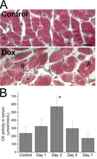
Histological examination of hearts extracted from rats treated with doxorubicin in our model revealed morphological alterations in the myocardium (Fig. 1.A), in accordance with those
described in the bibliography for doxorubicin-induced cardiomyopathy (Andreadou et al. 2007; Takemura & Fujiwara 2007; Sterba et al. 2013). At day 5 after drug administration, some cardiomyocytes with vacuolization and an abnormal morphology possibly due to myofibrillar disarrangement, could be observed in the stained heart sections. However, neither necrotic areas nor fibrosis were seen in Control or in doxorubicin–treated hearts at this stage of treatment.
Myocardial injury is known to cause an increase of CK level in serum (Rajappa 2005), and we measured higher values of CK activity in the serum of rats treated with doxorubicin, compared to Control animals (Fig. 1.B). It is important to note that the maximum CK activity in the serum was observed at day 3 after injection, and was followed by a decrease at days 5 and 7 of treatment, corroborating that there is a temporal window to detect heart lesion using this biomarker (Diotte et al. 2009).
3.1. Doxorubicin-activated cell death
Apoptosis of cardiomyocytes has been reported to be induced by doxorubicin in vitro, although the occurrence of this process in vivo and its relevance for the development of
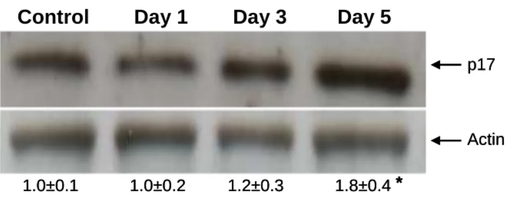
cardiomyopathy after treatment with the drug is a matter of debate (Takemura & Fujiwara 2007). We have experimentally assessed the induction of the apoptotic and necrotic cell death in the heart of rats treated with doxorubicin by measuring the level of active caspase-3 and the activity of calpains in heart homogenates. As shown in Fig. 2, a 1.8 fold increase of p17 cleaved fragment of caspase-3 was observed in samples obtained from animals 5 days after doxorubicin administration. Calpain activity of heart homogenates was measured using the fluorogenic substrate Suc-LY-AMC, as in a previous work by our laboratory (Lagoa et al. 2009), but no significant changes were
detected until day 7 of treatment compared to Control samples (results not shown).
3.2. Effect of doxorubicin on cellular bioenergetics biomarkers
Doxorubicin treatment has been shown to disturb several markers of the myocardial bioenergetics, but it is not clear if these are early changes or secondary in the process of cellular toxicity induced by the drug (Tokarska-Schlattner et al. 2006).
Mitochondria have been referred as a primary target of doxorubicin and a key factor in drug cardiotoxicity, reviewed in Berthiaume & Wallace, (2007). We initially studied the effect of
doxorubicin in the respiratory rate of mitochondria in vitro and we found no changes in state 4 and 3 respiratory rates, either using pyruvate/malate or succinate as substrates (results not shown). In spite of this result, as complexes I and II and CK are among the probably more sensitive targets of doxorubicin (Tokarska-Schlattner et al. 2006), we have measured the activity of these enzymes in mitochondria isolated from the heart of rats after drug administration (Fig. 3). While the enzyme activities at days 1 and 3 after injection were similar to those measured in Control samples, a significant reduction was detected at day 5. Complex I was the most heavily affected, with an almost 50% decrease in activity comparing Control and day 5 values (631±100 and 360±120 nmoles of NADH oxidized/min/mg protein, respectively). In addition, a loss of 35±10% of mitochondrial CK activity was also observed at day 5.
Another fraction of CK of high relevance for cardiomyocytes is that associated with myofibrils. As described in Materials and Methods, we prepared fractions of heart homogenates enriched in myofibrils and measured the activities of CK and myosin ATPase in these preparations. Doxorubicin treatment caused a gradual decline of both activities, with evident effects 5 days after drug injection (Fig. 4). At this time, CK and ATPase activities dropped 46 and 40% from Control values, respectively. The loss of myofibrillar CK activity seems to begin at an earlier time in the cardiotoxic process, since it was approximately 75% of the Control value already at day 1 after injection. However, total CK activity measured in heart homogenates, which includes the
mitochondrial, myofibrillar and cytosolic fractions of CK, did not show significant changes with doxorubicin treatment. Values in mol phosphocreatine hydrolyzed per min per mg of protein were: 18.2±4.0 in Control samples; and 14.2±2.2, 16.9±3.5 and 16.6±3.0 at days 1, 3 and 5 post-injection, respectively.
3.3. Effect of doxorubicin on markers of oxidatively generated damage
To evaluate the oxidatively generated damage elicited by doxorubicin treatment in rat heart, the following biomarkers were measured in heart homogenates: extent of lipid oxidation, levels of reduced thiol groups, of protein carbonyls and of protein nitrotyrosines.
The concentration of TBARS was found to increase two-fold after day 3 of treatment with doxorubicin compared to Control values (Fig. 5).
In contrast, the levels of reduced thiol groups in the samples of rats treated with doxorubicin were similar to Control values (Fig. S.1.A of Supplementary Data), in good agreement with the results obtained for GSH levels which were also unaffected by the treatment, as shown in the Table 1.
A study by other authors suggested an increase in protein carbonylation induced by doxorubicin in vivo (Andreadou et al. 2007). However, in our experimental conditions we did not detect an increase in the levels of protein carbonyls in heart homogenates until day 7 post-injection of doxorubicin using both the spectrophotometric and the Western blotting method with DNPH derivatization (Fig. S.1.B and C of Supplementary Data), nor in the level of protein nitrotyrosines, a marker of peroxynitrite and nitrosative stress (Fig. S.1.D of Supplementary Data).
3.4. Effect of doxorubicin on the activity of the major antioxidant cellular defense systems
The early increase in lipid oxidation caused by doxorubicin, noticed at the time in which the CK activity peaked to a maximum in blood serum and preceding the other effects of the drug in rat heart, strengthens the hypothesis that oxidative stress is a primary factor in doxorubicin cardiotoxic mechanism. However, this is not a generalized nitrosative/oxidative stress as pointed out by the results shown above. Then, it was important to check the activity of the major antioxidant systems to get a more in depth analysis of the oxidative stress phenomena induced by doxorubicin in rat heart. Previous studies pointed that disturbance of the GSH cellular homeostasis, the main
intracellular antioxidant, could play a major role in doxorubicin cellular toxicity (Wolf & Baynes 2006; Aluise et al. 2009; Diotte et al. 2009; Joshi et al. 2010). Thus we experimentally assessed the
possibility of GSH metabolism alterations after doxorubicin treatment in our model animals, and Table 1 summarizesthe effect of doxorubicin treatment on GSH levels and in the activity of the major GSH metabolizing enzymes glutathione reductase, glutathione S-transferase and glutathione peroxidase.
However, as shown in the Table 1 the concentrations of GSH measured in fresh
homogenates of heart samples from rats treated with the drug were similar to those measured in Control samples. In line with this observation, the activities of glutathione reductase, glutathione S-transferase and glutathione peroxidase did not show significant changes with doxorubicin treatment until day 5 post-injection. As the NADPH availability is a requirement for oxidized glutathione recycling by the reductase and NADPH-generation by G6PDH has been shown to play a key role in the maintenance of the redox status of cardiomyocytes and also to be implicated in other processes of cardiac dysfunction (Gupte et al. 2006; Santos et al. 2011), we measured this activity in heart homogenates and we found no significant differences between Control and doxorubicin-treated animals, see theTable 1. This enzyme was associated to endothelial oxidative stress and dysfunction induced by doxorubicin (Wolf & Baynes 2006), but seems to be not relevant in the cardiotoxic process.
The activity of the chief ROS scavenging enzymes SOD and catalase were also measured in heart samples and the results are also included in the Table 1. These results demonstrated a steady increase in catalase activity after doxorubicin injection, being significant at day 3 of treatment and later.
NQO1 has been reported to scavenge superoxide directly (Siegel et al. 2004) and, though it has a lower rate compared to dismutation catalyzed by superoxide dismutase (SOD), this property seems relevant in tissues/cells with lower SOD levels, like cardiovascular cells, where NQO1 inhibition was found to cause a decrease in superoxide scavenging capacity (Zhu et al. 2007). In addition, NQO1 is capable of reducing ubiquinone and probably also vitamin E (Beyer et al. 1996; Ross et al. 2000; Dinkova-Kostova & Talalay 2010), endogenous antioxidants whose regeneration
is critical for the protection of cell membranes against oxidatively generated damage. A previous work has pointed out the importance of NQO1 in prevention of lipid oxidation in artificial membranes and preservation of membrane integrity of rat hepatocytes exposed to doxorubicin (Beyer et al. 1996). Given that lipid oxidation was the dominant oxidative imbalance produced by doxorubicin in rat heart, we hypothesized that treatment with the drug affected the protective function of NQO1. The protein level and activity of this enzyme were measured in heart samples at days 1, 3 and 5 post-injection and found to be significantly lower only from day 3 post-injection, the time at which a significant lipid oxidation was also observed, and the results are presented in Fig. 6. Both NQO1 level and enzymatic activity showed an approximately 40% decrease in doxorubicin-treated rats compared to Control saline-treated animals.
3.5. Effect of doxorubicin on the activity of the major ROS producing systems
We then proceeded to investigate the sources of ROS stimulated by doxorubicin treatment and causing the oxidatively generated damage observed in rat heart. Mitochondrial enzymes, NADPH oxidases of the Nox family, uncoupled nitric oxide synthases and xanthine oxidase are the most important sources of O2·-/H2O2 reported for different cardiac pathophysiological conditions
(Santos et al. 2011; Sugamura & Keaney Jr 2011), but their specific role in doxorubicin cardiotoxicity is still not completely clarified.
In the first set of experiments, mitochondria isolated from rat hearts were incubated in vitro with doxorubicin and H2O2 release was measured by the Amplex Red method. Superoxide anion is
the primary ROS produced by mitochondrial electron transport chain (Murphy 2009), and can also be generated by redox cycling of doxorubicin quinone-semiquinone derivatives, catalyzed by mitochondrial NADH dehydrogenases (Doroshow & Davies 1986; Gille & Nohl 1997). It is well accepted that superoxide is rapidly converted to H2O2 by mitochondrial SOD and the fluorescent
probe Amplex Red gives a sensitive and robust method to quantify H2O2 generation (Zhou et al.
added in concentrations of 1 and 10 M to heart mitochondria respiring pyruvate and malate, had no significant effect on the rate of H2O2 release by the organelle.
Although a direct action of doxorubicin on heart mitochondria does not seem to account for mitochondrial H2O2 production, the in vivo treatment may potentiate mitochondrial ability to
produce ROS. We isolated heart mitochondria from rats treated with doxorubicin (day 3 post-injection) and compared the rate of ROS production with heart mitochondria from Control rats. As shown in Fig. S.2.B of Supplementary Data, the rates of H2O2 release by heart mitochondria from
the two experimental groups were identical, even when respiratory inhibitors that amplify mitochondrial H2O2 production were added to the assay medium. In addition, overall ROS
production was quantified by using dihydrodichlorofluorescein, a probe with a low specificity for different ROS (Martin-Romero et al. 2004), but again significant differences between mitochondria from doxorubicin-treated and Control animals were not detected (Fig. S.2.C of Supplementary Data).
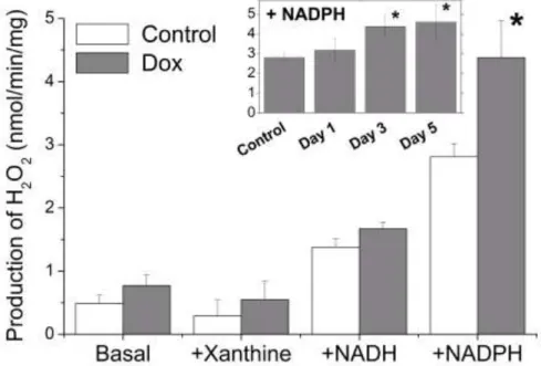
To investigate the possible participation of non-mitochondrial sources of ROS, a fraction of heart homogenates enriched in cytosol, microsomes and membrane fragments was prepared, as described in Materials and Methods (fraction supernatant of 12,500 x g), and the rate of H2O2
generation was measured in different experimental conditions (Fig. 7). The basal rates of H2O2
generation by the these fractions of heart homogenates from rats treated with doxorubicin (day 3 post-injection) were not significantly different from those of fractions obtained from Control rats. Similar results were obtained when the preparations were incubated with xanthine to stimulate the xanthine oxidase activity. The presence of NADH increased the capacity of homogenate fractions to produce H2O2, but in the same proportion with doxorubicin-treated and Control samples. However,
the stimulation by NADPH, in addition to being stronger, was significantly higher in the
preparations of hearts from doxorubicin-treated rats only from day 3 post-injection (Fig. 7). With these preparations, the rates of H2O2 production in the presence of NADPH after 3-5 days
Enzymes like nitric oxide synthases and the NADPH oxidases of the Nox family can produce O2·-/H2O2 in a NADPH-dependent manner and, besides from being implicated in several
cardiac pathological processes (Gupte et al. 2006; Akki et al. 2009; Sugamura & Keaney Jr 2011), are known to catalyze redox cycling of doxorubicin (Vásquez-Vivar et al. 1997; Deng et al. 2007). Moreover, Nox has been identified as the major source for NADPH-dependent production of superoxide anion in cardiac tissue (Nediani et al. 2007). As apocynin, the most used Nox inhibitor, interferes with H2O2 detection with Amplex Red in cell-free systems (Heumuller et al. 2008) and
can behave itself as an antioxidant acting as a scavenger of reaction products of hydrogen peroxide (Wind et al. 2010), we have experimentally assessed that apocynin affords a large inhibition of the coupled NADPH oxidase activity in our cardiac homogenates of doxorubicin-treated rats (day 3 post-injection). Our results showed that 250 and 500 μM apocynin inhibited 75±5% and >90%, respectively, of the total NADPH oxidase activity, which was 1.5±0.1 nmoles/min/mg of protein measured as indicated in the Materials and Methods section (average of data obtained with three different homogenates). Consistently, Nω-nitro-L-arginine methyl ester (L-NAME), a non-selective inhibitor of nitric oxide synthases, produced less than 10% inhibition of the NADPH oxidase activity and have no effect on the NADPH-stimulated production of H2O2 by the cardiac
homogenates at day 3 post-injection (average of data obtained with three different homogenates). Since Deng et al., (2007) have pointed to Nox2, composed of the catalytic subunit gp91phox and the membrane binding partner p22phox, as the most probable isoform activated in the cardiotoxic process, we have performed Western blotting analysis of the heart homogenates using
anti-gp91phox (cat. number 611415 from BD Transduction Laboratories, Lexington, KY, USA) and anti-p22phox (cat. number sc-20781 from Santa Cruz Biotechnology, Santa Cruz, CA, USA). Our results showed that at day 3 post-injection the expression levels of gp91phox and p22phox were not significantly different from those found in Control samples of heart homogenates (data not shown), therefore, excluding activation of Nox2 by up-regulation of its expression at the earlier stages of doxorubicin cardiotoxicity.
4. Discussion
In this work, we followed the temporal evolution of different markers of doxorubicin-induced early damage and possible targets of the drug in rat heart after an intraperitoneal injection of 20 mg of doxorubicin/kg body weight in adult Wistar rats. This treatment revealed the
involvement of the NQO1 and NADPH oxidase systems in the early stages of doxorubicin
cardiotoxicity in vivo. In parallel, several plausible effects and targets of the drug were found least relevant or secondary in the early stages of the cardiotoxic process.
Previous works have shown that treatment of rat and mouse with doxorubicin doses about 15-20 mg/kg body weight induces cardiac dysfunction (Mihm et al. 2002; Xiong et al. 2006; Ichihara et al. 2007; Diotte et al. 2009) and lesion evidenced by morphological changes (Sanchez-Quintana et al. 1994; Mihm et al. 2002; Andreadou et al. 2007) and increase of CK blood serum levels (Andreadou et al. 2007; Diotte et al. 2009), supporting the use of these treatments as
experimental models of doxorubicin cardiomyopathy (Sanchez-Quintana et al. 1994; Sadzuka et al. 1997; Mihm et al. 2002; Jang et al. 2004; Xiong et al. 2006; Andreadou et al. 2007; Ichihara et al. 2007; Diotte et al. 2009; Huelsenbeck et al 2011; Mokni et al. 2012). In good agreement with these results, we detected markers of myocardial lesion in the rats treated with doxorubicin in our model, specifically, cardiomyocyte degeneration and increase of CK in blood serum. Histological
observations excluded a widespread myocardial damage and a slight, but significant, activation of caspase-3 was detected at day 5 post-injection. Other authors have measured increases in the activity and level of cleaved form of caspase-3 in the heart of rodents following doxorubicin administration, with treatment protocols similar to our model, and the activation observed was also weak and time-dependent (Jang et al. 2004; Ichihara et al. 2007; Vitelli et al. 2007). On the other hand, activation of calpains, an event usually associated to disturbances in calcium homeostasis and ATP levels, and leading to rapid cell death, was not detected in our experimental assays of rat heart homogenates. Altogether, these results indicate the prevalence of a slow-developing apoptotic route
in our model of doxorubicin cardiotoxicity, while the necrotic route of cell death has little relevance in this animal model.
Lipid oxidation was found to be an early biomarker of heart lesion produced by doxorubicin, whereas statistically not-significant changes occurred in the level of reduced thiol groups, GSH, protein carbonyls and nitrotyrosines, suggesting that the oxidatively generated damage induced by the drug in vivo is not extensive, on the contrary, that the increased ROS production is very limited or focalized. Consistent with these experimental observations, the activities of the antioxidant enzymes glutathione reductase, glutathione S-transferase, glutathione peroxidase, G6PDH and SOD showed no significant alterations, but doxorubicin administration induced a stepwise and sustained increase in catalase activity beginning early after the intraperitoneal injections. The increase of catalase activity closely paralleled the increase in lipid oxidation, suggesting that this is a cellular response to counteract the increased H2O2 production in doxorubicin-treated rats. Noteworthy,
specific activation of catalase is also associated with human heart failure (Dieterich et al. 2000; Borchi et al. 2010). Upregulation of catalase, without significant changes in MnSOD, CuZnSOD and glutathione peroxidase, has been described in left ventricles from failing hearts due to dilated and ischemic cardiomyopathy (Dieterich et al. 2000). Interestingly, in other work, higher catalase activity values observed in right and left ventricles from failing hearts were positively correlated with the also increased rates of NADPH-dependent superoxide production (Borchi et al. 2010). Taken together, our data support the hypothesis of a restricted oxidatively generated damage in the heart of animals treated with doxorubicin, both regarding the ROS implicated and the intracellular extension, in the first steps of doxorubicin cardiotoxicity.
However, the increase of catalase activity is insufficient to afford full protection against the rise of lipid oxidation, probably because in the same period of time doxorubicin administration caused a significant decrease in NQO1 activity in rat heart, an enzyme that does not use
doxorubicin as substrate (Wallin R 1986; Cummings et al. 1992) but that significantly contributes to the superoxide scavenging capacity of cardiovascular cells (Zhu et al. 2007). This enzyme plays
important antioxidant and cytoprotective actions against toxic substances by reducing different quinones to the corresponding hydroquinones, thereby preventing the generation of semiquinone intermediates which have high tendency to react with oxygen producing superoxide anion, and promoting the elimination/excretion of cytotoxic compounds (Jaiswal 2000; Ross et al. 2000). Moreover, data from different studies point out that NQO1 can reduce oxidized forms of
ubiquinone and vitamin E, being implicated in maintenance of the reduced and active forms of these important lipid antioxidants (Beyer et al. 1996; Ross et al. 2000; Dinkova-Kostova & Talalay 2010). Our results show that, at day 3 post-injection of doxorubicin, the levels of NQO1 protein and activity in heart homogenates decreased to approximately 60% of Control values. This decrease should facilitate the accumulation of toxic quinonoid compounds in the heart of the animals and may slow down regeneration of ubiquinone and vitamin E, contributing to the increase of lipid oxidation observed at the same time of treatment. Noteworthy, the temporal decrease of NQO1 activity closely paralleled the temporal increase of lipid oxidation in our study. Indeed, it has been reported that co-administration of vitamins A and E attenuates lipid oxidation and tissue damage induced by doxorubicin in rabbit heart (Milei et al. 1986). In fact, it has been demonstrated that NQO1 is expressed in human heart (Jaiswal 2000) and also that NQO1 reduces ubiquinone
incorporated to lipid vesicles and inhibits lipid oxidation (Beyer et al. 1996). In this second study, it was observed that ubiquinone protects hepatocytes in vitro against doxorubicin and this protection was prevented by dicoumarol, an inhibitor of NQO1. More recently, Morrissy et al., (2012) have reported that NQO1 contributes to the protective effect of progesterone against doxorubicin-induced apoptosis of cardiomyocytes and that the induction of NQO1 by β-naphthoflavone attenuated
doxorubicin-induced apoptosis and enhanced the protective effect of progesterone, while NQO1 inhibition by dicoumarol potentiated doxorubicin toxicity to cardiomyocytes. On these grounds, our results suggest the development of clinical studies to further assess the implications of the decay of NQO1 in doxorubicin cardiopathology.
Studies in vitro have shown that doxorubicin can inactivate CK (Miura et al. 2000; Tokarska-Schlattner et al. 2002), and Mihm et al., (2002) reported that at day 5 after treatment of mice with doxorubicin, 20 mg/kg i.p., CK activity in heart myofibrillar fractions was decreased in approximately 30% comparing to Controls. Our results are in good agreement with this latter work since a 46% decrease of myofibrillar CK was also observed at day 5 post-injection. The loss of CK activity may be related with the increase in lipid peroxides in rat heart, given that hydroxynonenal, a secondary product of lipid oxidation, is known to inactivate the enzyme (Eliuk et al. 2007). Also the ATPase activity was lower in heart myofibrils from rats treated with doxorubicin, in line with previous studies by our laboratory that pointed out the high sensitivity of actin-myosin ATPase to oxidants in vitro (Tiago et al. 2006a; Tiago et al. 2006b). It is worth noting that the decrease in myofibril-associated CK activity we observed in doxorubicin cardiac pathology was not followed by changes in total CK activity, as also reported for human atrial fibrillation (Mihm et al. 2001), and a result that is consistent with different forms of CK (total cytosolic/myofibrillar and mitochondrial) having different susceptibilities to oxidative stress (Mihm et al. 2002; Tokarska-Schlattner et al. 2002).
The data obtained in the present work indicated that mitochondrial dysfunction is a later event in doxorubicin cardiotoxicity. Complexes I and II and CK are regarded as major targets of the drug at mitochondria and we found their activities decreased with treatment, but, equally to the myofibrillar enzymatic activities, these effects were significant only at day 5 post-injection. Mitochondrial and myofibrillar alterations are possibly a consequence of doxorubicin-induced oxidative stress that evolves previously, as evidenced by the increases in lipid oxidation and catalase activity. These biomarkers suggest doxorubicin-induced oxidative stress in rat heart
originates, at least initially, from an increase in H2O2 production in cardiac cells. The measurements
with mitochondria in vitro and with mitochondria isolated from treated rats indicated that
mitochondria are not the primary source of ROS induced by doxorubicin treatment. Other authors have reported even a decrease in H2O2 production rate by heart mitochondria isolated from rats
treated with doxorubicin (Jang et al. 2004). However, we cannot exclude that mitochondria contributes to ROS production in more advanced stages of doxorubicin cardiotoxic process.
Our study revealed that the NADPH oxidase activity of cardiac homogenates also increased with a temporal pattern close to that found for the increase of lipid oxidation, thus, suggesting that NADPH oxidases afford a significant contribution to doxorubicin-induced oxidative stress in rat heart in the first steps of the cardiotoxic process. Heart samples from doxorubicin-treated rats (day 3 post-injection) showed an increased rate of NADPH-dependent production of H2O2, while xanthine
and NADH-stimulated production rates were similar in treated and Control animals, excluding xanthine oxidase and NADH dehydrogenases as relevant sources of O2·-/H2O2 in this model. As
indicated previously, nitric oxide synthases and Nox isoforms are the major cardiac NADPH oxidases that catalyze doxorubicin redox cycling coupled with O2·- production. As the rate of
NADPH-dependent production of H2O2 by cardiac homogenates was found to be unaltered by
L-NAME and apocynin largely inhibited the coupled NADPH oxidase activity of cardiac homogenates of doxorubicin-treated rats (3 days post-injection), our results pointed out that activation of NADPH oxidases of the Nox family is the most likely cause of the increase of NADPH oxidase activity induced by doxorubicin. NADPH oxidase activation may afterwards contribute to mitochondrial dysfunction, including complex I inhibition as we observed at day 5 in our model, since cross talk between mitochondria and NADPH oxidases is now being unravelled (Khan et al. 2011). As pointed out in the introduction, cardiotoxic secondary effects of clinical treatment with doxorubicin consists on the development of dilated cardiomyopathy that progresses to heart failure, and an increased NADPH-dependent production of superoxide anion was also observed in heart homogenates from an experimental model of heart failure in dog (Gupte et al. 2006) and in human failing hearts (Heymes et al. 2003; Nediani et al. 2007; Borchi et al. 2010). Consistent with our results, in human heart failure, the mitochondrial complex I, xanthine oxidase and nitric oxide synthases were attributed minor importance as pathological ROS sources, because the respective inhibitors rotenone, oxypurinol and L-NAME had no evident effect on superoxide
production by heart samples, while a significant contribution of NADPH oxidases of the Nox family has been highlighted (Heymes et al. 2003; Nediani et al. 2007; Borchi et al. 2010). Lipid oxidation is also increased in human falling hearts and a significant correlation was found with the rate of NADPH-dependent production of superoxide anion (Nediani et al. 2007). Moreover, the results obtained in our animal model are also in line with the active implication of NADPH oxidases in the apoptotic death induced by doxorubicin to cardiomyocytes in vitro reported by Gilleron et al., (2009). Nox2 and Nox4 are the major isoforms of Nox expressed in cardiac tissue (Santos et al. 2011), but previous works have pointed out that Nox2 is the isoform activated in doxorubicin cardiotoxicity (Deng et al. 2007; Yoshida et al. 2009; Huelsenbeck et al. 2011) as well as in cardiac hypertrophy (Santos et al., 2011; Takemoto et al., 2001). Our results exclude up-regulation of the expression of the main Nox2 subunits gp91phox and p22phox, but it is to be noted that recruitment of cytosolic subunits of Nox2 expressed in heart tissue can lead to the observed activation of this enzyme by doxorubicin treatment, as pointed out by Yoshida et al., 2009 and Huelsenbeck et al., 2011. In addition, as exposure of Nox2 to H2O2 elicits a 50% increase of its NADPH oxidase
activity (Li et al. 2001), an alternate and simple possibility is that doxorubicin-induced increase of H2O2 can account for most of the 55-65% increase of the NADPH oxidase activity observed in heart
homogenates of doxorubicin-treated rats at 3 days post-injection. Owing to its relevance, the molecular mechanism(s) underlying the stimulation of the NADPH oxidase activity in the heart of doxorubicin-treated rats deserve to be further studied.
5. Conclusion
Our work points to the key role of NADPH oxidases of the Nox family in production of ROS during the initial stages of doxorubicin cardiotoxicity. In addition, our results also implicate deficiencies in the NQO1 system in the cardiac alterations induced by this important therapeutic agent. The observed oxidative imbalance is initially focalized in the cell membrane without a significant alteration of GSH metabolism, and the lack of increase of protein nitrotyrosines (a widely accepted marker of oxidatively generated damage by peroxynitrite) suggests a low incidence of nitric oxide-mediated tissue inflammation in the early events in this process. The survey of possible cardiotoxic effects of doxorubicin in our model indicated the first signs of cardiac
oxidative stress induced by the drug are the increases in lipid oxidation and catalase activity. These changes occur previous to other alterations in cellular energetics, mitochondrial function and apoptosis activation, which certainly assist the cardiotoxic process but seem secondary to the early oxidative imbalance.
Declaration of Interest
The authors declare that there are no conflicts of interest.
This work has been supported by Grants GR10092 (to CGM) and GR10067 (to VGM)ofthe Junta de Extremadura, with FEDER co-financing.
References
Aebi H. (1984). [13] Catalase in vitro. Methods Enzymol 105:121-126.
Akki A, Zhang M, Murdoch C, Brewer A, Shah AM. (2009). NADPH oxidase signaling and cardiac myocyte function. J Mol Cell Cardiol 47:15-22.
Aluise CD, St.Clair D, Vore M, Butterfield DA. (2009). In vivo amelioration of adriamycin induced oxidative stress in plasma by gamma-glutamylcysteine ethyl ester (GCEE). Cancer Letters 282:25-29. Andreadou I, Sigala F, Iliodromitis EK, Papaefthimiou M, Sigalas C, Aligiannis N, Savvari P, Gorgoulis V,
Papalabros E, Kremastinos D. (2007). Acute doxorubicin cardiotoxicity is successfully treated with the phytochemical oleuropein through suppression of oxidative and nitrosative stress. J Mol Cell Cardiol 42:549-558.
Bast A, Haenen G, Bruynzeel A, Van der Vijgh W. (2007). Protection by flavonoids against anthracycline cardiotoxicity: from chemistry to clinical trials. Cardiovasc Toxicol 7:154-159.
Berthiaume J, Wallace K. (2007). Adriamycin-induced oxidative mitochondrial cardiotoxicity. Cell Biol
Toxicol 23:15-25.
Beyer RE, Segura-Aguilar J, Di Bernardo S, Cavazzoni M, Fato R, Fiorentini D, Galli MC, Setti M, Landi L, Lenaz G. (1996). The role of DT-diaphorase in the maintenance of the reduced antioxidant form of coenzyme Q in membrane systems. Proc Natl Acad Sci USA 93:2528-2532.
Borchi E, Bargelli V, Stillitano F, Giordano C, Sebastiani M, Nassi PA, d'Amati G, Cerbai E, Nediani C. (2010). Enhanced ROS production by NADPH oxidase is correlated to changes in antioxidant enzyme activity in human heart failure. Biochim Biophys Acta - Mol Basis Dis 1802:331-338.
Carlberg I, Mannervik B. (1985). [59] Glutathione reductase. Methods in Enzymology - Glutamate,
Glutamine, Glutathione, and Related Compounds 113:484-490.
Chicco AJ, Hydock DS, Schneider CM, Hayward R. (2006). Low-intensity exercise training during doxorubicin treatment protects against cardiotoxicity. Journal of Applied Physiology 100:519-527. Cummings J, Allan L, Willmott N, Riley R, Workman P, Smyth JF. (1992). The enzymology of doxorubicin
quinone reduction in tumour tissue. Biochem Pharmacol 44:2175-2183.
Deng S, Kruger A, Kleschyov AL, Kalinowski L, Daiber A, Wojnowski L. (2007). Gp91phox-containing NAD(P)H oxidase increases superoxide formation by doxorubicin and NADPH. Free Radical Biol Med 42:466-473.
Dieterich S, Bieligk U, Beulich K, Hasenfuss G, Prestle J. (2000). Gene expression of antioxidative enzymes in the human heart: Increased expression of catalase in the end-stage failing heart. Circulation 101:33-39.
Dinkova-Kostova AT, Talalay P. (2010). NAD(P)H:quinone acceptor oxidoreductase 1 (NQO1), a multifunctional antioxidant enzyme and exceptionally versatile cytoprotector. Arch Biochem Biophys 501:116-123.
Diotte NM, Xiong Y, Gao J, Chua BHL, Ho YS. (2009). Attenuation of doxorubicin-induced cardiac injury by mitochondrial glutaredoxin 2. Biochim Biophys Acta - Mol Cell Res 1793:427-438.
Doroshow JH, Davies KJA. (1986). Redox cycling of anthracyclines by cardiac mitochondria. II. Formation of superoxide anion, hydrogen peroxide, and hydroxyl radical. J Biol Chem 261:3068-3074.
Dudka J, Burdan F, Korga A, Iwan M, Madej-Czerwonka B, Cendrowska-Pinkosz M, Korobowicz-Markiewicz A, Jodlowska-Jedrych B, Matysiak W. (2012). Intensification of doxorubicin-related oxidative stress in the heart by hypothyroidism is not related to the expression of cytochrome P450 NADPH-reductase and inducible nitric oxide synthase, as well as activity of xanthine oxidase. Oxidative
Medicine and Cellular Longevity, doi:10.1155/2012/139327.
Eliuk SM, Renfrow MB, Shonsey EM, Barnes S, Kim H. (2007). Active Site Modifications of the Brain Isoform of Creatine Kinase by 4-Hydroxy-2-nonenal Correlate with Reduced Enzyme Activity: Mapping of Modified Sites by Fourier Transform-Ion Cyclotron Resonance Mass Spectrometry. Chemical
Research in Toxicology 20:1260-1268.
Ernster L. (1967). [56] DT diaphorase. Methods in Enzymology - Oxidation and Phosphorylation 10:309-317.
Fang C, Gu J, Xie F, Behr M, Yang W, Abel ED, Ding X. (2008). Deletion of the NADPH-cytochrome P450 reductase gene in cardiomyocytes does not protect mice against doxorubicin-mediated acute cardiac toxicity. Drug Metab Dispos 36:1722-1728.
Flohé L, Gunzler WA. (1984). [12] Assays of glutathione peroxidase. Methods in Enzymology - Oxygen
Radicals in Biological Systems 105:114-120.
Gille L, Nohl H. (1997). Analyses of the molecular mechanism of adriamycin-induced cardiotoxicity. Free
Radical Biol Med 23:775-782.
Gilleron M, Marechal X, Montaigne D, Franczak J, Neviere R, Lancel S. (2009). NADPH oxidases
participate to doxorubicin-induced cardiac myocyte apoptosis. Biochem Biophys Res Commun 388:727-731.
Gupte SA, Levine RJ, Gupte RS, Young ME, Lionetti V, Labinskyy V, Floyd BC, Ojaimi C, Bellomo M, Wolin MS, Recchia FA. (2006). Glucose-6-phosphate dehydrogenase-derived NADPH fuels superoxide production in the failing heart. J Mol Cell Cardiol 41:340-349.
Habig WH, Pabst MJ, Jakoby WB. (1974). Glutathione S transferases. The first enzymatic step in mercapturic acid formation. J Biol Chem 249:7130-7139.
Heumüller S, Wind S, Barbosa-Sicard E, Schmidt HHHW, Busse R, Schröder K, Brandes RP. (2008). Apocynin is not an inhibitor of vascular NADPH oxidases but an antioxidant. Hypertension 51:211-217. Heymes C, Bendall JK, Ratajczak P, Cave AC, Samuel JL, Hasenfuss G, Shah AM. (2003). Increased
myocardial NADPH oxidase activity in human heart failure. J Am Coll Cardiol 41:2164-2171.
Huelsenbeck J, Henninger C, Schad A, Lackner KJ, Kaina B, Fritz G. (2011). Inhibition of Rac1 signaling by lovastatin protects against anthracycline-induced cardiac toxicity. Cell Death Dis 2:e190;
doi:10.1038/cddis.2011.65.
Ichihara S, Yamada Y, Kawai Y, Osawa T, Furuhashi K, Duan Z, Ichihara G. (2007). Roles of oxidative stress and Akt signaling in doxorubicin cardiotoxicity. Biochem Biophys Res Commun 359:27-33. Il'yasova D, Mixon G, Wang F, Marcom PK, Marks J, Spasojevich I, Craft N, Arredondo F, Digiulio R.
(2009). Markers of oxidative status in a clinical model of oxidative assault: A pilot study in human blood following doxorubicin administration. Biomarkers 14:321-325.
Jaiswal AK. (2000). Regulation of genes encoding NAD(P)H:quinone oxidoreductases. Free Radical Biol
Med 29:254-262.
Jang YM, Kendaiah S, Drew B, Phillips T, Selman C, Julian D, Leeuwenburgh C. (2004). Doxorubicin treatment in vivo activates caspase-12 mediated cardiac apoptosis in both male and female rats. FEBS
Letters 577:483-490.
Joshi G, Aluise CD, Cole MP, Sultana R, Pierce WM, Vore M, St Clair DK, Butterfield DA. (2010). Alterations in brain antioxidant enzymes and redox proteomic identification of oxidized brain proteins induced by the anti-cancer drug adriamycin: implications for oxidative stress-mediated chemobrain.
Neuroscience 166:796-807.
Kebieche M, Lakroun Z, Lahouel M, Bouayed J, Meraihi Z, Soulimani R. (2009). Evaluation of epirubicin-induced acute oxidative stress toxicity in rat liver cells and mitochondria, and the prevention of toxicity through quercetin administration. Experimental and Toxicologic Pathology 61:161-167.
Khan SA, Nanduri J, Yuan GX, Kinsman B, Kumar GK, Joseph J, Kalyanaraman B, Prabhakar NR. (2011). NADPH Oxidase 2 Mediates Intermittent Hypoxia-Induced Mitochondrial Complex I Inhibition: Relevance to Blood Pressure Changes in Rats. Antioxid Redox Signaling 14:533-542.
Lagoa R, Lopez-Sanchez C, Samhan-Arias AK, Ganan CM, Garcia-Martinez V, Gutierrez-Merino C. (2009). Kaempferol protects against rat striatal degeneration induced by 3-nitropropionic acid. J Neurochem 111:473-487.
Lagoa R, Graziani I, Lopez-Sanchez C, Garcia-Martinez V, Gutierrez-Merino C. (2011). Complex I and cytochrome c are molecular targets of flavonoids that inhibit hydrogen peroxide production by mitochondria. Biochim Biophys Acta - Bioenerg 1807:1562-1572.
Li WG, Miller FJ Jr, Zhang HJ, Spitz DR, Oberley LW, Weintraub NL. (2001). H(2)O(2)-induced O(2) production by a non-phagocytic NAD(P)H oxidase causes oxidant injury. J Biol Chem 276:29251-29256.
Martin-Romero FJ, Gutierrez-Martin Y, Henao F, Gutierrez-Merino C. (2004). Fluorescence measurements of steady state peroxynitrite production upon SIN-1 decomposition: NADH versus
dihydrodichlorofluorescein and dihydrorhodamine 123. J Fluoresc 14:17-23.
Mihm MJ, Yu F, Carnes CA, Reiser PJ, McCarthy PM, Van Wagoner DR, Bauer JA. (2001). Impaired myofibrillar energetics and oxidative injury during human atrial fibrillation. Circulation 104:174-180. Mihm MJ, Yu F, Weinstein DM, Reiser PJ, Bauer JA. (2002). Intracellular distribution of peroxynitrite
during doxorubicin cardiomyopathy: Evidence for selective impairment of myofibrillar creatine kinase.
Br J Pharmacol 135:581-588.
Milei J, Boveris A, Llesuy S, Molina H, Storino R, Ortega D, Milei S. (1986). Amelioration of adriamycin-induced cardiotoxicity in rabbits by prenylamine and vitamins A and E. American Heart Journal 111:95-102.
Miura T, Muraoka S, Fujimoto Y. (2000). Inactivation of creatine kinase by Adriamycin(R) during interaction with horseradish peroxidase. Biochem Pharmacol 60:95-99.
Mokni M, Hamlaoui-Guesmi S, Amri M, Marzouki L, Limam F, Aouani E. (2012). Grape seed and skin extract protects against acute chemotherapy toxicity induced by doxorubicin in rat heart. Cardiovasc
Toxicol 12:158-165.
Morrissy S, Strom J, Purdom-Dickinson S, Chen Q. (2012). NAD(P)H:quinone oxidoreductase 1 is induced by progesterone in cardiomyocytes. Cardiovasc Toxicol 12:108-114.
Murphy MP. (2009). How mitochondria produce reactive oxygen species. Biochem J 417:1-13.
Nediani C, Borchi E, Giordano C, Baruzzo S, Ponziani V, Sebastiani M, Nassi P, Mugelli A, d'Amati G, Cerbai E. (2007). NADPH oxidase-dependent redox signaling in human heart failure: Relationship between the left and right ventricle. J Mol Cell Cardiol 42:826-834.
Panis C, Herrera AA, Victorino VJ, Campos FC, Freitas LF, De Rossi T, Colado AN, Cecchini AL, Cecchini R. (2012). Oxidative stress and hematological profiles of advanced breast cancer patients subjected to paclitaxel or doxorubicin chemotherapy. Breast Cancer Research and Treatment 133:89-97.
Rajappa M, Sharma A. (2005). Biomarkers of cardiac injury: An update. Angiology 56:677-691.
Robinson CE, Keshavarzian A, Pasco DS, Frommel TO, Winship DH, Holmes EW. (1999). Determination of Protein Carbonyl Groups by Immunoblotting. Anal Biochem 266:48-57.
Ross D, Kepa JK, Winski SL, Beall HD, Anwar A, Siegel D. (2000). NAD(P)H:quinone oxidoreductase 1 (NQO1): Chemoprotection, bioactivation, gene regulation and genetic polymorphisms.
Sadzuka Y, Sugiyama T, Shimoi K, Kinae N, Hirota S. (1997). Protective effect of flavonoids on doxorubicin-induced cardiotoxicity. Toxicology Letters 92:1-7.
Sanchez-Quintana D, Climent V, Garcia-Martinez V, Macias D, Hurle JM. (1994). Extracellular matrix arrangement in the papillary muscles of the adult rat heart. Alterations after doxorubicin administration and experimental hypertension. Basic Research in Cardiology 89:279-292.
Santos CXC, Anilkumar N, Zhang M, Brewer AC, Shah AM. (2011). Redox signaling in cardiac myocytes.
Free Radical Biol Med 50:777-793.
Siegel D, Gustafson DL, Dehn DL, Han JY, Boonchoong P, Berliner LJ, Ross D. (2004). NAD(P)H:quinone oxidoreductase 1: role as a superoxide scavenger. Mol Pharmacol 65:1238-1247.
Singal PK, Iliskovic N. (1998). Doxorubicin-Induced Cardiomyopathy. N Engl J Med 339:900-905. Spitz DR, Oberley LW. (1989). An assay for superoxide dismutase activity in mammalian tissue
homogenates. Anal Biochem 179:8-18.
Stanton RC, Seifter JL, Boxer DC, Zimmerman E, Cantley LC. (1991). Rapid release of bound glucose-6-phosphate dehydrogenase by growth factors: Correlation with increased enzymatic activity. J Biol Chem 266:12442-12448.
Sterba M, Popelova O, Vavrova A, Jirkovsky E, Kovarikova P, Gersl V, Simunek T. (2013). Oxidative stress, redox signaling, and metal chelation in anthracycline cardiotoxicity and pharmacological cardioprotection. Antioxid Redox Signaling 18:899-929.
Sugamura K, Keaney Jr J. (2011). Reactive oxygen species in cardiovascular disease. Free Radical Biol Med 51:978-992.
Takemura G, Fujiwara H. (2007). Doxorubicin-Induced Cardiomyopathy: From the Cardiotoxic Mechanisms to Management. Progress in Cardiovascular Diseases 49:330-352.
Takemoto M, Node K, Nakagami H, Liao Y, Grimm M, Takemoto Y, Kitakaze M, Liao JK. (2001). Statins as antioxidant therapy for preventing cardiac myocyte hypertrophy. J Clin Invest 108:1429-1437. Tiago T, Simao S, Aureliano M, Romero FJ, Gutierrez-Merino C. (2006a). Inhibition of skeletal muscle
S1-myosin ATPase by peroxynitrite. Biochem 45:3794-3804.
Tiago T, Ramos S, Aureliano M, Gutierrez-Merino C. (2006b). Peroxynitrite induces F-actin
depolymerization and blockade of myosin ATPase stimulation. Biochem Biophys Res Commun 342:44-49.
Tokarska-Schlattner M, Wallimann T, Schlattner U. (2002). Multiple interference of anthracyclines with mitochondrial creatine kinases: Preferential damage of the cardiac isoenzyme and its implications for drug cardiotoxicity. Mol Pharmacol 61:516-523.
Tokarska-Schlattner M, Zaugg M, Zuppinger C, Wallimann T, Schlattner U. (2006). New insights into doxorubicin-induced cardiotoxicity: The critical role of cellular energetics. J Mol Cell Cardiol 41:389-405.
Vásquez-Vivar J, Martasek P, Hogg N, Masters BSS, Pritchard J, Kalyanaraman B. (1997). Endothelial nitric oxide synthase-dependent superoxide generation from adriamycin. Biochem 36:11293-11297.
Ventura-Clapier R, Saks VA, Vassort G, Lauer C, Elizarova GV. (1987). Reversible MM-creatine kinase binding to cardiac myofibrils. American Journal of Physiology - Cell Physiology 253:C444-C455. Vitelli MR, Piegari E, Rodolico G, Leone L, Rossi F. (2007). Cardiac apoptosis and variations of the
exposure times to the same cumulative dose of doxorubicin. J Mol Cell Cardiol 42:S83-S84. Wallin R. (1986). Adriamycin and DT-diaphorase. Cancer Lett 30:97-101.
Wind S, Beuerlein K, Eucker T, Müller H, Scheurer P, Armitage ME, Ho H, Schmidt HHHW, Wingler K. (2010). Comparative pharmacology of chemically distinct NADPH inhibitors. Br J Pharmacol 161:885-898.
Wolf MB, Baynes JW. (2006). The anti-cancer drug, doxorubicin, causes oxidant stress-induced endothelial dysfunction. Biochim Biophys Acta - Gen Subj 1760:267-271.
Xiong Y, Liu X, Lee CP, Chua BHL, Ho YS. (2006). Attenuation of doxorubicin-induced contractile and mitochondrial dysfunction in mouse heart by cellular glutathione peroxidase. Free Radical Biol Med 41:46-55.
Yoshida M, Shiojima I, Ikeda H, Komuro I. (2009). Chronic doxorubicin cardiotoxicity is mediated by oxidative DNA damage-ATMp53-apoptosis pathway and attenuated by pitavastatin through the inhibition of Rac1 activity. J Mol Cell Cardiol 47:698–705.
Zhang Y, El-Sikhry H, Chaudhary KR, Batchu SN, Shayeganpour A, Jukar TO, Bradbury JA, Graves JP, DeGraff LM, Myers P, Rouse DC, Foley J, Nyska A, Zeldin DC, Seubert JM. (2009). Overexpression of CYP2J2 provides protection against doxorubicin-induced cardiotoxicity. Am J Physiol - Heart Circ
Physiol 297:H37-H46.
Zhou M, Diwu Z, Panchuk-Voloshina N, Haugland RP. (1997). A stable nonfluorescent derivative of resorufin for the fluorometric determination of trace hydrogen peroxide: applications in detecting the activity of phagocyte NADPH oxidase and other oxidases. Anal Biochem 253:162-168.
Zhu H, Jia Z, Mahaney JE, Ross D, Misra HP, Trush MA, Li Y. (2007). The highly expressed and inducible endogenous NAD(P)H:quinone oxidoreductase 1 in cardiovascular cells acts as a potential superoxide scavenger. Cardiovasc Toxicol 7:202-211.
Table 1
Effect of doxorubicin treatment on the activity of the major antioxidant systems in rat heart. Heart homogenates from n= 3-4 animals for each time of treatment were used for measurements and means±SE of results are presented. * p<0.05 versus Control group.
Control (saline-treated)
Doxorubicin- treated
Day 1 Day 3 Day 5
GSH level (nmol/mg) 39.1±10.2 39.5±9.8 34.4±10.1 36.2±8.2
Glutathione reductase activity
(nmol/min/mg) 22±4 18±3 23±5 20±4
Glutathione S-transferase activity
(nmol/min/mg) 69±10 55±8 56±7 84±6
Glutathione peroxidase activity
(nmol/min/mg) 530±37 480±89 528±56 469±44
G6PDH activity (nmol/min/mg) 5.3±0.7 5.4±0.9 6.0±0.6 5.8±0.5
SOD activity (U/mg) 440±105 531±79 563±88 492±91
Figure 1. Myocardial lesion induced by doxorubicin treatment in our rat model. Panel A shows
micrographs of myocardium sections from Control and doxorubicin-treated rats (Dox group, day 5 after injection) stained by Masson trichrome staining procedure. Myocytes with an altered
morphology (*) and signs of vacuolization (#) were observed in heart samples from Dox group. Scale bar: 10µm. Panel B presents CK activity values measured in serum samples from Control and doxorubicin-treated rats at different times after drug injection. Results are means±SE of
measurements with samples from n= 6 rats of each experimental group or treatment time. *p<0.05
Figure 2. Quantification of p17 active fragment of caspase-3 (p17 kDa band) by
Western blotting analysis of heart homogenates from rats treated with doxorubicin (n= 3 for each time of treatment). β-Actin was used as housekeeping protein. Means±SE of blot intensities relative to Control are indicated. * p<0.05 versus Control group.
Control Day 1 Day 3 Day 5
1.0±0.1 1.0±0.2 1.2±0.3 1.8±0.4 *
p17
Actin
Control Day 1 Day 3 Day 5
1.0±0.1 1.0±0.2 1.2±0.3 1.8±0.4 *
p17
Figure 3. Activity of CK (A) and of complexes I and II (B) in sub-mitochondrial
particles from the heart of rats treated with saline (Control) and with doxorubicin 1, 3 and 5 days after injection. Results presented are means±SE of measurements with heart preparations from n= 3 animals for each time of treatment. * p<0.05 versus Control group.
Figure 4. Activity of CK (A) and of ATPase (B) in myofibrillar fractions from the heart
of rats treated with saline (Control) and with doxorubicin 1, 3 and 5 days post-injection. Results shown are means±SE of measurements with preparations from n= 3 animals for each time of treatment. * p<0.05 versus Control group.
Figure 5. Effect of doxorubicin treatment on the extent of lipid oxidation in rat heart.
The concentration of TBARS in heart homogenates from n= 4 animals for each time of treatment was measured and means±SE of results are presented. * p<0.05 versus Control group.