Preparation and Characterization of Nanoscale Cobalt Blue Pigment for Ceramic Inkjet
Printing by Sol-Gel Self-Propagating Combustion
Qi Tanga,b, Haixiang Zhub,c, Cheng Chenb, Yanxiang Wangc, Zhigang Zhub*, Jianqing Wua,
Weiheng Shihd
Received: March 27, 2017; Revised: May 25, 2017; Accepted: July 03, 2017
Cobalt blue pigments were prepared by self-propagating combustion followed by sol-gel method
using aluminum nitrate and cobalt nitrate as the raw material. X ray difraction (XRD), scanning electron microscopy (SEM), iber optic spectrometer and colorimetric analysis were used to investigate the efect
of reaction temperature, metal ion concentration and dispersant upon the gel formation. The results show that the formation of gel network could be promoted by increasing the concentration of metal ion
(0.01~0.2 mol/L) and sol-gel reaction temperature (80~90°C), and the disper-sibility and stability of
the pigment powder could be improved with dispersing agent. The dried gel precursor was calcined at
1250 °C to form 200~500 nm cobalt aluminum with spinel structure with highly negative value of b*.
Keywords: sol-gel method, self-propagating combustion, cobalt aluminate spinel, cobalt blue pigment
* e-mail: [email protected]
1. Introduction
Inkjet printing technology has been rapidly developed
for the ceramic tile decoration. It allows lexible design and
control of printing images, inks and substrates with higher
surface coverage, and high number of relectance points1. One of the urgent problems for ceramic inkjet printing is the development of the ceramic ink due to the demand for ceramic tile products with high-resolution images. Ceramic cobalt blue pigments such as Co2SiO4 (olivine), Co2SiO4
(willemite) and Co3−sAlsO4 (cobalt spinel, s = 0, 1, 2 and 3)
have received signiicant attention due to their superior properties such as high refractive index, refractory, color and chemical stability. For example, CoAl2O4 is the most stable structure of the spinel-type crystal family, wherein the tetrahedral sites are occupied by Co2+ and the octahedral sites by the Al3+2-4.
Several methods have been developed for the synthesis
of cobalt-blue based systems. The most common methods include sol-gel method, solid-state reaction, micro-emulsion, co-precipitation and polymeric precursor method5,6. Sol-gel is high reproducibility and low cost method, which utilize high active compound as precursor, and the raw materials
are well mixed in the liquid which undergo hydrolysis to
form transparent and stable sol system. The sol system is able to form three-dimensional network structure after aging, and thus produce uniform nano powder through drying and heat treatment. Chemlal et al. prepared cobalt aluminate CoAl2O4 by utilizing sol-gel method, where HNO3, Al2O3 and CoCl2·6H2O were selected as raw materials, and discussed
the inluence of pH value upon the crystallization and surface
properties of CoAl2O47. Cui et al. synthesized a series of spinel nanoparticles through sol-gel route and studied their particle size and size distributions8.
In this study, cobalt blue pigment precursor was prepared by a sol-gel method using aluminum nitrate and cobalt nitrate as raw materials. It was shown that the spinel cobalt aluminate CoAl2O4 with strong blue color were prepared
without any dopants. The efects of calcination temperature,
the concentration of metal ion, selection of dispersant on microstructure and colorimetric data of the pigments were investigated and used to improve the physical properties. In addition, it is shown that the sol-gel method produces CoAl2O4 powders that have better particle distribution and dispersity than the solid-state reaction method.
aSchool of Materials Science and Engineering, South China University of Technology, 510641, Guangzhou, China.
bSchool of Environmental and Materials Engineering, College of Engineering, Shanghai Polytechnic University, 201209, Shanghai, China.
cSchool of Materials Science and Engineering, Jingdezhen Ceramic Institute, 333001, Jingdezhen, Jiangxi, China.
2. Experimental
2.1 Chemicals and materials
Cobalt nitrate (Co(NO3)2·6H2O), aluminum nitrate
(Al(NO3)3·9H2O), ethylene glycol (EG, C2H6O2), glycerol
(GC, C3H8O3), ammonia (NH3·H2O) and citric acid (C6H8O7)
were purchased from Sinopharm Chemical Reagent Co., Ltd. and used as received without further puriication. Ultra-pure water was used in all experiments (18.2 MΩ·cm).
2.2 Sample preparation
Firstly, a precursor solution including cobalt nitrate and aluminum nitrate with molar ratio of 1:2 was prepared at
75 °C, a mixture of citric acid and dispersant (ethylene glycol or glycerol) with molar ratio of 1:1 was then added, and the
precursor solution was gently stirred for 2h. An ammonia solution was added dropwise to adjust the pH value to 6.5,
and the solution was allowed to evaporate in a 40~90 °C
water bath. After the solution was gelated, a transparent gel
was obtained, and the resultant was iltered of, washed and dried overnight at 110 °C. Then, the dry gel was placed in an oven at 300 °C for 5h, and black precursor (loose powder)
was obtained after the self propagating combustion. The
resulting powder was calcined at 900~1250 °C in a mule
furnace for 60 minutes, and the cobalt blue pigment was obtained after the thermal treatment.
2.3 Characterization
Automatic colorimeter (SC-80C, Beijing Kangguang Optical Instrument Co., Ltd.) was used to measure the
color factors, L*, a* and b*. The CIELab colorimetric
method, recommended by the Commission Internationale
de l’Eclairage, was used. Two mutual orthogonal axes, a*
and b*, represent the hue or color dimensions. The third
axis, lightness (L*), is perpendicular to the a* and b* plane and has a value of 0 for black and 100 for white. Positive
a* value corresponds to red color, while negative value to green color. At the meantime, Positive b* value corresponds to yellow color, while negative value to blue color9. Fiber
optic spectrophotometer (USB4000- XR1-ES, Ocean Optics) was used to determine the relection intensity, spectra were recorded in the wavelength range between 350 and 900 nm. X-ray difraction (XRD, D8 Advance, Bruker) with Cu Kα radiation (λ=1.5418 Å) was conducted from 20° to 70° for the qualitative phase analysis of the pigment. Structural and morphological characterizations were performed using a ield emission scanning electron microscope (FESEM, S-4800, Hitachi), operating at an acceleration voltage of 3 - 5 kV.
3. Results and Discussion
3.1 Efect of sol-gel temperature
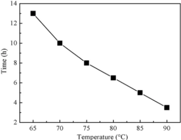
The temperature dependence of the gel forming time
was irst studied, as shown in Fig. 1. The gel formation started once the temperature reached to 65 °C and the
whole process could be completed after 13.5 h, and the
complexation reaction between the metal ion and citric
acid was not able to occur if the temperature is lower than
65 °C. The gelation time was greatly reduced as the reaction
temperature increased, and the whole gelation process could be shortened to 3.5 h while the temperature was
90 °C. However, further increasing the temperature led to
form uniform gel, which is mainly due to fast evaporation of the water. Therefore, the optimized sol-gel reaction
temperature is 80~90 °C.
Figure 1. Efect of temperature on the gel forming time.
3.2 Efect of metal ion concentration
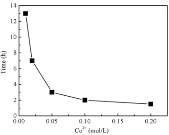
The efect of metal ion concentration on gelation time is
shown in Fig. 2. The Al3+:Co2+ molar ratio was ixed at 1:2 and the concentration of Co2+ ions varied from 0.01 mol/L
to 0.2 mol/L during the preparation. It is obvious that the
gelation time was dramatically shortened from 13 h to 1.5 h, as the Co2+ concentration increased from 0.01 mol/L to
0.2 mol/L. This suggests that the increase of total content of
metal ions promoting the gel formation. It is speculated that the presence of Co2+ reduces the intermolecular repulsion and reduces the molecular hydration, which facilitates the formation of three-dimensional network structures. These
samples were calcined at 1150 °C and the resulting blue
Figure 2. Efect of metal ion concentration on gelation time.
The colorimetric data in Table 1 summarized the color performance of the pigments prepared with varied metal ion content. It can be found that the b* value increased from -21.73 to -25.45, while a* value decreased from -20.53 to -19.18,
as the increase of Co2+ concentration. This indicates that the
pigment with 0.2 mol/L Co2+ corresponded more to blue color
(b* = -25.45), and less to green color (a* = -19.18). On the
other hand, the lightness of the pigment is governed by the coordinate parameter L*, the higher Co2+ concentration also resulted in a higher L* value, i.e. the pigment was lighter.
Table 1. The colorimetric data for cobalt blue pigments with diferent
metal ions concentration.
Samples Co2+ concentration (mol/L) L* a* b*
B1 0.01 43.69 -20.53 -21.73
B2 0.05 44.79 -20.45 -23.03
B3 0.10 44.70 -19.56 -24.19
B4 0.20 45.55 -19.18 -25.45
The efects of metal ions on color performance could be mainly attributed to the complexation reaction between
Co2+, Al3+and citric acid during preparation. Small amount
of metal ion resulted in lower complexes content and the gelation was thus not homogenous, which afected the
self-propagating combustion and the formation of the
spinel structure. Furthermore, the relection spectra of the
blue pigments were depicted in Fig. 3. The intensity of the
relection peak at 436 nm (blue) increased with the increasing
of Co2+ content, and the maximum value is 3530.70. This result is consistent with the colorimetric data in Table 1.
3.3 Efect of the dispersants
The ethylene glycol (EG) and glycerol (GC) were used
as dispersant during sol-gel process to avoid the aggregation.
Fig. 4 shows the morphology of the pigments using diferent
dispersants and being calcined at 1150 °C. It could be found that the use of organic dispersants, like EG and GC, resulted in homogeneous distribution of cobalt powder, while GC performed better than EG. One possible reason is that the EG and GC form hydrogen bonds with –COOH groups of citric acid, and it can be further sufered an esteriication reaction during high temperature, which is beneit to the formation of the gel network. Another reason is that −OH groups in EG and GC could efectively retard the colloidal particles
gathering together and thus greatly reduce aggregation.
The −OH group in GC is 1.5 times more than that in EG, therefore, the addition of dispersant GC produce uniform pigment particles than EG. The colorimetric data of the pigments prepared with diferent dispersants was presented
in Table 2. It is observed that for b* coordinate, it kept highly negative value for both samples used dispersants, and the
better performance of the sample with GC is resulted from
the uniform cobalt powder particle.
Figure 4. SEM images of the cobalt blue pigment samples calcined at 1150 °C using diferent dispersants: (A) EG and (B) GC. The
scale bars are 500 nm in length.
Table 2. The colorimetric data for cobalt blue pigments with
diferent dispersant.
Samples Dispersant L* a* b*
A-1150 EG 47.15 -16.01 -22.08
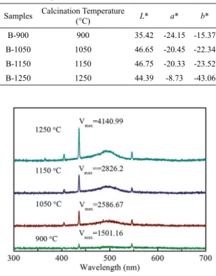
B-1150 GC 46.75 -20.33 -23.52
Figure 3. Relective intensity of the samples with diferent metal
3.4 Efect of calcination temperature
The XRD pattern are presented in Fig. 5, and it demonstrated that the powder synthesized at diferent temperature both
have spinel CoAl2O4 structure, according to JCPDS card (No.
44-0160). However, the crystallinity of specimen calcined at 900 °C spinel is much less than that calcined at 1150 °C.
Table 3. The colorimetric data for cobalt blue pigments calcined
at diferent temperature.
Samples Calcination Temperature (°C) L* a* b*
B-900 900 35.42 -24.15 -15.37
B-1050 1050 46.65 -20.45 -22.34
B-1150 1150 46.75 -20.33 -23.52
B-1250 1250 44.39 -8.73 -43.06
Figure 5. XRD patterns of the pigments calcined at diferent
temperature.
Figure 6. SEM micrographs of pigments calcined at (A) 900 °C, (B) 1050 °C and (C) 1250 °C. The scale bars are 500 nm in length.
Figure 7. Relective intensity of the pigments calcined at diferent
temperature.
Fig. 6 shows the SEM micrographs of cobalt blue pigments calcined at 900 °C, 1050 °C, 1150 °C (see Fig. 4B) and 1250 °C. As can be seen, the crystallization of the pigment particles initialed from 900 °C and the particle size was 40~50 nm, and with the increase of calcining temperature,
the product revealed more uniform crystal structure, and the grain grew gradually from tens to hundreds of nm.
We studied the inluence of the calcination temperature on
the color performance of the pigments. After self-propagating combustion, the black powder was separated and calcined
for 60 mins at 900 °C, 1050 °C, 1150 °C and 1250 °C,
respectively. The experimental results were listed in Table 3. It is obvious that diferent calcination temperatures resulted in diferent color performances of the pigments. At 900 °C,
the coordinate a* was greater than the coordinate b*, and the pigment was dark green. With the increase of calcination temperature, the coordinate b* value increased and a* gradually decreased, and the color of pigment changed from dark green through dark blue to bright blue. It is found in
Fig. 7, the intensity of the relection peak at 436 nm (blue)
increased with the increasing of calcination temperature which is consistent with the colorimetric data. It revealed
that the calcination temperature remarkably afects the color
of the product and the coordinate b* reached -43.06 as the
calcination temperature was raised to 1250 °C. This result is
better than previous reported works, such as Co0.95Zn0.05Al2O4
system (b* = -29.54)4, Co
0.5Zn0.5Al2O4 system (b* = -36.24) 6 and Mg0.8Co0.2Al2O4 pigment (b* = -41.67)10.
4. Conclusions
In conclusion, high purity cobalt blue pigments with spinel structure were prepared by sol-gel method followed by self-propagating combustion. The results show that the formation of gel network could be promoted by increasing
the concentration of metal ion (0.01~0.2 mol/L) and sol-gel reaction temperature (80~90 °C). The dispersibility and stability
glycerol as dispersant. The particle size of the pigment increases with the increasing of calcination temperature, and the b* value of the pigment could be reached as high
as -43.06 at 1250 °C, which is higher than previous results.
This is a facile route to produce CoAl2O4 powders without introducing any dopants, and it has better particle distribution and dispersity than traditional solid-state reaction.
5. Acknowledgments
This work was supported by the National Natural Science Foundation of China (61471233, 21504051), the Program for Professor of Special Appointment (Eastern Scholar) at Shanghai Institutions of Higher Learning, the Shuguang and YangFan Project from Science and Technology Commission of Shanghai Municipality (14SG52, 14YF1410600).
6. References
1. Derby B. Inkjet printing ceramics: From drops to solid. Journal
of the European Ceramic Society. 2011;31(14):2543-2550.
2. Chen Z, Shi E, Li W, Zheng Y, Zhong W. Hydrothermal synthesis
and optical property of nano-sized CoAl2O4 pigment. Materials Letters. 2002;55(5):281-284.
3. Nakatsuka A, Ikeda Y, Yamasaki Y, Nakayama N, Mizota T. Cation distribution and bond lengths in CoAl2O4 spinel. Solid
State Communications. 2003;128(2-3):85-90.
4. Chen YX, Hu Q, Cao CE, Lu XL, Hong C, Shen HR. Efects of
Zn2+ and Cr3+ doping on nano-sized CoAl
2O4 spinel pigments
by hydrothermal processing. Journal of Inorganic Materials.
2012;27(12):1317-1320.
5. Srisawad N, Chaitree W, Mekasuwandumrong O, Praserthdam
P, Panpranot J. Formation of CoAl2O4 nanoparticles via
low-temperature solid-state reaction of ine gibbsite and cobalt
precursor. Journal of Nanomaterials. 2012;2012:108369.
6. de Souza LKC, Zamian JR, da Rocha Filho GN, Soledade LEB, dos Santos I MG, Souza AG, et al. Blue pigments based on CoxZn1-xAl2O4 spinels synthesized by the polymeric precursor method. Dyes and Pigments. 2009;81(3):187-192.
7. Chemlal S, Larbot A, Persin M, Sarrazin J, Sghyara M, Raiq M.
Cobalt spinel CoAl2O4 via sol-gel process: elaboration and surface properties. Materials Research Bulletin.
2000;35(14-15):2515-2523.
8. Cui H, Zayat M, Levy D. Sol-Gel Synthesis of Nanoscaled Spinels Using Propylene Oxide as a Gelation Agent. Journal
of Sol-Gel Science and Technology. 2005;35(3):175-181.
9. Eliziário SA, de Andrade JM, Lima SJG, Paskocimas CA, Soledade LEB, Hammer P, et al. Black and green pigments based on chromium–cobalt spinels. Materials Chemistry and Physics. 2011;129(1-2):619-624.
10. Ianoş R, Lazău R, Barvinschi P. Synthesis of Mg1−xCoxAl2O4 blue
pigments via combustion route. Advanced Powder Technology.