Regioselective Binding of Spermine, N
1,N
12-Bismethylspermine,
and N
1,N
12-Bisethylspermine to tRNA
Pheas Revealed by 750 MHz
1
H-NMR and its Possible Correlation with Cell Cycling and
Cytotoxicity
Benjamin Frydman
a, William M. Westler
b, Aldonia Valasinas
a,
Debora L. Kramer
c, and Carl W. Porter
caSLIL Biomedical Corp., 535 Science Drive, Suite C, Madison, WI 53711-1066 USA
bNational Magnetic Resonance Facility, University of Wisconsin-Madison, Madison,
WI 53706, USA
cGrace Cancer Drug Center, Roswell Park Cancer Institute, Elm and Carlton Streets,
Buffalo, NY 14263 USA
Neste trabalho foi feito o estudo de ligação de espermina (SPM), N1, N12- bismetilespermina (BMS) e N1, N12-bisetilespermina (BES) ao tRNAfen usando RMN de 1H a 750 MHz. As poliaminas foram enriquecidas com 13C nos resíduos 5-CH2 e 8-CH2, sendo obtidos os picos cruzados por efeito
de Overhauser nuclear entre as ressonâncias dos hidrogênios dos metilenos marcados com 13C e vários hidrogênios de grupos imino de pares de bases do tRNAfen através de espectros 1D filtrados por 13C. Foi encontrado que, enquanto SPM e BMS se ligam ao N(3)-H dos pares de bases T54-m1A58, U50-A64 e U52-A62, BES liga-se só nos pares T54-m1A58 e U50-A64. Esta regiosseletividade de ligação das três poliaminas ao tRNA foi correlacionada com seus efeitos biológicos no crescimento celular. Usando células de câncer de melanoma humano (MALME-3M), foi encontrado que SPM e BMS não tem efeito e é citostático, respectivamente, enquanto que BES é claramente citotóxica. Esta última poliamina também afeta o ciclo celular e, contrário ao comportamento de SPM e BMS, acarreta a uma clara parada do ciclo celular G1 /S.
The binding of spermine (SPM), N1,N12-bismethylspermine (BMS) and N1,N12 -bisethylsper-mine (BES) to tRNAPhe was studied using 1H-NMR at 750 MHz. The polyamines were enriched in
13C at the 5-CH
2 and 8-CH2 residues and the nuclear Overhauser enhancement (NOE) cross peaks
connecting the 1H-NMR resonances of the 13C-methylenes and several base paired imino protons of tRNAPhe were obtained using 1D 13C-half filtered spectra. It was found that while SPM and BMS bind to the N(3)-H of base pairs T54-m1A58, U50-A64 and U52-A62, BES binds only to T54-m1A58 and U50-A64. This regioselectivity in the binding of the three polyamines to tRNA was correlated with their biological effects on cell growth. Using human melanoma cancer cells (MALME-3M), we found that SPM and BMS were without effect and cytostatic, respectively, while BES was distinctly cytotoxic. The latter also affected cell cycling and, at variance with SPM and BMS, lead to a distinct G1/S cell cycle arrest.
Keywords:1H-NMR of polyamines, tRNAPhe, melanoma, spermine
Introduction
Spermine 1 (SPM) (Fig. 1) and spermidine (SPD) are polyamines widely distributed in biological systems where they are absolutely required for cell proliferation and also
to support carcinogenesis1-3. Of the many hypotheses ad-vanced to explain their biological effects4, the most solid one concerns their interaction with nucleic acids5-7. Polyamines bind to ribosomal subunits, to DNA, and to
Article
aPhone: 608-231-3854; Fax: 608-231-3892.
tRNA1. When binding to DNA they can cause DNA to condense and aggregate and can induce both B-to-Z and B-to-A transitions in certain DNA sequences8-10. The inter-action of both SPM and SPD with tRNA has been exten-sively studied using solution 1H-NMR11a, 13C-NMR7, and
15N-NMR11b,11c techniques. The polyamines are strong
bases protonated at physiological pH and the aforemen-tioned NMR studies showed that they bind to the nucleo-tides preferentially through hydrogen bonds generated by the protonated –NH2+– residues (N4 and N9) internally
present in the polyamine backbone (Fig. 1). The primary –NH3+ groups bind to tRNA much more weakly than the
secondary –NH2+– groups, thus casting doubt on the
sim-plistic notion that polyamines are just “organic cations” analogous to Mg2+ or K+. The 15N-NMR experiments sug-gest that the specificity with which nitrogens in polyamines bind to tRNA is a consequence of the different hydrogen bonding modes that can be established between both types of molecules. More recently11a we have shown, using high
field 1H-NMR analysis, that SPM binds in solution to the TΨC loop of tRNAPhe, at the corner of the L-shaped tRNA molecule where the TΨC and D loops meet. The impor-tance of this part of the tRNA molecule for its correct in vivo function is known from random mutagenesis experi-ments12.
Synthetic analogs of polyamines have been shown to be promising anticancer agents. They have the potential of killing cells and inhibiting cell growth both in vivo and in vitro3,13. A decade ago13a Bergeron and coworkers showed that N1,N12-bisethylspermine (BES) 3 (Fig. 1) was a pow-erful antineoplastic agent; these results led to the synthesis of a large number of highly cytotoxic Nα,Nω-bisethyl polyamine analogs3,13b,13c, two of which reached Phase I
clinical trials. The former disclosure13a also reported that N1,N12-bismethylspermine (BMS) 2 (Fig. 1) was a much weaker antineoplastic agent than BES. This divergence between the inhibitory effects on cell growth of BES and BMS remained unexplored until very recently when their effect was evaluated in vitro in MALME-3M human mela-noma cells. It was found that while BES is cytotoxic, BMS is cytostatic14. These results suggest that the antineoplastic effects of the Nα,Nω-bisalkyl derivatives of spermine could be due, at least in part, to their effects on the protein synthesis process of the cell cycle rather than on the DNA
strongest hydrogen bonds with the nucleotides (as is the case with SPM or SPD), while the terminal –NH2+C2H5
groups (N1 and N12) formed the weaker bonds. The same was true for the Nα,Nω-bismethyl derivatives; the strongest hydrogen bonds between the polyamine and tRNA were those formed by the central nitrogens (N4 and N9), inde-pendent of the alkyl substituent (methyl or ethyl) present at the terminal nitrogen groups. Binding of polyamines to tRNA has been shown to be relevant to the fidelity of the translation process16-19, as well as to codon-anticodon rec-ognition and ribosomal binding20-21. Polyamines also stimulate aminoacylation in general and their effects on the structure of tRNA compelled attention to the possible role of these compounds for protein synthesis22. We therefore decided to examine the binding regiospecificity of sper-mine (SPM) 1, bisethylspermine (BES) 3 and bismethyl-spermine (BMS) 2 to tRNA using high field 1H-NMR, and to attempt to correlate these results with their effect on the essential cell cycle processes and on the cell growth inhibi-tion of a tumor cell line.
Results and Discussion
Regioselective binding to tRNAPhe of spermine (SPM) 1, bismethylspermine (BMS) 2, and bisethylspermine (BES) 3
To identify the binding domains of 2 and 3 with tRNA we pursued the chemical and spectroscopic approach we devised to establish the binding of spermine 1 with tRNAPhe.11a The strategy is based on the use of 1H-1H nuclear Overhauser enhancement (NOE) spectra where the spatial proximity between the polyamine analog and the tRNA protons could be determined by the appearance of peaks resulting from the 1H-NMR resonances. In order to use this approach it is necessary to have a set of clearly resolved 1H-NMR resonances of the tRNA assigned to well-defined regions within the structure. A valuable link between the structure of tRNA and its 1H-NMR spectrum can be found in the 9.5-15.0 ppm region, where only the hydrogen bonded imino protons of the paired nucleotide bases resonate; these imino resonances have been assigned to individual bases in E. coli23 and yeast tRNAPhe24 (Fig. 2). We used these assignments to monitor the sites of binding, by measuring NOE cross peaks between the 1 H-imino resonances from the macromolecule and the polyamine analog protons. The upfield region of the tRNA spectrum (1.0-3.0 ppm) also allows the identification of the resonances of the methyl groups present in the tRNAPhe bases and was used for measuring NOE cross peaks with Figure 1. Structures of [13C2]-enriched spermine 1 (R=H), N1,N12
the spermine protons.11a The aromatic region of the tRNA
1H-NMR spectrum (6.0-9.0 ppm) is less useful for these
determinations since the non-exchangeable aromatic pro-tons have not been unequivocally assigned.
Ideally, labeling the –NH2+–, –NH2+R, and –NH3+
pro-ton resonances of N1,N12-bisalkylspermines 2 and 3 or spermine 1 would be necessary, as these groups are directly involved in the binding process. Fast exchange with the aqueous solvent, however, precludes the observation of these resonances. This leaves the CH2-protons of the
methylenes α to the amino groups as the only practical choice for implementing this study. These protons resonate between 2.8-3.1 ppm; for the relative polyamine/tRNA concentrations that we use they will therefore be only marginally visible, due to the severe overlap with the tRNA sugar resonances starting at ca. 3.1 that integrate for hun-dreds of sites. In order to alleviate this problem we used polyamines enriched with 13C in the carbon bonded to the –NH2+– groups (Fig. 1). This allowed the acquisition of 13C-half-filtered 1H-NMR spectra where only protons
bound to 13C-heteroatoms appear along one of the fre-quency dimensions. The outline of the pulse sequences on which our tRNA/polyamine binding studies were based can be seen in Fig. 3. The sequence used in the 1D, 13 C-half-fil-tered, proton-proton NOE spectra is summarized in se-quence (1):
D1 − 90H− ∆± 90C−180H− 90C− ∆ −90H−
− Tmix− 90H−δ−(−) 90H−± acquisition (1)
The imino proton region of tRNAPhe (9.0-15.0 ppm) shown in Fig. 4A was recorded in cacodylate buffer in the absence of Mg2+. It is known24,25 that the structure of tRNAPhe in the absence of Mg2+ is basically the same as in the presence of Mg2+, and that the 1H-NMR spectrum in the region of 9.0-15.0 ppm is essentially independent of its presence. When the 1D 13C half-filtered NOE spectrum of the imino region of the 1:8 tRNAPhe:[5,8-13C2]-spermine
complex was recorded, a strong NOE signal at 12.48 ppm could be measured (Fig. 4C). We had already shown 11a that this interaction corresponds to the NOE cross peak of the
[5,8-13CH2] methylenes of spermine and the N(3)-H of the
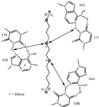
T54-m1A58 base pair (Fig. 5). Base T54 on the TΨC loop is hydrogen bonded through its N(3)H to m1A58 and this Figure 2. tRNAPhe and the 1H-NMR of its imino region recorded at
750 MHz.
Figure 3. Pulse sequence used to obtain one-dimensional 13C half-filtered proton-proton NOE spectra. The narrow and wide lines represent 90° and 180° pulses, respectively. Gz represents a pulse field gradient. Broadband
decoupling of 13C during the acquisition period was achieved by GARP modulation. Details of the timing, phases, and frequencies are given in Experimental.
Figure 4. 750 MHz 1D 13C half-filtered NOE spectra of the imino region of 8:1 complexes of [5,8-13C2]-enriched polyamines with tRNAPhe. (A)
base stacks on top of the three bases G57, G18, and G19 (the latter two of loop D) to form a stack of four purines. This interaction is critical for the stabilization of the TΨC loop at the juncture with the D loop (Fig. 2). Two weaker NOE cross peaks were also observed in the interaction of [5,8-13C2]-spermine and the imino protons of other
tRNAPhe base pairs. The NOE cross peak at 13.43 ppm (Fig. 4C) corresponds to the imino proton linking U50 to A64 on the T stem (Fig. 5). We confirmed Hilber’s assignment of this resonance (resonance H)25 to this base pair using
2D-NOE correlations. The second cross peak at 13.93 ppm corresponds to the interaction of spermine with an imino proton of a second UA base pair; namely U52 A62 (reso-nance D in Hilber’s assignment25). Both U50 A64 and U52 A62 are base pairs of the T-stem and help locate the spermine molecule at the juncture of the TΨC loop and the D loop (Fig. 2). The importance of this part of the tRNA molecule for its correct in vivo function is well known26. When the interaction of [5,8-13C2]-N1,N12
-bismethylsper-mine (BMS) 2 with tRNAPhe was examined using an analo-gous procedure to that used with [5,8,13C2]-spermine, the
same pattern of NOE cross-peaks was obtained (Fig. 4D). Thus, the binding domain of BMS to tRNAPhe is similar to
that found for spermine. A different NOE cross peak pattern was obtained when the binding of [5,8-13C
2]-N1,N12
-bi-sethylspermine (BES) 3 with tRNAPhe was examined. While strong NOE signals were obtained with the imino protons of the T54 A58 and U50 A64 base pairs, the interaction with the U52 A62 imino proton could not be detected (Fig. 4B). It is therefore very likely that BES docks
tRNAPhe at the juncture of the TΨC and D loops will affect the kink of the tRNAPhe and therefore the specificity of aminoacyl-tRNAPhe formation22. A linkage of this hypothe-sis with cell growth was sought using human melanoma cancer cells (MALME-3M).
Effects of spermine (SPM) 1, bismethylspermine (BMS) 2, and bisethylspermine (BES) 3 on MALME-3M cell growth, cell cycle and polyamine pools.
MALME-3M cells treated with 10 µM SPM grew simi-larly to untreated cells for 120 h (Fig. 6). In the presence of BMS the cells grew for 48 h and then completely ceased growing.. There was significant loss in cell number during BMS treatment to suggest a cytotoxic response. Compared to BMS, BES inhibited growth by 60% afer 48 h which was followed by a cytotoxic response as indicated by a steady loss in cell number to below the initial seeding density. Also with BES treatment, there was a significant number of detached cells previously characterized to be apoptotic14.
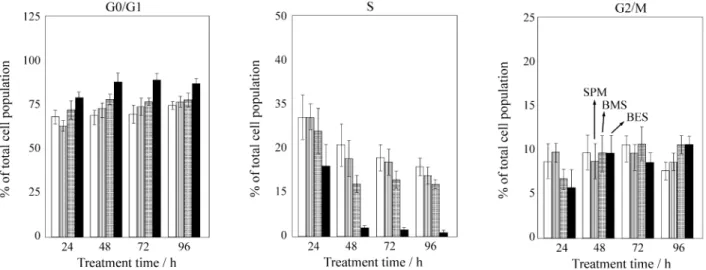
Samples taken for cell cycle analysis at 24 h intervals up to 96 h treatment (Fig. 7) showed that SPM and BMS treat-Figure 5. Outline of the 1H-1H cross peaks of 13C-half-filtered NOE
spectra of [13C]-enriched spermines 1-3 with TΨC base pairs in tRNAPhe.
ments only marginally altered cell cycling and the cell population profile remained similar to that of untreated cells, with ~65% of cells in G0/G1, ~27% in S, and ~11%
in G2/M phase. In contrast, BES-treated cells showed a 50%
decrease in S phase cells accompanied by an increase in G1
cells only at 24 h. By 48 h, only 2% of the cells remained in S phase indicating a full G1/S arrest. No significant
change was observed for G2/M phase cells, and the fact that
10% of the cells remained in this phase suggests the possi-bility of an additional block here.
The melanoma cells were also analyzed for polyamine and polyamine analog pools (Table 1). Decreases occurred in all three of the intracellular polyamines by 48 h and these natural pools were replaced with comparable amounts of BMS and BES. Thus, the distinct differences observed in effects on growth and cell cycle between these two analogs are independent of their abilities to accumulate intracellu-larly or to deplete polyamine pools. The results basically show three separate effects of SPM, BMS and BES on MALME-3M cell growth; namely, no effect by SPM, a cytostatic effect by BMS, and a cytotoxic effect by BES. Only the cytotoxic analog, BES, had affected growth and cell cycling prior to 48 h. The latter resulted in a significant
loss in S phase cells by 24 h with a concomitant increase in G1. Interestingly, it also binds to tRNA with a different
regioselectivity than that of SPM or BMS. Thus, the effect on cell cycle by BES implies that there is some distinct gain or loss of biological action for this analog compared to SPM and BMS. One possibility has been demonstrated here that results from the different binding mode of BES to tRNA which could lead to a lack of fidelity in the translation mechanism, cell cycle arrest, and ultimately to a cytotoxic effect.
Experimental
1H-NMR determinations
tRNAPhe from brewers yeast was from Sigma. It was
dissolved in 0.5 mL of a buffer containing 50 mM sodium cacodylate, 2 mM EDTA, 400 mM NaCl (pH 7.2), and dialyzed for 20 h against the same buffer in double-sided biodialysers (Sialomed). The resulting tRNAPhe solution
was polyamine-free (assayed by HPLC). The isotopically enriched spermines (Fig. 1) were added to the dialyzed tRNA solution to give final, 1 mM tRNAPhe, 8 mM
polyamine mixtures. 1H-NMR spectra were recorded at 750
MHz at 5 °C. The water signal was suppressed by the jump
Table 1. Polyamine analysis of MALME-3M cells treated with SPM, BMS, and BES.
Treatment Polyamine Pools*
(10 µM, 48 h) % Control growth
Put Spd Spm Analog
(pmol/106 cells)
Control 100 995 1835 2090
--SPM 120 < 5 410 2730
--BMS 70 < 5 < 5 65 5950
BES 37 < 5 < 5 100 6140
*Pool values were based on averages of at least three separate determinations with S.D. < 15%.
placed at 41 ppm. The 1H spectra was 22,522 Hz (30 ppm)
and for each acquisition 2,048 complex data points were collected. The number of acquisitions for these spectra was 100,000 (41 h collection time). The solvent composition was 95% H2O/5% 2H2O. All chemical shifts are referenced
to internal DSS.
Cell growth
MALME-3M cells obtained from the American Type Culture Collection (Rockville, MD) were grown and main-tained as described14. Cells were seeded 24 h prior to treatment with 10 µM of SPM and each analog for an additional 120 h. Cell numbers were determined electroni-cally (Coulter Counter; Coulter Electronics, Hialeah, FL).
Flow cytometry
Cell analysis was performed on attached cells following treatments of 10 µM analog or SPM for 24 h intervals up to 96 h. Briefly, cells were stained using a propidium iodide buffer and 10,000 events were recorded by FACScan flow cytometer (Becton Dickinson, San Jose, CA) to generate DNA histograms as described14.
Polyamine pool determinations
Following treatment of the MALME-3M cells for 48 h with 10 µM SPM, DMSPM, and DESPM, the intracellular polyamine and analog pools were determined on acid ex-tracts using a high-performance liquid chromatography system described previously27.
Materials
[5,8-13C2]-Spermine, [5,8-13C2]-N1,N12
-bismethyl-spermine, and [5,8-13C2]-N1,N12-bisethylspermine were
prepared by Prof. Keijiro Samejima (Josai University, Sai-tama, Japan) following published procedures28. N1,N12 -bis-methyl and N1,N12-bisethylspermines were prepared as follows according to our published procedures3. 1,4-Bis(mesitylenesulfonyloxy) butane was condensed with either N-methyl or N-ethyl-N’-(3-(mesitylenesulfony-lamino)propyl)-mesitylene sulfonamide. Deprotection in acid media of the resulting tetramides gave the tetrahydro-chlorides of either 2 or 3.
Conclusions
A) When spermine (SPM), N1,N12-bismethylspermine (BMS) and N1,N12-bisethylspermine (BES) enriched with
13C at 5-CH
2 and 8-CH2 were added to tRNAPhe it was
m1A58 and U50-A64. All these base pairs are located at the junction of the TΨC loop and D loop.
C) When the effects of SPM, BMS, and BES on the growth of human melanoma cancer cells (MALME-3M) were compared, it was found that SPM and BMS behaved in a similar manner. They were minimally growth inhibi-tory up to 48 h, they did not alter the cell cycle whose profiles remained similar to the control cells in both cases, and they were not cytotoxic. BES, on the contrary, was rapidly cytotoxic after 48 h and caused a distinct G1/S cell
cycle arrest. These BES effects were not attributed to specific decreases in the intracellular polyamine pools. We, therefore, propose that the growth effects of BES could be correlated with its binding to tRNA that differs from that of SPM, the natural polyamine involved in cell growth. The cytotoxic action of BES could be related to its interference with the fidelity or efficiency of the cellular translation process.
Acknowledgments
NMR studies were carried out with support from the NIH Biomedical Technology Program (RR02301) and funding from the University of Wisconsin, NSF Academic Infrastructure Program (BIR-9214394), NIH Shared In-strumentation Program (RR02781, RR08438), NSF Bio-logical Instrumentation Program (DMB-8415048), and U.S. Department of Agriculture.
References
1. Cohen, S.S. A Guide to the Polyamines; Oxford Uni-versity Press, Oxford, 1998.
2. Marton, L.J.; Pegg, A.E. Annu. Rev. Pharmacol. Toxi-col.1995, 33, 55.
3. Reddy, V.K.; Valasinas, A.; Sarkar, A.; Basu, H.S.; Marton, L.J.; Frydman, B. J. Med. Chem.1998, 41, 4723; and references therein.
4. Tabor, C.W.; Tabor, H. Annu. Rev. Biochem.1984, 53, 749.
5. Sakai, T.T.; Cohen, S. Progr. Nucl. Acid. Res. Mol. Biol. 1975, 17, 15.
6. Feuerstein, B.G.; Marton, L.J. In The Physiology of Polyamines; Bachrach, U.; Heimer, Y.M., eds., CRC Press, Boca Raton, Florida, v. I, 109, 1989.
7. Frydman, B.; de los Santos, C.; Frydman, R.B. J. Biol. Chem.1990, 265, 20874.
9. (a) Chen, H.-H.; Behe, M.J.; Rau, D.C. Nucl. Acid. Res.1984, 12, 2381; (b) Russell, W.C.; Precious, B.; Martin S.R.; Bayley, P.M. EMBO J.1983, 2, 1647. 10. Feuerstein, B.G.; Pattabiraman, N.; Marton, L.J. Nucl.
Acid. Res.1990, 17, 6883.
11. (a) Frydman, B.; Westler, W.M.; Samejima, K. J. Org. Chem.1996, 61, 2588; (b) Fernandez, C.O.; Frydman, B.; Samejima, K. Cell. Mol. Biol.1994, 40, 933; (c) Frydman, L.; Rossomando, P.C.; Frydman, V.; Fer-nandez, C.O.; Frydman, B.; Samejima, K. Proc.Natl. Acad. Sci. USA1992, 89, 9186.
12. Hou, Y.M.; Schimmel, P. Biochemistry 1992, 31, 4157.
13. (a) Bergeron, R.J.; Neims, A.H.; McMannis, J.S.; Hawthorne, T.R.; Vinson, J.R.T.; Bartell, R.; Ingeno, M.J. J. Med. Chem.1988, 31, 1183; (b) Bergeron, R.J.; McMannis, J.S.; Liu, Ch. Z.; Feng, Y.; Weimar, W.R.; Luchetta, G.R.; Wu, Q.; Ortiz-Ocasio, J.; Vinson, J.R.T.; Kramer, D.; Porter, C. J. Med. Chem.1994, 37, 3464; (c) Basu, H.S.; Pellarin, M.; Feuerstein, B.G.; Shirahata, A.; Samejima, K.; Deen, D.F.; Marton, L.J.
Cancer Res. 1993, 53, 3948; (d) McClosky, D.E.; Casero, R.A., Jr.; Woster, P.M.; Davidson, N.E. Can-cer Res. 1995, 55, 3233; (e) Li, Y.; Eiseman, J.L.; Sentz, D.L.; Rogers, F. A.; Pan, S.-S.; Hu, L.-T.; Egorin, M.J.; Callery, P.S. J. Med. Chem.1996, 39, 339; (f) Huber, M.; Poulin, R. Cancer Res.1995, 55, 934; (g) Aizencang, G.; Frydman, R.B.; Giorgeri, S.; Sambrotta, L.; Guerra, L.; Frydman, B. J. Med. Chem.
1996, 38, 4337; (h) Frydman, J.; Ruiz, O.; Robetto, E.; Dellacha, J.M.; Frydman, R.B. ; Mol. Cell. Biochem.
1991, 100, 9; (i) Moyano, N.; Frydman, B.; Buldain, G.; Ruiz, O.; Frydman, R.B. J. Med. Chem.1990, 33, 1969; (j) Aizencang, G.; Harari, P.; Buldain, G.; Guerra, L.; Pickart, M.; Hernandez, P.; Frydman, B.
Cell. Mol. Biol.1998, 44, 615.
14. Kramer, D.L.; Fogel-Petrovic, M.; Diegelman, P.; Cooley, J.M.; Bernacki, R.J.; McMannis, J.S.;
Ber-geron, R.J.; and Porter, C.W. Cancer Res.1997, 57, 5521.
15. Basu, H.S.; Wright, W.D.; Deen, D.F., Roti-Roti, J.; Marton, L.J. Biochemistry1993, 32, 4073.
16. Kusama-Eguchi, K.; Irisawa, M.; Watanabe, S.I.; Watanabe, K.; Igarashi, K. Arch. Biochem. Biophys.
1991, 288, 495.
17. Snyder, R.D.; Edwards, M.L. Biochem. Biophys. Res. Commun.1991, 176, 1383.
18. McMurry, L.; Algranati, I.D. Eur. J. Biochem.1986,
155, 383.
19. Lotfield, R.B.; Eigner, E.A.; Patuszyn, A. J. Biol. Chem.1981, 256, 6729.
20. Algranati, I.D., Goldenberg, S.H. Trends Biochem. Sci.1977, 2, 272.
21. Abraham, A.K.; Phil, A. Trends Biochem. Sci.1981,
6, 106.
22. (a) Rould, M.A.; Perona, J.J.; Soll, D. and Steita, T.A.
Science 1989, 246, 1135; (b) Ruff, M.; Krish-naswamy, S.; Boeglin, M.; Poterszman, A.; Mitschler, A.; Podjarny, A.; Rees, B.; Thiery, J.C. and Moras, D.
Science1991, 252, 1682.
23. Hyde, E.I.; Reid, B.R. Biochemistry 1985, 24, 4315. 24. Heerschap, A.; Walters, J.A.L.I.; Hilbers, C.W. Nucl.
Acids Res.1986, 14, 983.
25. Hilbers, C.W.; Heerschap, A.; Haasnot, C.A.G.; Wal-ters, J.A.L.I. J. Biomol. Struct. Dyn.1983, 4, 1983. 26. Dirheimer, G.; Keith, G.; Dumas, P.; Westhof, E. In
tRNA: Structure, Biosynthesis andFunction, Söll, D.; RajBhandary, U.L., eds., ASM Press, Washington, DC, 93, 1995.
27. Kramer, D.L.; Miller, J.T.; Bergeron, R.J.; Khomutov, R.; Khomutov, A.; Porter, C.W. J.Cell. Physiol.1993,
155, 300.
28. Samejima, K.; Matsushima, N.; Niitsu, M.; Beppu, T.; Frydman, B. Chem. Pharm. Bull.1995, 43, 2001.
![Figure 4. 750 MHz 1D 13 C half-filtered NOE spectra of the imino region of 8:1 complexes of [5,8- 13 C 2 ]-enriched polyamines with tRNA Phe](https://thumb-eu.123doks.com/thumbv2/123dok_br/18988402.459727/3.918.101.443.105.280/figure-filtered-spectra-imino-region-complexes-enriched-polyamines.webp)