The Use of
13C and
1H-NMR in the Structural Elucidation of a New
Nor
-Lupane Triterpene
Mário J. Junges
a, João B. Fernandes
b*; Paulo C. Vieira
a, Maria Fátima das
G. Fernandes da Silva
b, and Edson Rodrigues Filho
ba
Departamento de Química, Universidade Federal do Rio Grande,
Rua Eng. Alfredo Huch 475, C.P. 474, 96201-900 Rio Grande - RS, Brazil
b
Departamento de Química, Univesidade Federal de São Carlos,
Rod. Washington Luis km 235, 13565-905 São Carlos - SP, Brazil
Um triterpeno nor-lupânico, inédito, foi isolado das folhas de Eugenia florida DC (Myrtaceae). Tomando-se por base as evidências espectroscópicas, RMN 1H, 13C, experimentos de RMN 2D, em especial correlações homonuclear 1H-1H, heteronuclear 1H-13C e correlações a várias ligações (HMBC), sua estrutura foi elucidada como: 29-hidroxiplatan-28-ato de β-D-glicosila.
The new nor-lupane triterpene, isolated from the leaves of Eugenia florida DC (Myrtaceae) was identified on the basis of its spectroscopic data, 13C and 1H-NMR-2D, specially COSY 1H-1H, COSY 1H-13C and HMBC correlations, as 28-O-β-D-glucopyranosyl ester of 29-hydroxyplatanic acid.
Keywords: Nor-lupane triterpene, 13C and 1H-NMR, HMBC, COSY, Eugenia florida, Myrtaceae
Introduction
The triterpenes are a large group of plant substances with a wide spectrum of biological activities, such as anti-HIV1. The study of the Eugenia florida DC (Myrtaceae) leaves, which occur in the areas of tropical and subtropical2 climate, is underway in our laboratories. In this work an unpublished triterpene with skeleton nor-lupane was iden-tified. The identification of its structure was made through the use of spectroscopic methods. The homonuclear 1H-1H and one bond and multiple bond heteronuclear 1H-13C correlations experiments were used as the main tools to identify this new compound.
Results and Discussions
The structural determination of 1 was done on the basis of a comparative analysis of its 1H and 13C-NMR 1D and 2D data with those reported in the literature for platanic acid1,3,4. In the 1H-NMR spectrum of 1, it was observed the presence of 5 methyl signals, δ 0.77, 0.85, 0.94, 0.96 and 1.07 (Table 1). The signal at δ 2.21 of the CH3-29, which is characteristic of platanic acid1 was not detected. At δ
3.26, it was observed a triplet (J = 7.6 Hz) of the Hax bounded to C-3, which was coupled with H-2. The
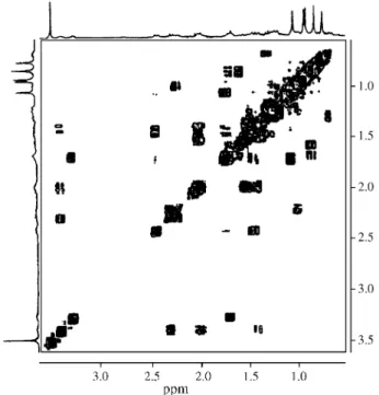
multi-plet appearing at δ 3.36-3.43 corresponds to H-19, coupled with H-18 and H-21. The COSY 1H-1H spectrum of 1 (Fig. 1) also exhibited several important correlations to establish the triterpene skeleton and the attribution of the 1H chemi-cal shifts (Table 1) was supported by these data. From the DEPT 135 experiment it was possible to determine that 1 was a primary alcohol, which justifies the fact that 1 showed only 5 methyl groups, instead of 6 as in platanic acid. The location of the hydroxyl group at CH2-29 (δ 68.6), whose 1H signals appeared as two doublets at δ 4.40 and 4.32 (J = 18.8 Hz) and the attribution of the chemical shifts of all methyl groups, was carried out by analysis of the HMBC spectrum (Figs. 2 and 3). It was noticed the un-equivocal correlation of the two methyl groups, H-23 (δ
1.07) and H-24 (δ 0.85) with C-3 at δ 78.1 (3J, Table 1), and with the carbons C-5 at δ 55.7, and C-4 at δ 39.2 (2J). It was also noticed the correlation of the methyl H-25 (δ
0.77) with C-10 at δ 37.4 (2J) and of two methyl groups, H-26 and H-27 with C-8 at δ 40.9 (respectively 2J and 3J). H-29 showed correlation with the carbonyl at δ 214.2 (C-20)3.
The presence of the sugar moiety in the molecule was verified by the signal of the anomeric hydrogen detected at Article
J. Braz. Chem. Soc., Vol. 10, No. 4, 317-320, 1999. © 1999 Soc. Bras. Química
δ 5.95 (d, J = 8.4 Hz), confirmed by other signals between
δ 3.70 and 4.36. The inspection of the COSY 1H-1H spec-trum (Fig. 1), relatively to the glucosyl unit, allowed to establish the correlation between the hydrogens H-1’ to H-6’. The inspection of the HMQC spectrum of 1 (Fig. 2, Table 1) showed correlation of H-1’-H-6’ signals with the signals at δ 95.5, 73.6, 78.3, 70.9, 78.2 and 62.0, C-1’ to C-6’ respectively. These signals and these correlations are the same registered in the literature for β-D-glucose7. After having assigned the 1H and 13C chemical shifts of the sugar moiety, it was still remaining to locate this unit in the nor-lupane skeleton at the position C-3, C-28 or C-29. The 13C-NMR spectrum showed a carbonyl carbon signal at δ
174.7, shielded by 4.0 ppm, which occurs when C-28 is an ester function5,6. This functionality was confirmed by the HMBC spectrum (Table 1, Fig. 3), whose 1H signal at δ
5.95 was correlated to the carbonyl carbon at δ 174.7 (3J). This unequivocally confirmed that the position C-28 was indeed esterified with β-D-glucose.
Finally, the mass spectrum of 1, obtained by FAB/MS, revealed the peak corresponding to the molecular ion (m/z 637, M+1, 3%), and the peaks 636 (5), 473 (20), 275 (32), 183 (100) Daltons.
These data allowed to attribute the structure of 1 as being 28-O-β-D-glucopyranosyl ester of 29-hydroxypla-tanic acid which is now described for the first time in the literature.
Experimental
General experimental procedure
The 1H and 13C-NMR experiments were recorded in a Bruker spectrometer ARX, operating respectively at 400 MHz for 1H and 100 MHz for 13C, using deutero pyridin
Figure 1.1H x 1H-COSY spectrum of 1.
Figure 2. HMQC spectrum of 1.
Table 1.1H and 13C-NMR data of 1 and platanic acid.1,3 The H-1 and C-13 chemical shift assignments were obtained from 2D spectra (HMBC, HMQC and COSY 1H-1H) and 1 D (fully decoupled from 1H and DEPT). The spectra were recorded in pyridine, and the chemical shifts referenced to internal TMS.
1
H or 13C 1 (δ) HMBC 1 (δ) platanic
acid (δ)
1 ax. 0.82-0.91 (m) 39.1 39.3
1 eq. 1.50-1.60 (m)
2 1.63-1.73 (m) 27.9 28.2
3α 3.26 (t, J = 7.6) 78.1 77.8
4 39.2 39.5
5 0.69 (d, J = 9.6) 55.7 55.7
6 1.25-1.38 (m) 21.0 21.1
7 1.30* 34.4 34.6
8 40.9 40.9
9 1.27* 50.7 50.7
10 37.4 37.7
11 1.40* 18.6 18.8
12 1.00* 27.6 27.7
13 2.22-2.28 (m) 37.2 37.7
14 42.5 42.5
15 ax. 1.04-1.12 (m) 29.9 30.2
15 eq. 1.70-1.77 (m)
16 ax. 1.40-1.50 (m) 31.4 32.3
16 eq. 2.44 (dl, J = 12.4)
17 56.6 56.4
18 2.30 (t, J = 6.7) 49.9 49.7
19 3.36-3.43 (m) 46.3 52.0
20 214.2 211.6
21 ax. 1.38-1.44 (m) 29.0 28.7
21 eq. 1.95-2.03 (m)
22 ax. 1.45-1.56 (m) 36.8 37.4
22 eq. 1.95-2.03 (m)
23 αCH3 1.07 (s) C-3 and C-4, (3J/2J) /C-5, (3J) 28.5 28.6 24 βCH3 0.85 (s) C-3, (3J) / C-4 andC-23, (2J/3J) 16.1 16.3 25 βCH3 0.77 (s) C-10, (2J) /C-5, (3J) 16.4 16.3
26 βCH3 0.94 (s) C-8, (2J) 16.1 16.3
27 αCH3 0.96 (s) C-8, (3J) 14.9 14.8
28 174.7 178.7
29 a 4.40 (d, J = 18.8) C-20, (2J) 68.1 29.6
29 b 4.32 (d, J = 18.8) C-20, (2J)
1’ ax 5.95 (d, J = 8.4) C-28, (3J) 95.0 95.0
2’ ax 3.81 (t, J = 8.4) 73.6 73.6
3’ ax 3.91 (t, J = 8.4) 78.3 78.3
4’ ax 3.95 (t, J = 8.4) 70.9 70.9
5’ ax 3.70-3.79 (m) 78.2 78.2
6’ a 4.15 (dd, J = 2.8/12) 62.0 62.0
6’ b 4.07 (dd, J = 4.4/12)
*Center of the signal correlated.
(C5D5N) as solvent, with TMS as internal standard. HMQC and HMBC data were acquired using the microprogram invbtp (J = 145 and 9 Hz respectively). Melting point: Kofler block (Rochester) with one non calibrated ther-mometer. FT/IR: Bomen spectrometer, model M 102, KBr (1% of sample). For the chromatographic analysis a droplet countercurrent chromatograph Eyela DCC-300 model, equipped with 300 columns (42.0 x 0.3 cm) and also a R-HPLC Shimadzu, model CR4A equipped with UV de-tector Shimadzu model SPD-6AV were used.
Plant material
Leaves of Eugenia florida DC, were collected in Feb-ruary of l991, in the Ecological reserve of the Luís Antônio - São Paulo, Brazil and identified by Drs. José Rubens Pirani (USP) and Marcos Sobral (UFRGS). The respective voucher was deposited in the herbarium of the Instituto de Biociências of University of São Paulo-SP (SPF 75745). The leaves were dried separately in a stove with circulating air at 40 °C and grounded in a Willey mill.
Extraction and isolation of the chemical constituents
The leaves (541 g) were extracted consecutively with hexane (3 x 1 L), dichloromethane (3 x 1 L) and methanol (3 x 1 L) with intervals of 3 days between each extraction at room temperature. After distillation of the solvents, it was obtained in each extract 3.9 g, 4.7 g and 5.1 g of solid residues. The residue obtained from the MeOH extraction (5.1 g) was submitted to droplet countercurrent chromatog-raphy, using CHCl3:MeOH:H2O (5:5:3 v/v) as solvent, with the organic layer being the mobile one. After 48 h of analysis, 200 fractions, 13 mL each, were collected and combined into 8 groups based on the results from analytical TLC. The fraction 67-74 (15 mg) was crystallized in metha-nol:acetone (9:1) and submitted to recycling-HPLC. Solu-tions of 7.5 mg mL-1 in MeOH were prepared and 2 mL of
these solutions were chromatographed on polymeric pack-ing column (Asahipak GS-310 P, 21.5 cm ID x 50.0 cm L) using MeOH for elution with a flow rate of 8 mL min-1. The UV detector was set at 215 nm. Three cycles of 60 min afforded compound 1 (9 mg, 28-O-β-D-glucopyranosyl ester of 29-hydroxyplatanic acid), solid, mp 240-241 °C. IR νmax KBr disc cm-1: 3398, 2944, 2869, 1744, 1714, 1459, 1378, 1065, 589. 1H-NMR (400 MHz) and 13C-NMR (100 MHz): Table 1. FAB/MS, m/z (relative intensity, %): 637 (M+1, 3), 636 (5), 473 (20), 275 (32), 183 (100).
Acknowledgement
The authors are grateful to Fundação de Amparo à Pesquisa do Estado de São Paulo (FAPESP), Conselho Nacional de Desenvolvimento Científico e Tecnológico (CNPq), Fundação Coordenação de Aperfeiçoamento de Pessoal de Ensino Superior (CAPES), MCT/PRONEX Fi-nanciadora de Estudos e Projetos (FINEP) for financial support and NPPN/UFRJ by FAB/MS.
References
1. Toshihiro, F.; Yoshiki, K.; Kilkuskie, R.; Consentino, L.M.; Ballas, L.M.; Jiang, J.B.; Janzen, W.P.; Chen, I.S.; Lee, K.H. J. Nat. Prod. 1994, 57, 243.
2. Kawasaki, M.L. A família Myrtaceae na Serra do Cipó, Minas Gerais, Brasil, M.Sc. Thesis in Botany, USP, 1984.
3. Carpenter, R.; Sotheeswaran, S.; Sultanbawa, M.U. Org. Magn. Reson. 1980, 14, 462.
4. Roque, N.F.; Olea, G.S.R. Química Nova 1990, 13, 278. 5. Ikuta, I.; Itokawa, H. Phytochemistry 1988, 27, 2813. 6. Janeczko, Z.; Sendra, J.; Kmiec, K.; Brieskorn, C.H.
Phytochemistry, 1990, 29, 3885.
7. Kundu, A.P.; Mahato, S.B. Phytochemistry 1993, 32, 999.
Received: March 29, 1999
FAPESP helped in meeting the publication costs of this article HO O HO O O O OH OH HO HO H 1' 27 28 20 21 19 29 26 25 10 8 3 23 24
Figure 3. Main correlations detectedin the HMBC spectrum of 28-O-β-D-glucopyranosyl ester of 29-hydroxyplatanic acid (1).