Early experience with the New Cardia
Ultrasept
TM
Device for Patent Foramen
Ovale Occlusion: Still a Good Option
Francisco Chamié
1, Luiz Carlos do Nascimento Simões
2, Renata Mattos
3, Daniel Chamié
4,
João Carlos Tress
5, Maximiliano Otero Lacoste
6ABSTRACT
Background: The purpose of this study was to report an early
experience with the use of the new CARDIA UltraseptTM device for the percutaneous occlusion of patent foramen ovales. Methods: Patients of any age or weight with a patent foramen ovale and previous embolic events or migraines with poor clinical control were selected by transesophageal echocardiography. The efficacy criteria included a lack of residual shunt on the transesophageal echocardiogram with microbubble testing after six months and no recurrence of neurologic events. No patients were excluded based on the morphological patent foramen ovale criteria. Results:
From April of 2011 to May of 2012, 22 patients (six males and 16 females) ranging in age from 16 to 68 years were submitted to the occlusion procedure. Only one patient had no history of stroke or transient ischaemic attack, instead having migraines with a poor clinical response. No patient had an atrial septal aneurysm. The device was implanted in all cases, for a total of 23 devices; the 25 mm device was most often used. Only one patient presented a residual shunt at the one-month follow-up echocardiogram and re-mains under observation. There have been no recurrences to date. There were two minor complications and no deaths.
Conclusions: The CARDIA UltraseptTM device is safe,
effec-tive and easy to use. It must be further evaluated in more complex cases, such as larger patent foramen ovales and in the presence of atrial septal aneurysms.
DESCRIPTORS: Foramen ovale, patent. Heart catheterisation.
Prostheses and implants.
© 2012 Elsevier Editora Ltda. and Sociedade Brasileira de Hemodinâmica e Cardiologia Intervencionista. All rights reserved. 1 Master’s Degree. Head of Interventional Cardiology Sector of
Con-genital Structural Defects of the Hospital Federal dos Servidores do Estado – MS/RJ. Rio de Janeiro, RJ, Brazil.
2 Master’s Degree. Head of the Child and Adolescent Clinical-Surgical Service of the Instituto Nacional de Cardiologia. Rio de Janeiro, RJ, Brazil. 3 Physician; interventional cardiologist at Serviço Clínico-Cirúrgico da Criança e do Adolescente of the Instituto Nacional de Cardiologia. Rio de Janeiro, RJ, Brazil.
4 Physician; interventional cardiologist at the Instituto Dante Pazzanese de Cardiologia. São Paulo, SP, Brazil.
5 Ph.D. Head of do Sector of Echocardiography of the Hospital de Clínicas de Niterói. Niterói, RJ, Brazil.
6 Physician; echocardiographist at the Hospital Quinta d’Or. Rio de Janeiro, RJ, Brazil.
Correspondence to: Francisco Chamié. Av. Borges de Medeiros, 3.501/103 – Lagoa – Rio de Janeiro, RJ, Brasil – CEP 22470-001
E-mail: [email protected]
Received on: 06/16/2012 • Accepted on: 8/20/2012
Original Article
RESUMO
Experiência Inicial com a Nova Prótese CARDIA UltraseptTM para Fechamento do Forame Oval
Patente: Ainda uma Boa Opção
Introdução: Neste trabalho os autores visam a apresentar sua
experiência inicial com o uso da nova prótese CARDIA Ultra-septTM para fechamento percutâneo do forame oval. Métodos:
Foram selecionados por ecocardiograma transesofágico pacientes portadores de forame oval com eventos embólicos prévios, de qualquer idade e peso, ou enxaqueca de difícil controle clínico. Os critérios de eficácia incluíram ausência de shunt residual
pelo ecocardiograma transesofágico com teste de microbolhas após seis meses e ausência de recidiva de eventos neurológicos. Não foram excluídos pacientes com base em dados morfológicos do forame oval. Resultados: De abril de 2011 a maio de 2012 foram submetidos ao procedimento de oclusão 22 pacientes (6 do sexo masculino e 16 do sexo feminino) com idades entre 16 e 68 anos. Apenas uma paciente não tinha antecedente de acidente vascular cerebral ou ataque isquêmico transitório e sim enxaqueca de difícil controle clínico. Nenhum paciente apresentou aneurisma de septo atrial. Foi possível o implante em todos os casos, sendo utilizados 23 dispositivos. O dispositivo mais utilizado foi o de 25 mm. Apenas um paciente ficou com
shunt residual ao ecocardiograma de um mês e continua em
seguimento. Não houve recidivas de eventos até o momento. Houve duas complicações menores e não houve óbitos.
Con-clusões: O dispositivo CARDIA UltraseptTM é seguro, de fácil
manuseio e eficaz. Ainda necessita ser mais bem avaliado em casos mais complexos, como os forames ovais maiores e com aneurisma do septo atrial.
DESCRITORES: Forame oval patente. Cateterismo cardíaco.
T
he foramen ovale, which is a crucial structure for foetal circulation, remains patent in 25% to 30% of the adult population.1,2 By allowing the passageof the interatrial flow from right to left, the patent fo-ramen ovale is now recognised to be associated with cerebral thromboembolic events3–9 and migraines with
aura.10–12 Several case series studies suggest that
occlu-sion by the percutaneous technique may be indicated in these situations,13–17 and may even be more effective
than medical therapy in the secondary prevention of cerebrovascular event recurrence in patients with a pat-ent foramen ovale-related ischaemic stroke or transipat-ent ischaemic attack (TIA).16
Several devices have been developed for patent foramen ovale (PFO) occlusion. The CARDIA prostheses (Cardia Inc. – Eagan, MN, USA) have been available since 1998 and have been redesigned several times,18–20
although the redesigned prostheses preserve the same basic structural concept of two IvalonTM (polyvinyl
alcohol) discs supported by nitinol struts.
The seventh-generation CARDIA prostheses, termed Ultrasept™, maintains the basic concepts described above, but now both discs have rounded edges to minimise the risk of erosion and/or perforations.
The objective of this study was to report an initial experience using this prosthetic device for PFO occlusion.
METHODS
Study Project
This prospective, single-centre single-arm study included all patients who underwent foramen ovale occlusion with the CARDIA UltraseptTM prosthesis.
Patients of any age and weight who had the follow-ing symptoms were selected: one or more neurological events with no definite causes (cryptogenic) and a diag-nosis based on clinical and/or neurological imaging; and migraine with aura, with an unsatisfactory response to traditional medication or prophylactic clinical indication, as formalised by the attending physician in risk PFO with the passage of large bubbles during normal breathing.
The cases were selected by a transesophageal echocardiogram with a positive bubble test, with an agitated saline solution while breathing spontane-ously or after the Valsalva manoeuver. No type of foramen was eliminated based on its morphological characteristics.
All procedures were performed with the prior ad-ministration of 200 mg of acetylsalicylic acid.
Device description
The new version, as did the previous versions, consists of two round discs of Ivalon™ but is supported by six rounded nitinol loops instead of struts (Figure 1).
The prostheses are available in Brazil in sizes of 20 mm, 25 mm and 30 mm, which correspond to the length of the nitinol loops. The device size was selected on a case-by-case basis, according to the length and opening of the produced tunnel, the stability and consistency of the septum primum, and the presence or absence of lipomatous degeneration of the septum secundum.
Along with the device, a flexible bioptome was offered, with a safety lock, to capture the contact pin in the centre of the right disc. This bioptome allows for the introduction of the prosthesis into a transparent cylinder carrier, which has a distal end that can be dilated to enable better adaptation to the flexed disc carrier. The other end of the cylinder fits into a short sheath and is transparent, single-sized, and features a haemostatic valve (Figure 2). The prosthesis slides easily from the carrier to the short sheath and thus can be transferred to the long sheath (Mullins), which will lead it to the heart, pushed by the long bioptome cable. For the aforementioned prosthesis sizes, Mullins sheaths of 10-F to 11-F are required.
The CARDIA UltraseptTM prosthesis can be easily
recaptured and repositioned while attached to the delivery system.
Figure 1 – Details of the new version of the CARDIA prosthesis. A, Left surface of the disc. Note the metal completely covered by the Ivalon™ sponges. B, Surface of the right disc, which is smaller than the left one, with the nitinol loops and the central capture pin clearly visible. C, Prosthesis profile view, held by the bioptome.
Figure 2 – Details of the carrier system. A, Carrier with a dilated distal extremity to better guide the device disks, thereby minimis-ing the trauma to the Ivalon™ sponges. The short sheath, also transparent, fits perfectly into the carrier, facilitating the prosthesis delivery. B, Detail of the steps of prosthesis delivery, from left to right: observe the prosthesis attached to the bioptome, the carrier and the short sheath.
A B C
Implantation technique
The technique employed in all cases was the same used in previous versions and has already been described.20
Follow-up
All patients were maintained on dual antiplatelet therapy with 200 mg/day of acetylsalicylic acid and 75 mg/day of clopidogrel for three months, followed by the isolated use of acetylsalicylic acid at the same dose between the third and sixth months after the procedure. The follow-up was conducted by clinical evaluations and transthoracic echocardiograms immediately after the pro-cedure and after one and three months. A transesophageal echocardiography with the bubble test using an agitated saline solution was performed after six months.
Statistical Analysis
Continuous variables were expressed as means
± standard deviations, and categorical variables were expressed as numbers and percentages.
RESULTS
From April of 2011 to May of 2012, 22 patients underwent the percutaneous occlusion of a patent fo-ramen ovale with the CARDIA UltraseptTM prosthesis.
Only six patients were males. Their ages ranged from 16 to 68 years (45.7 ± 13.1 years). All patients had a prior clinical picture of ischaemic cerebral episodes (CVA/TIA), except one, in whom the indication was the difficult clinical management of migraines (Table).
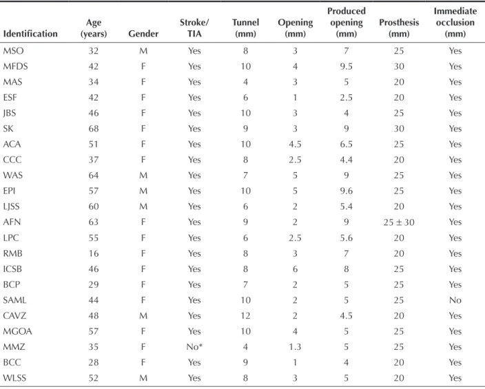
TABLE Patient characteristics
Identification
Age
(years) Gender
Stroke/ TIA
Tunnel (mm)
Opening (mm)
Produced opening
(mm)
Prosthesis (mm)
Immediate occlusion
(mm)
MSO 32 M Yes 8 3 7 25 Yes
MFDS 42 F Yes 10 4 9.5 30 Yes
MAS 34 F Yes 4 3 5 20 Yes
ESF 42 F Yes 6 1 2.5 20 Yes
JBS 46 F Yes 10 3 4 25 Yes
SK 68 F Yes 9 3 9 30 Yes
ACA 51 F Yes 10 4.5 6.5 25 Yes
CCC 37 F Yes 8 2.5 4.4 20 Yes
WAS 64 M Yes 7 5 9 25 Yes
EPI 57 M Yes 10 5 9.6 25 Yes
LJSS 60 M Yes 6 2 5.4 20 Yes
AFN 63 F Yes 9 2 9 25 ± 30 Yes
LPC 55 F Yes 6 2.5 5.6 20 Yes
RMB 16 F Yes 8 3 7 20 Yes
ICSB 46 F Yes 8 6 8 25 Yes
BCP 29 F Yes 7 2 5 25 Yes
SAML 44 F Yes 10 2 5 25 No
CAVZ 48 M Yes 12 2 4.5 20 Yes
MGOA 57 F Yes 10 4 5 25 Yes
MMZ 35 F No* 4 1.3 5 25 Yes
BCC 28 F Yes 9 1 4 20 Yes
WLSS 52 M Yes 8 3 5 20 Yes
* Migraine with poor clinical control
The lengths of the tunnels ranged from 4–12 mm (8.13 ± 2 mm) with non-induced openings of 1–6 mm (2.94 ± 1.30 mm). The variation of the produced opening obtained with the rigid guidewire positioned behind the foramen was 4–9.5 mm (6.13 ± 2.01 mm).
The implant was feasible in all cases. A total of 23 devices were used in 22 patients. The size used most often was 25 mm (47.8% of patients), followed by the 20-mm size (39.1%) and the 30-mm size (Figure 3).
The patient who required two prostheses (AFN) had a thickened septum secundum due to lipomatous degeneration and a hypermobile septum primum. A 25-mm device was implanted, which was fixed only in the septum primum, keeping the foramen open with the passage of large bubbles from right to left. In the same procedure, a second 30-mm device was implanted without difficulties; this device was able to capture both the lipomatous septum and the first device, completely occluding the residual defect. The bubble test conducted after implantation showed no passage of bubbles into the left atrium.
In one patient (JBS), the prosthesis right disk was trapped in the Eustachian valve, which was quite
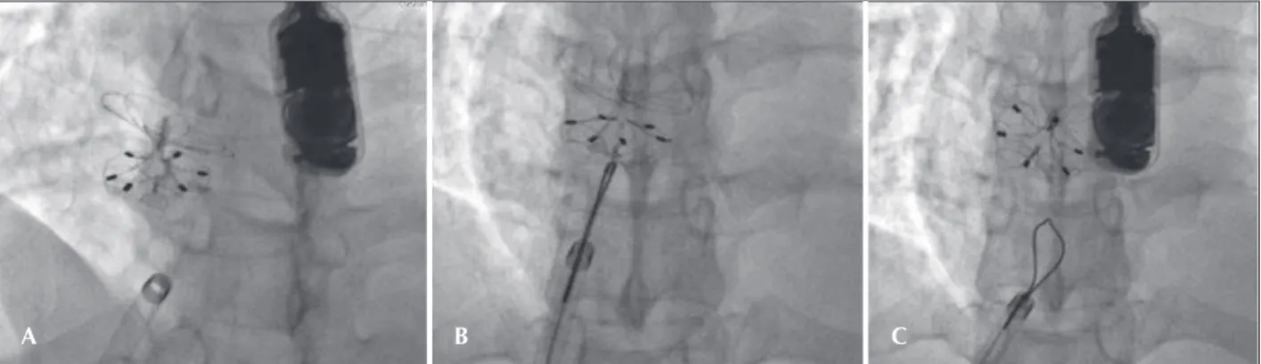
large, and could not be configured properly, keeping the prosthesis open with a large shunt inside it. After several unsuccessful attempts to loosen the right disc, the device was released from the delivery cable on the assumption that it would become free and then achieve the desired configuration, thus reducing the prosthesis profile. As that outcome did not occur, the right disc was recaptured with a 15-mm snare catheter and re-introduced into the long sheath. The set was advanced through the septum into the left atrium roof, and the prosthesis was repositioned with the right disk always configured near the extremity of the long sheath to prevent it from being arrested again in the large valve. The prosthesis was released, and the bubble test con-ducted after the release showed no passage of bubbles through the interatrial septum (Figure 4).
One patient (SAML) presented a minimal additional orifice near the inferior vena cava. The foramen was closed with a 25-mm prosthesis. The bubble test showed no subsequent passage of bubbles from right to left through the second orifice, which was allowed to remain open. There were two minor immediate complications. One patient (AFN) had bleeding in the endotracheal tube after implantation, presumably due to an entrance laceration
Figure 4 – Details of the implant in the patient JBS. A, Prosthesis released after the first implant, with the right disk fixed in the Eustachian valve, holding it open with a large residual shunt. B, Right disk captured by the loop catheter (Amplatz Goose Neck snare, 15 mm) and retracted to be pulled again into the long sheath for a second implant. C, Repositioned prosthesis. After being released from the snare, it reached the correct posi-tion. In this profile, the residual shunt is no longer observed. The control bubble test was negative.
Figure 3 – Details of the implant. A, Prosthesis positioned in the atrial septum, completely configured but still attached to the delivery system. B, Prosthesis after release. Note that the prosthesis changes its spatial position and adapts perfectly to the plane of the septum, free of the weight of the delivery system. C, Released prosthesis, observed through the left atrium, on three-dimensional echocardiography.
A B C
of the left superior pulmonary vein caused by the long sheath. She was under observation in the Intensive Care Unit, but transfusion or any other surgical measure was not required. The patient was discharged the next day in good status. The second patient (BCP) had a fresh thrombus that was unorganised, adhered to the left prosthesis disc, and was visualised by transesophageal echocardiography during the procedure. The heparin dose was increased to 10,000 U. The prosthesis and the long sheath were withdrawn slowly and thoroughly washed with heparinised saline solution. Then, after careful in-spection, the sheath and prosthesis were reintroduced, and the new implant procedure was uneventful. The patient showed no signs of neurological involvement and was discharged in good status.
Over half of the patients (57.1%) reached the six-month follow-up. There has been no recurrence of neurological events in any of the cases to date. The patient with prior migraine (MMZ) had headache with aura in the first month after implantation, although less severe than the previous episodes. Another pa-tient (CCC) showed an allergy to clopidogrel and was treated with aspirin only. One patient (SAML) showed a residual shunt through the interior of the prosthesis on the control transthoracic echocardiogram after one month. This patient is being monitored and has had no complications. The others were adequately occluded (Figure 5). There were no deaths in the present series.
DISCUSSION
The percutaneous occlusion of patent foramen ovales is currently performed in most haemodynamics services worldwide. It has proved to be a safe, effec-tive, and easily reproducible procedure with excellent results, especially with the new generations of prostheses developed specifically for this purpose.1,12,16,21–28
There are few studies in the literature using the CARDIA prosthesis. In this new version, the round shape of the two discs has less risk of erosion or perforation. Notably, the left disc nitinol loops are located underneath the Ivalon™ sponge, reducing the risk of thrombosis and promoting more homogeneous endothelialisation.
The configuration of the prosthesis is facilitated by the use of the carrier and the short transparent sheath, which enables the easy detection of the presence of air within the system.
Despite the introduced changes, high-profile sheaths remain more necessary than desired. However, as they are introduced by venous access, the risk of complications is low, even in children. By always using the sheaths with the adequate gauge recommended by the manufacturer, there was no difficulty in loading the prosthesis, which slid easily through the interior of the sheath in all cases.
The implantation was performed without difficulty, and the technique does not differ from those described for other prostheses with the same purpose. Compared with previous models of the device, slightly more space might be needed for the right disk configuration, which could explain the case (JBS) in which it was trapped by the large Eustachian valve.
A point to be considered is the possibility of the bioptome becoming entrapped in the disk loops im-mediately after prosthesis release from the delivery system, as previously observed. It is important to always keep the bioptome open during withdrawal until its introduction into the long sheath. If any resistance or the movement of the prosthesis along with it is noticed, the right disc should not be pulled, which would increase the risk of prosthesis dislocation; instead, the open bioptome should be carefully rotated until it is released from the captured snare. Another suggestion is to keep the long sheath near the bioptome at the time of the opening, in order to prevent an excessive lateral displacement of the cable, which could increase the risk of the bioptome jaws becoming trapped in the right disc loops.
The visibility on the transesophageal echocardiog-raphy is quite satisfactory, although a less experienced echocardiographist might occasionally be confused re-garding the contour of the left disc, which may appear to be maladapted to the septum, especially while the prosthesis is attached to the delivery system. Normally, after release, it adapts best to the plane of the septum and its correct positioning then becomes clear. A low device profile was observed in all cases and good adaptability to the defect, with good rates of immediate closure, either by colour Doppler echocardiogram or through the bubble test performed after implantation.
CONCLUSIONS
The CARDIA UltraseptTM device is durable, easy to
use, fully repositionable and reimplantable, has a low profile and has good occlusion rates compared with similar prostheses.
The short time of the follow-up did not allow for drawing conclusions regarding the effectiveness of the endothe lialisation on the rates of event recurrence or long-term complications.
Figure 5 – Details of the control transesophageal echocardiography, six months after implantation. A, The prosthesis is well-positioned in the atrial septum. B, Bubble test, showing no bubbles passing into the left atrium, confirming the effectiveness of the device.
A B
BUBBLE TEST
The initial results are encouraging in all cases. Although this new device was used in septa with great mobility, its use in more complex scenarios (e.g., cases with large septal aneurysms) has yet to be tested.
CONFLICTS OF INTEREST
Francisco Chamie is a technical consultant and proctor of Neomex Hospital Ltda. (representative in Brazil of Cardia Inc. – Minneapolis, MN, USA). The remaining authors declare no conflicts of interest.
REFERENCES
1. Brasselet C, Duval S. Which patent foramen ovale needs to be closed, and how?. Ann Cardiol Angeiol. 2011;60(6): 366-72.
2. Surmely JF, Meier B. Percutaneous closure of the patent forâ-men ovale. Minerva Cardioangiol. 2007;55(5):681-91. 3. Lechat P, Mas JL, Lascault G, Loron P, Theard M, Klimczac M,
et al. Prevalence of patent foramen ovale in patients with stroke. N Engl J Med. 1988;318(18):1148-52.
4. Webster MW, Chancellor AM, Smith HJ, Swift DL, Sharpe DN, Bass NM, et al. Patent foramen ovale in young stroke patients. Lancet. 1988;2(8601):11-2.
5. Di Tulio M, Sacco RL, Gopal A, Mohr JP, Homma S. Patent foramen ovale as a risk factor for cryptogenic stroke. Ann Intern Med. 1992;117(6):461-5.
6. Overell JR, Bone I, Lees KR. Interatrial septal abnormalities and stroke: a meta-analysis of case-control studies. Neurology. 2000;55(8):1172-9.
7. Mas JL, Arquizan C, Lamy C, Zuber M, Cabanes L, Derumeaux G, et al. Recurrent cerebrovascular events associated with patent foramen ovale, atrial septal aneurysm, or both. N Engl J Med. 2001;345(24):1740-6.
8. Lamy C, Giannesini C, Zuber M, Arquizan C, Meder JF, Trys-tram D, et al. Clinical and imaging findings in cryptogenic stroke patients with and without patent foramen ovale: the PFO-ASA study. Stroke. 2002;33(3):706-11.
9. Handke M, Harloff A, Olschewski M, Hetzel A, Geibel A. Patent foramen ovale and cryptogenic stroke in older patients. N Engl J Med. 2007;357(22):2262-8.
10. Butera G, Biondi-Zoccai GG, Carminati M, Caputi L, Usai S, Bussone G, et al. Systematic review and meta-analysis of currently available clinical evidence on migraine and patent foramen ovale percutaneous closure: much ado about nothing? Catheter Cardiovasc Interv. 2010;75(4):494-504.
11. Khessali H, Mojadidi MK, Gevorgyan R, Levinson R, Tobis J. The effect of patent foramen ovale closure on visual aura without headache or typical aura with migraine headache. JACC Cardiovasc Interv. 2012;5(6):682-7.
12. Rigatelli G, Dell’Avvocata F, Cardaioli P, Giordan M, Braggion G, Aggio S, et al. Improving migraine by means of primary trans-catheter patent foramen ovale closure: long-term follow-up. Am J Cardiovasc Dis. 2012;2(2):89-95.
13. Bridges ND, Hellenbrand W, Latson L, Filiano J, Newburger JW, Lock JE. Transcatheter closure of patent foramen ovale after presumed paradoxical embolism. Circulation. 1992;86(6): 1902-8.
14. Kutty S, Sengupta PP, Khandheria BK. Patent foramen ovale: the known and the to be known. J Am Coll Cardiol. 2012; 59(19):1665-71.
15. Spies C, Hijazi ZM. Indications, techniques and results of percutaneous patent foramen ovale closure. Minerva Cardio-angiol. 2012;60(1):111-23.
16. Wahl A, Jüni P, Meier B. Long-term propensity score-matched comparison of percutaneous closure of patent foramen ovale with medical treatment after paradoxical embolism. Circula-tion. 2012;125(6):803-12.
17. Luermans JG, Post MC, Schräder R, Sluysmans T, Vydt T, Vermeersch P, et al. Outcome after percutaneous closure of a patent foramen ovale using the intrasept device: a multicentre study. Catheter Cardiovasc Interv. 2008;71(6):822-8.
18. Spies C, Strasheim R, Timmermanns I, Schraeder R. Patent foramen ovale closure in patients with cryptogenis throm-boembolic events using the Cardia PFO occluder. Eur Heart J. 2006;27(3):365-71.
19. Silva JM, Pedra S, Fontes V, Arnoni D, Reyes R, Pontes S, et al. Experiência inicial com a utilização do dispositivo Cardia Intrasept no fechamento percutâneo do forame oval patente. Rev Bras Cardiol Invasiva. 2007;15(4):386-93.
20. Chamié F, Chamié D, Ramos S, Simões LC, Rossi Filho RI, Tress JC, et al. Oclusão da comunicação interatrial com a nova prótese Atriasept-CARDIA: experiência inicial. Rev Bras Cardiol Invasiva. 2009;17(1):94-101.
21. Khairy P, O’Donnel CP, Landzberg MJ. Transcatheter closure versus medical therapy of patent foramen ovale and presumed paradoxical thromboemboli: a systematic review. Ann Intern Med. 2003;139(9):753-60.
22. Ewert P, Kretschmar O, Peters B, Nuernberg JH, Khaliq HA, Nagdyman N, et al. Preliminary experience with a new 18 mm Amplatzer PFO Occluder for small persistente foramen ovale. Catheter Cardiovasc Interv. 2003;59(4):518-21. 23. Chatterjee T, Petzsch M, Ince H, Rehders TC, Körber T, Weber F,
et al. Interventional closure with Amplatzer PFO occluder of patent foramen ovale in patients with paradoxical cerebral embolism. J Interv Cardiol. 2005;18(3):173-9.
24. Billinger K, Ostermayer S, Carminati M, De Giovanni JV, Ewert P, Hess J, et al. Helex Septal Occluder for transcatheter closure of patent foramen ovale: multicentre experience. EuroInterven-tion. 2006;1(4):465-71.
25. Spies C, Timmermanns I, Reissmann U, van Essen J, Schräder R. Patent foramen ovale closure with the Intrasept Occluder: complete 6-56 months follow up of 247 patients after pre-sumed paradoxical embolism. Catheter Cardiovasc Interv. 2008;71(3):390-5.
26. Chamié F, Simões LCN, Chamié D, Mattos R, Ramos S, Tress JC, et al. Utilização de prótese bioabsorvível para oclusão de defeitos do septo atrial: um passo em direção ao futuro. Rev Bras Cardiol Invasiva. 2010;18(4):448-55.
27. Collins NJ, Hatton R, Ng K, Bhagwandeen R, Attia J, Oldmeadow C, et al. Percutaneous device closure of patente foramen ovale using the premere occlusion device: initial experience, pro-cedural and intermediate-term results. J Invasive Cardiol. 2012;24(4):164-8.