Morphological Basis for the Study of the Interatrial Septum in the
Human Fetus
Hugo Becker Amaral, Paulo Zielinsky, Aron Ferreira da Silveira, Ijoni Costabeber, Luiz Henrique Nicoloso, Olmiro
Cezimbra de Souza Filho, Marcelo Salum, João Luiz Manica, Juliana Silveira Zanettini, Ane Micheli Costabeber
Fetal Cardiology Unit of the Cardiology Institute of Rio Grande do Sul, and the Morphology Department of the Health Sciences Center of the Federal University of Santa Maria – Porto Alegre, RS, Brazil
Summary
Objective: To describe morphological features of the interatrial septum in normal fetuses, especially foramen ovale (FO) and septum primum (SP), in order to compare septum primum excursion with foramen ovale diameter.
Methods: Septum primum excursion (SPE) toward the left atrium (LA) and foramen ovale diameter (FOD) were measured
LQWKHKHDUWVRIWHQIRUPDOGHK\GHIL[HGKXPDQIHWXVHVUDQJLQJIURPWRZHHNVRIJHVWDWLRQ+LVWRORJLFDOVHFWLRQV
were obtained from the foramen ovale (FO), septum primum (SP), septum secundum (SS), left atrium (LA), and right atrium (RA).
Results:)2'DQG63(PHDVXUHPHQWVZHUHWKHIROORZLQJ)2'PPDQG63(PPLQWKUHHIHWXVHVZLWK SUHVXPHGJHVWDWLRQDODJH*$RIZHHNV)2'PPDQG63H[FXUVLRQPPLQIRXUIHWXVHVZLWKSUHVXPHG *$RIZHHNVSOXV)2'PPDQG63(LQWKUHHIHWXVHVZLWKSUHVXPHG*$RIZHHNV&DUGLDFPXVFXODU
fibers were identified in both the septum primum and secundum.
Conclusion: Based on its muscular components, it may be suggested that SP is active in character, influencing blood flow through the FO, SP mobility, and its excursion into the LA.
Key words: Fetal heart, heart septum/anatomy & histology, heart septal defects, atrial.
0DLOLQJDGGUHVV/XL]+HQULTXH1LFRORVR
Av. Princesa Isabel, 370 - 90620-001 – Porto Alegre, RS - Brazil E-mail: [email protected]
Manuscript received June 18, 2006; revised manuscript received October 18, 2006; accepted November 10, 2006.
Introduction
Embryonic development of the human heart depends on cell-cell interaction, particularly myocardial and mesenchymal cells1-6. This interaction accounts for the
differentiation and morphogenesis of specific areas in the heart, among them cardiac septa7-10. The endocardial
cushion, a clustering of endocardial and mesenchymal cells, plays a direct role in cardiac septa and atrioventricular valve formation7,11.
Partitioning of the primitive atrium begins at the end of the fourth gestational week through formation, modification, and subsequent fusion of the septum primum and septum secundum12. The primitive atrium
division process is complex not only because the septus primum fuses with endocardial cushions, but also because permanent communication must exist to allow blood flow from the right to the left atrium12,13. The septum primum
edge is attached to endocardial cushions, delimiting an interatrial communication called ostium or foramen primum, which disappears after fusion of the septum primum to the endocardial cushions12,13.
Before the ostium primum disappears, numerous
perforations caused by programmed cell death develop at the center of septum primum, forming the ostium or foramen secundum, which serves as a shunt for oxygenated blood from the ductus venosus to pass from the right to the left atrium12.
The septum secundum develops more slowly, to the right of the septum primum, migrating as the ostium primum closes and gradually overlapping the ostium secundum. Septum secundum separates the atria incompletely, thereby forming the foramen ovale13.
During intrauterine life, the foramen ovale allows most oxygenated blood entering the right atrium from the inferior vena cava and ductus venosus to pass into the left atrium. It also prevents blood from flowing in the opposite direction, because the septum primum closes against a relatively rigid septum secundum. After birth, the foramen ovale usually closes, and the interatrial septum completely separates atrial chambers12.
Cell analysis has demonstrated that, during the first developmental stages, septum primum has a muscular central portion in which myocytes are in close contact14. In addition
to myocytes, there is also the aforementioned presence of mesenchymal tissue in the septum primum14-16. The septum
primum is covered by the endocardium, which is made up of elongated and flattened cells17.
allowed identification of cardiac muscular fibers in both structures and, hence, the presence of contractility. It also enabled visualization of blood vessels supplying these sites, in addition to intramural nerves and neurons of the neurovegetative system. Figures 1 through 7 show the macroscopic and microscopic sections and their descriptions.
Discussion
During fetal life, septum primum acts as a valve that closes the foramen ovale when atrial contractions occur. During diastole, septum primum bulges into the left atrium, allowing maximal opening and right-to-left flow22,23.
In previous studies, our team evaluated septum primum echocardiographic behavior during fetal life. The relationship between septum primum mobility and the presence of atrial extrasystoles was assessed in a study comparing a group of fetuses with atrial extrasystoles and a control group of normal fetuses. The redundancy index (RI), defined as the ratio between septum primum maximal excursion and left atrial maximal diameter on fetal echocardiogram (four-chamber view), showed that septum primum is more redundant in fetuses with atrial extrasystoles during intrauterine life16.
Septum primum mobility has proved to be an indicator of left diastolic function. Taking into account that septum primum mobility may reflect changes related to left atrial pressure, alterations in left ventricular relaxation and/or compliance may affect the behavior of the former. With the use of groups of fetuses of diabetic mothers with septal myocardial hypertrophy as a model for decreased left ventricular compliance and relaxation it was noted that, in this situation, septum primum excursion is reduced. Septum primum may also develop hypertrophy. Therefore, the greater the myocardial hypertrophy in the interventricular In earlier studies, our team analyzed septum primum
mobility, its development during intrauterine life18, and
its relationship with the presence of atrial extrasystoles19,20
and left ventricular diastolic function21. To evaluate septum
primum excursion quantitatively at every cardiac cycle, its maximal excursion toward the left atrium is measured in a four-chamber view. An “excursion index” of septum primum was created to correlate its maximal excursion to left atrial diameter20,21.
This study aimed at describing morphological features of the interatrial septum in normal fetuses, especially foramen ovale and septum primum, to compare septum primum excursion with foramen ovale diameter.
Methods
For anatomical evaluation, both septum primum maximal excursion toward the left atrium and foramen ovale diameter were measured in the hearts of ten formaldehyde-preserved human fetuses (gestational ages between 28 and 36 weeks) from the Laboratório de Anatomia do Feto e do Recém-nascido do Departamento de Morfologia do Centro de Ciências da Saúde da Universidade (Fetal and New-born Anatomy Laboratory of the Morphology Department of the University’s Health Sciences Center).
Anatomical dissection was performed according to standard technique, with en bloc resection of the heart and lungs. The lungs were then resected at the level of the pulmonary hila, and the heart was cut transversely to the interventricular septum from the apex to the base, allowing ventricular cavities to be visualized by opening the atrial wall and exposing the septum primum, septum secundum, and foramen ovale. Septum primum maximal excursion and foramen ovale diameter were measured under a colposcope with 13x magnification using an adapted measurement device (a caliper with two sharp metal ends).
Septum primum excursion was measured by fixing one of the ends of the measurement device in the septum secundum and the other in the middle of the free edge of the septum primum, which was pushed toward the left atrium as far as its maximal excursion, without deforming neighboring structures.
Foramen ovale diameter was measured at a 90° angle in relation to the septum primum excursion measurement, corresponding to a line between septum primum implantation sites.
Histological sections were obtained from the foramen ovale, septum primum, septum secundum, left atrium, and right atrium. Subendocardial and subepicardial tissue slides were stained using the hematoxylin-eosin method and Goldner’s technique, in light green solution.
Results
The anatomical measurements of the formaldehyde-preserved fetal hearts are presented in Table 1.
Table 1 – Foramen ovale diamenter and septum primum excursion measured in millimeters (mm)
Presumed gestational
age in weeks FOD (mm) SPE (mm)
28 3.5 2.8
28 3.1 2.9
28 3.5 3.1
34 3.5 4.0
34 3.3 4.5
34 3.5 4.9
34 3.5 5.0
36 3.3 6.0
36 3.5 8.0
36 4.5 9.0
suggested that septum primum excursion may be a more suitable index for ventricular diastolic function than those traditionally accepted, since it shows an inverse relationship with interventricular septal hypertrophy and is easier to perform than the atrioventricular valve flow analysis.
In an earlier study, it was also found that septum primum maximal excursion is increased during fetal breathing, a finding that was related to an increase in left ventricular distensibility due to respiratory movements24.
The most recent hypothesis raised by our team attempts to explore the notion that the morphological composition of septum primum would directly influence its echocardiographic
behavior, most notably its mobility, thereby reflecting its excursion index.
Each stage during septum primum formation is directly related to its role in fetal heart circulation. Its histological structure directly affects its behavior on blood flow that cross the foramen ovale25,26.
Thus, it may be suggested that septum primum is active in character, and this behavior may influence the blood flow through the foramen ovale. Moreover, septum primum mobility itself, as well as its excursion into the left atrium, depends not only on left atrial pressure, but also on its muscle fibers.
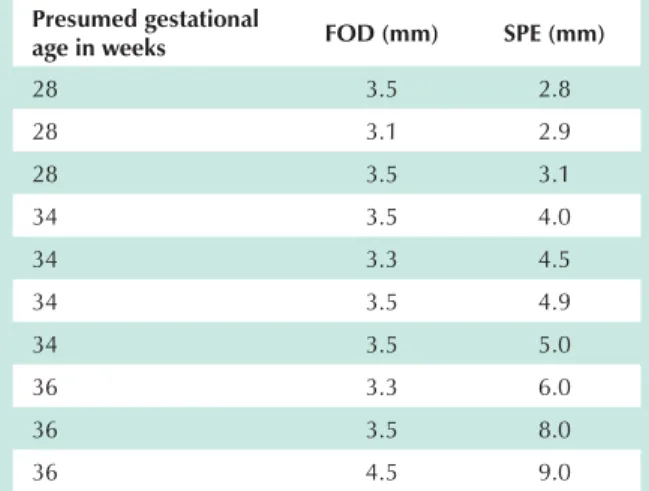
Fig. 1 -Foramen ovale seen from the right atrium to the left atrium.
Crista dividens
Septum primum
Septum secundum Right atrial
wall
Interatrial septum
Free edge of the septum primum
Inferior
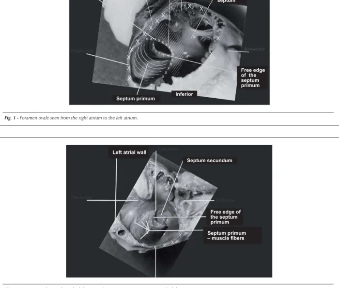
Fig. 2 -Foramen ovale seen from the left atrium. Septum primum excursion into the left atrium.
Septum secundum Left atrial wall
Free edge of the septum primum
Septum primum
Fig. 3 -Foramen ovale (A). Septum primum (B). Septum secundum (C). Right
DWULXP'/HIWDWULXP([PDJQLILFDWLRQ*ROGQHUVWDLQLQJ Fig. 4 -5LJKWDWULXP&/HIWDWULXP'[PDJQLILFDWLRQ*ROGQHUVWDLQLQJSeptum primum origin on the left atrium wall (A). Septum primum (B).
Fig. 6 -6HSWXPSULPXPHQGRFDUGLXPVKRZLQJWKHHQGRWKHOLXP$DQGVXEHQGRWKHOLXPZLWKFRODJHQRXVILEHUV%DQGILEUREODVWV&[PDJQLILFDWLRQ*ROGQHU
staining.
Fig. 7 -%DVHRIWKHVHSWXPVHFXQGXPVKRZLQJYDVFXODUL]DWLRQ$DQGDQHXUDOJDQJOLRQ9HJHWDWLYHQHXURQ%[PDJQLILFDWLRQ*ROGQHUVWDLQLQJ
Acknowledgements
We would like to thank Sérgio Oliveira Silveira, laboratory technician, Department of Morphology, CCS, UFSM.
Supported by: FAPERGS – Fundação de Amparo à Pesquisa
do Estado do RS, CAPES and CNPq.
Potential Conflict of Interest
References
1. De Haan RL. Morphogenesis. Annu Rev Physiol. 1964; 26: 15-46.
2. De Haan RL. Morphogenesis of the vertebrate heart. In: Haan RL, Ursprung H, ed. Organogenesis. New York: Holt, Rinehart and Winston; 1965. p. 377-419.
3. De Haan RL, Fujii S, Satin J. Cell interactions in cardiac development. Dev Growth Differ. 1990; 32: 233-41.
4. Manasek FJ. The extracellular matrix: a dynamic component of the developing embryo. In: Moscona A, Monroy A, eds. Current topics in development biology. New York: Academic Press; 1975. p. 35-102.
5. Icardo JM. Cardiac morphogenesis and development. Experientia. 1988; 44: 11-2.
6. Icardo JM. Development biology of the vertebrate heart. J Exp Zool. 1996; 275: 144-61.
7. Markwald R, Fitzharris T, Manasek F. Structural development of endocardial cushions. Am J Anat. 1977; 148: 85-120.
8. Hay DA. Development and fusion of the endocardial cushion. In: Rosenquist GC, Bergfma D, eds. Birth defects, original articles. New York: Allan R Liss Inc.; 1978. p. 69-90. (Series XIV, nº 7).
9. Bolender DL, Markwald RR. Epithelial-mesenchymal transformation in atrioventricular cushion tissue morphogenesis. Scanning Electron Microsc. 1979; 3: 313-22.
10. Steding G, Seidl W. Contribution to the development of the heart. Part I: normal development. Thorac Cardiovasc Surg. 1980; 28: 386-409.
11. Argüelo C, De La Cruz M, Sanchez M. Ultrastructural and experimental evidence of myocardial cell differentiation into connective tissue cell in embryonic chick heart. J Mol Cell Cardiol. 1978; 10: 307-15.
12. Moore K, Persaud TVN. Sistema cardiovascular. In: Moore KL, Persaud TVN. Embriologia clínica. 6ª ed. Rio de Janeiro: Guanabara Koogan; 2000. p. 332-82.
13. Anderson RH, Webb S, Brown NA. Clinical anatomy of the atrial septum with reference to its developmental components. Clin Anat. 1999; 12: 362-74.
14. Patten BM. The formation of the cardiac loop in the chick. Am J Anat. 1922;
30: 373-97.
15. Patten BM, Kramer TC, Barry A. Valvular action in the embryonic chick heart by localized apposition of endocardial masses. Anat Rec. 1948; 102: 299-312.
16. Arrechedera H, Alvarez M, Strauss M, Ayesta C. Origin of mesenchymal tissue in the septum primum: a structural and ultrastructural study. J Mol Cell Cardiol. 1987; 19: 641-51.
17. Wessels A, Anderson RH, Markwald RR, Webb S, Brown NA, Viragh S, et al. Atrial development in the human heart: na immunohistochemical study with emphasis on the role of mesenchymal tissues. Anat Rec. 2000; 259 (3): 288-300.
18. Zielinsky P, Firpo C, Martha VF, Silva ES. Estudo ecocardiográfico pré-natal da redundância do septum primum e sua relação com a gênese de extra-sístoles atriais no feto. Arq Bras Cardiol. 1995; 65: 153-7.
19. Zielinsky P, Firpo C, Martha VF, Silva ES. Papel da membrana da fossa oval no desencadeamento de arritmias cardíacas fetais. Rev Bras Ginec Obstet. 1995; 17: 711-9.
20. Firpo C, Zielinsky P. Mobility of the flap valve of the primary atrial septum in the developing human fetus. Cardiol Young. 1998; 8 (1): 67-70.
21. Firpo C, Zielinsky P. Behavior of septum primum mobility in third-trimester fetuses with myocardial hypertrophy. Ultrasound Obstet Gynecol. 2003; 21: 445-50.
22. Puerta Fonolla AJ, Orts Llorca F. Origin and development of the septo primeiro. Acta Anat (Basel). 1978; 100 (2): 250-7.
23. Morse DE, Rogers CS, McCann PS. Atrial septation in the chick and rat: a review. J Submicrosc Cytol. 1984; 16: 259-72.
24. Miyague NI, Ghidini A, Miyague LLT. Fetal breathing movements are associated with changes in compliance of the left ventricle. Fetal Diagn Ther. 1997; 12: 72-5.
25. Anderson RH, Webb S, Brown NA. Clinical anatomy of the septum primum with reference to its developmental components. Clin Anat. 1999; 12: 362-74.