528 528
Diabetic patients with and without peripheral
neuropathy reveal different hip and ankle
biomechanical strategies during stair descent
Pacientes diabéticos com e sem a neuropatia periférica mostram diferentes
estratégias biomecânicas de quadril e tornozelo ao descer escada
Andreja P. Picon1, Cristina D. Sartor1, Maria I. Roveri1, Anice C. Pássaro1, Neli R. Ortega2, Isabel C. N. Sacco1
Abstract
Background: The progression of diabetes and the challenge of daily tasks may result in changes in biomechanical strategies.
Descending stairs is a common task that patients have to deal with, however it still has not been properly studied in this population.
Objectives: We describe and compare the net joint moments and kinematics of the lower limbs in diabetic individuals with and without
peripheral neuropathy and healthy controls during stair descent. Method: Forty-two adults were assessed: control group (13), diabetic group (14), and neuropathic diabetic group (15). The flexor and extensor net moment peaks and joint angles of the hip, knee, and ankle were described and compared in terms of effect size and ANOVAs (p<0.05). Results: Both diabetic groups presented greater dorsiflexion [large effect size] and a smaller hip extensor moment [large effect size] in the weight acceptance phase. In the propulsion phase, diabetics with and without neuropathy showed a greater hip flexor moment [large effect size] and smaller ankle extension [large effect size]. Conclusion: Diabetic patients, even without neuropathy, revealed poor eccentric control in the weight acceptance phase, and in the propulsion phase, they showed a different hip strategy, where they chose to take the leg off the ground using more flexion torque at the hip instead of using a proper ankle extension function.
Keywords: biomechanics; diabetic polyneuropathy; kinematics; kinetics; motion.
Resumo
Contextualização: A progressão do Diabetes Mellito e as atividades desafiadoras do dia a dia podem resultar em mudanças da
estratégia biomecânica adotada. Descer escadas é uma tarefa comum do dia a dia, vivenciada pelos pacientes, mas ainda não foi satisfatoriamente estudada nessa população. Objetivos: Descrever e comparar os momentos articulares e a cinemática de membros inferiores em indivíduos diabéticos com e sem a neuropatia periférica e controles saudáveis durante o descer escadas. Método:
Quarenta e dois adultos foram avaliados: grupo controle (13), grupo diabético (15) e grupo de diabéticos neuropatas (14). Os picos flexores e extensores dos momentos articulares e os ângulos articulares de quadril, joelho e tornozelo foram comparados e descritos por análise do tamanho do efeito e ANOVAs (p<0,05). Resultados: Na fase de aceitação do peso, ambos os grupos diabéticos apresentaram maior ângulo de dorsiflexão de tornozelo [tamanho de efeito grande] e menor momento extensor de quadril [tamanho de efeito grande]. Na fase de propulsão, diabéticos com e sem a neuropatia apresentaram maior momento flexor de quadril [tamanho de efeito grande] e menor ângulo de extensão de tornozelo [tamanho de efeito grande]. Conclusão: Pacientes diabéticos, mesmo antes da neuropatia instalada, revelaram um pobre controle excêntrico na fase de aceitação do peso e, na fase de propulsão, esses pacientes mostraram uma estratégia diferente ao levar o membro inferior à frente a partir de um maior torque flexor de quadril ao invés de usar uma função extensora apropriada de tornozelo.
Palavras-chave: biomecânica; polineuropatias diabéticas; cinemática; cinética; movimento.
Received: 06/25/2012 – Revised: 06/29/2012 – Accepted: 07/05/2012
1Physical Therapy, Speech and Occupational Therapy Department; School of Medicine; Universidade de São Paulo (USP), São Paulo, SP, Brazil 2Medical Informatics Department, School of Medicine, USP, São Paulo, SP, Brazil
Introduction
In the diabetic population, biomechanical alterations during level walking have been extensively discussed in the literature and they show important changes that are related to balance and sensory-motor impairments. However, investiga-tion of other activities of daily living in this populainvestiga-tion, such as stair negotiation, remains insuicient. In daily life, diabetic individuals have to manage slopes, change directions during locomotion, and ascend and descend steps. hese activities play an important role in the functionality and independence of diabetic individuals, and the capacity to perform daily tasks is an important factor of a good quality of life.
It has been shown that diabetic individuals have functional deicits of the knee and ankle extensors during gait and other daily living activities1-4. he stair descent task requires greater
eccentric control of these lower limb extensor muscles and well preserved and eicient balance, since the body is moving with gravity5. If the mechanical demands of the task required are not
well managed, they become risk factors for falls in diabetic neu-ropathic individuals6-8. A better comprehension of the
biome-chanics of this motor task in diabetic individuals can contribute to preventive and rehabilitative actions in this population9.
Stair descent is characterized mainly by energy absorption mechanisms performed by the knee and ankle joints, which show greater angular excursion as opposite to what the hip joint performs5,10,11. he ankle plays an important role12,13, as its
full range of motion allows a suitable distribution of the me-chanical energy absorption at the initial foot contact with the step and a proper propulsion at the end of the stance5.
Ankle function is dramatically afected in diabetic patients since they present a limited range of motion (ROM), lower and delayed triceps surae activity2,14-16 combined with progressive
loss of foot sensitivity. During stair descent, normal ankle ROM and proper muscle eccentric control are even more neces-sary, and if ankle function is impaired, compensations and adaptations in the kinetics and kinematics of the knee and hip would be expected, as Mueller et al.17 suggested in level gait.
Additionally, stair descent involves greater external forces and consequently more complex musculoskeletal and balance re-sponses that have to be performed by individuals with severe motor and sensorial deicits. hese individuals have to adapt their neuromuscular responses to a situation that requires dif-ferent limb coordination patterns and higher eccentric muscle demands, mainly in the weight acceptance phase, compared to a more common task such as walking.
A few biomechanical descriptions of other daily mo-tor tasks in diabetic subjects have been shown in the lit-erature, but none discussed kinetics and kinematics while
descending steps. Maluf et al.18 observed higher peak
pres-sures in diabetic patients during level walking, ramp climb-ing, stair climbclimb-ing, and changing direction compared to level walking. Onodera et al.16 observed a mechanical
disad-vantage of the vastus lateralis and gastrocnemius medialis muscles in stair negotiation by diabetic individuals.
No previous study has investigated lower limb net joint moments in diabetic individuals during the performance of challenging daily tasks such as stair descent, nor has previ-ous research determined net joint moments in patients with diferent severities of diabetes and its chronic complication – peripheral neuropathy. herefore, it has not been possible to identify diferences in kinetic patterns between early and ad-vanced stages of diabetes. Whether lower limb kinetic patterns change during stair descent along with the progression of dia-betes remains unclear. he net joint moments can potentially show how diabetic and diabetic neuropathic patients deal with their pathological condition and with the mechanical and bal-ance demands of stair descent.
Taking into account that stair negotiation is a challenging and diicult situation for diabetic and neuropathic patients because of their deicits, we hypothesized that the greater the severity of the disease (progression of diabetes marked by the onset of peripheral neuropathy), the greater the ki-netic and kinematic changes during stair descent, mostly in the knee and ankle since these joints are essential to the task of descending stairs19,20 and are compromised in diabetic
indi-viduals. herefore, the aim of this study was to describe and compare the sagittal net joint moments and kinematics of the main joints of the lower limbs in diabetic individuals with and without peripheral neuropathic and non-diabetic control indi-viduals during stair descent.
Method
Subjects
Forty-two adults (20 men, 22 women) were divided into three groups: a control group composed of 13 non-diabetic asympto-matic individuals (CG, 54.7±7.6 years, 72.1±12.2 kg, 1.69±0.1 m, BMI 25±5 kg/m2); 15 individuals diagnosed with diabetes (DG,
55±6.9 years, 81.6±16.4 kg, 1.69±0.1 m, BMI 30±6 kg/m2, 7.1±1.4 years
of duration of diabetes, 135.8±39.1 mg/dL of glycaemia, Hb1Ac 6.91%); and 14 diabetic individuals clinically diagnosed with pe-ripheral diabetic neuropathy (DNG, 60.2±4.0 years, 74.7±9.7 kg, 1.66±0.1 m, BMI 27±7 kg/m2, 13±4.3 years of duration of diabetes,
530
statistical diferences (ANOVA) among the groups (at mean values) in sex (p=0.501, chi square test), height (p=0.507), body mass (p=0.123), age (p=0.060) or BMI (p=0.07). he diabetic groups were statistically diferent with respect to neuropathy scores (Michigan Neuropathy Screening Instrument ques-tionnaire – MNSI)21 (p<0.001), duration of diabetes (p<0.001),
glycaemic levels (p<0.001), and Hb1Ac levels (p<0.001), as expected, since neuropathic status comes from worse control and/or a longer duration of diabetes.
he inclusion criteria for the diabetic groups were as fol-lows: more than 5 years since the diagnosis of diabetes mel-litus; a score equal to or higher than 6 out of 13 on the MNSI; a score equal to or higher than 3 out of 8 on the foot Physical Assessment for the DNG; and a score equal to or lower than 3 out of 13 and 2 out of 8 for the DG (DG, median of 2.5 in the MNSI questionnaire and 2 in the Physical Assessment; DNG, median of 7 in the MNSI questionnaire and 3 in the Physical Assessment). he exclusion criteria for all groups were: over 65 years of age; partial or total amputation; Charcot arthropathy (or any other major orthopaedic foot alteration conirmed by radiography); presence of retinopathy or nephropathy; plantar ulcers at the time of the evaluation; presence of any other mus-culoskeletal disorder or pain; and inability to descend stairs without the use of a handrail.
All procedures were approved by the Ethics Committee of Hospital das Clínicas da Faculdade de Medicina da Universi-dade de São Paulo, city of São Paulo, state of São Paulo, Brazil (protocol number 0305/08) (n. 0305/08), and all participants gave written informed consent.
Procedures
Before data acquisition, all subjects were interviewed using the Activities-Speciic Balance Conidence Scale (ABC)22. his
scale was used to better characterize activities related to stair negotiation and stair descent and determine whether they are indeed a challenging motor situation to both diabetic and neu-ropathic patients. Among the available validated scales, the ABC scale was the most speciic to address the task of stair negotiation and related activities. he higher the score is, the higher the subject’s conidence. hese scores were statistically diferent among groups (ANOVAs, p<0.01). he CG reached a total score of 98.9 (2.5)%, the DG scored 93.9 (4.8)%, and the DNG scored 82 (10.7)%, indicating a progressive loss in con-idence during daily activities, including stair negotiation. In tasks not related to stair negotiation (i.e. “walking around the house”, “getting in/out of car”, “walking across parking lot”), we did not ind any statistical diferences among groups. However,
in speciic activities related to slope negotiation, the groups were statistically diferent: stair negotiation [CG=99(4)%; DG=90(17)%; DNG=69(22)%, p<0.001], escalator negotiation [CG=99(2)%; DG=83(28)%; DNG=76(28)%, p<0.001], and ramp negotiation [CG=99(2)%; DG=87(7)%; DNG=79(25)%, p=0.047].
Passive-relective markers (20 mm in diameter) were aixed to the skin with VHB tape (3M®
) using a standard Cleveland Clinic marker set23. Extra markers were placed bilaterally at the
medial knee joint line, medial malleolus, and irst metatarsal joint for the static standing trial, in order to determine relative joint centers of rotation. hree non-collinear relective markers were ixed at two squares (technique clusters) placed over the lateral thigh and over the shank. he laboratory coordinate system was established at one corner of the force plate. Lower limb segment translations and rotations were reported relative to neutral posi-tions deined during the initial standing static trial.
The three-dimensional kinematics was evaluated with six infrared cameras (Optitrack FLEX: V100, Natural Point, OR, USA). The automatic digitizing process, the 3D recon-struction of the markers’ positions, and the filtering of kin-ematic data were performed using Arena software (Natural Point, OR, USA). Ground reaction forces were acquired by a force plate (AMTI OR-6–1000, Watertown, MA, USA) em-bedded in the floor at the end of the last stair step. Data acquisition was synchronized and sampled by an A/D card (AMTI, DT 3002, 12 bits) at 100 Hz.
Before data acquisition, all participants received the same instructions: to descend barefoot the last three steps of a ive-step staircase, without using the handrail, beginning the task with the opposite limb to the one being evaluated, and posi-tioning one foot on each step during the descent. hey were also instructed to continue walking after the last descent step. he subjects were instructed to descend the staircase as they would do on a daily basis, but the cadence was controlled by a digital metronome at 96 steps/minute, which was rigorously followed by the subjects in order to reduce the inluence of cadence variation within trials and subjects24. Each step of
the staircase was 32 cm deep, 60 cm wide, and 20 cm high, and the staircase had a 32-degree slope.
Numerical and statistical analysis
he 3D inverse dynamic bottom-up method was employed to calculate the net moments of hip, knee, and ankle joints in the sagittal plane, using Visual3D software (C-motion, Water-loo, ON, Canada). he inertial properties were based on Demp-ster’s standard regression equations25. A negative net joint
moment was considered an extensor moment, and a positive one was considered a lexor moment. Forward motion of the lower segment was regarded as lexion (positive values) and backward motion as extension (negative values)26.
he results were interpreted considering three periods of the stance phase of descending stairs, as proposed by McFadyen and Winter19: (i) weight acceptance phase, double
stance phase (0-19% of the cycle); (ii) continuance forward phase, horizontal displacement, and body elevation (19-53% of the cycle); and (iii) controlled lowering phase, vertical dis-placement of the body, and simple stance phase (53-100% of the cycle). In this study, the third stage of the stance phase (controlled lowering) was considered the propulsion phase of level walking, as the subjects stepped on the force plate dur-ing the transition to the loor plane.
The vertical (y) and horizontal (x) mean velocities were calculated from the displacement of the pelvic centre dur-ing the whole stance phase of stair descent, and compared among groups.
We calculated the maximal flexion and extension angles of each joint, the hip extensor net joint moment peak at the weight acceptance phase (~15% of the stance), the hip flexor net joint moment peak at the propulsion phase (~80% of the stance), the knee flexor net joint moment peak at the forward continuance phase (~30% of the stance), the knee extensor net joint moment peak at the propulsion phase (~60% of the stance), the first ankle flexor net joint moment peak at the weight acceptance phase (~20% of the stance), and the second ankle flexor net joint moment peak at the propulsion phase (~80% of the stance). Data of only one randomly selected lower limb per subject was analyzed and
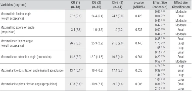
Table 1. Mean values (standard deviations) of the sagittal kinematics (degrees), effect size and its classification, and p-value of the comparisons among control (CG, 1), diabetic (DG, 2) and diabetic neuropathic (DNG, 3) groups for the hip, knee, and ankle joints of stair descent.
Variables (degrees) CG (1) (n=13)
DG (2) (n=15)
DNG (3) (n=14)
p-value (ANOVA)
Effect Size (cohen’s d)
Effect size Classification
Maximal hip flexion angle
(weight acceptance) 27.2 (9.1) 24.4 (6.4) 24.7 (8.0) 0.423
0.62 (1-2)
0.04 (2-3)
0.45 (1-3)
Moderate Small Moderate
Maximal hip extension angle
(propulsion) 3.4 (7.8) 1.0 (3.6) 1.0 (2.2) 0.735
0.42 (1-2)
0.00 (2-3)
0.44 (1-3)
Moderate Small Moderate
Maximal knee flexion angle
(weight acceptance) 26.5 (3.6) 25.3 (2.9) 21.0 (2.0) 0.145
0.38 (1-2)
1.78 (2-3)
1.98 (1-3)
Small Large Large
Maximal knee extension angle (propulsion) 14.2 (8.9) 12.9 (14.5) 10.8 (4.0) 0.264
0.11 (1-2)
0.20 (2-3)
0.52 (1-3)
Small Small Moderate
Maximal ankle dorsiflexion angle (weight acceptance) 13.7 (0.1)* 16.4 (0.8) 17.4 (3.7) 0.036
4.74 (1-2)
0.39 (2-3)
1.44 (1-3)
Large Small Large
Maximal ankle plantarflexion angle (propulsion) -17.3 (5.4)* -10.9 (7.1) -9.2 (1.6) 0.005
1.04 (1-2)
0.34 (2-3)
2.15 (1-3)
Large Small Large *represents the different group compared to the others (ANOVA).
Figure 1. Mean profile of the ankle sagittal angular excursion during stance phase of stair descent for Control (CG), diabetic (DG), and diabetic neuropathic (DNG) groups.
Time (% Stance phase)
CG
Ankle Sagittal Angle (degrees)
plantarflexion dorsiflexion
DG DNG
0 20 40 60 80 100
532
compared. Five valid steps performed by the selected limb were used for statistical purposes.
Biomechanical, anthropometric, and demographic vari-ables followed a normal distribution (Shapiro-Wilk Test), and variances were homogeneous (Levene’s Test). Statistical tests to compare variables included an analysis of variance (ANOVA), followed by a Newman-Keuls post hoc test. In order to verify the size of the diference between groups of net joint moments and kinematic variables, we calculated the efect size, which quantiies the size of the diference between groups and may be a true measure of the signiicance of the diference between the groups (Browner, 2006; halheimer and Cook, 2002).
Results
he vertical and horizontal mean velocities were difer-ent between diabetic groups and CG. he DNG showed a signiicantly greater vertical velocity than other groups and presented a large efect size [CG=-0.19 (0.06) m/s, DG=-0.21 (0.05) m/s, and DNG=-0.26(0.08) m/s*]. he DG and DNG showed a signiicantly lower horizontal velocity (x) than the CG and presented a large efect size [CG=0.82 (0.14) m/s*, DG=0.64 (0.08) m/s, and DNG=0.76 (0.10) m/s].
he DG and DNG showed greater dorsilexion and lesser plantarlexion compared to the CG, which can be conirmed by the large efect size values (Table 1, Figure 1). he DNG indi-viduals showed less knee lexion (large efect size) in the weight acceptance phase (Table 1).
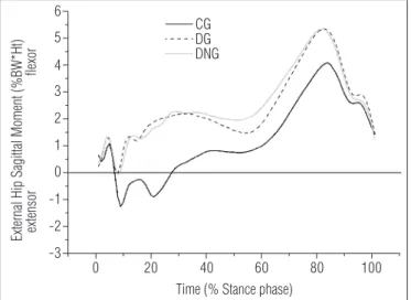
he efect size calculation for the net moments of the hip revealed large efects among groups, characterized by a smaller extensor moment in the weight acceptance phase and a greater lexor moment at propulsion for both diabetic groups (Table 2, Figure 2).
Discussion
his study aimed at describing and comparing lower limb kinetics and kinematics during stair descent in diabetics pa-tients with and without neuropathy and in healthy individuals. he only diference observed between early (DG) and advanced
Table 2. Mean values (standard deviations) of the sagittal net joint moment variables, effect size and its classification, and p-value of the comparisons among control (CG, 1), diabetic (DG, 2) and diabetic neuropathic (DNG, 3) groups for the hip, knee, and ankle joints of stair descent.
Variable (% BW. Height) CG (1) (n=13)
DG (2) (n=15)
DNG (3) (n=14)
p-value (ANOVA)
Effect Size (cohen’s d)
Effect size Classification
Hip extension moment peak
(weight acceptance ~15% of the stance) -2.5 (1.2) -1.1 (1.4) -1.2 (1.9) 0.066
1.11 (1-2)
0.06 (2-3)
0.84 (1-3)
Large Small Large
Hip flexion moment peak
(propulsion ~80% of the stance) 4.3 (2.0) 5.9 (1.7) 5.9 (2.1) 0.059
0.90 (1-2)
0.00 (2-3)
0.81 (1-3)
Large Small Large
Knee flexion moment peak
(forward continuance ~30% of the stance) 3.0 (1.8) 3.9 (2.7) 4.1 (2.5) 0.443
0.40 (1-2)
0.08 (2-3)
0.52 (1-3)
Moderate Small Moderate
Knee extension moment peak
(propulsion ~60% of the stance) -1.1 (0.7) -1.4 (1.5) -1.0 (1.5) 0.795
0.26 (1-2)
0.28 (2-3)
0.09 (1-3)
Small Small Small
1st ankle flexion moment peak
(weight acceptance ~20% of the stance) 8.1 (2.1) 8.4 (1.5) 8.8 (1.9) 0.630
0.17 (1-2)
0.24 (2-3)
0.36 (1-3)
Small Small Small
2nd ankle flexion moment peak
(propulsion ~80% of the stance) 6.8 (1.2) 7.6 (1.1) 7.5 (1.0) 0.259
0.72 (1-2)
0.10 (2-3)
0.66 (1-3)
Moderate Small Moderate
Figure 2. Mean profile of the hip sagittal net moment during stance phase of stair descent for Control (CG), diabetic (DG), and diabetic neuropathic (DNG) groups.
Time (% Stance phase) CG
External Hip Sagittal Moment (%BW*Ht) extensor flexor
DG DNG
0 20 40 60 80 100
stages of the disease (DNG) was in the knee kinematics; how-ever, both diabetic groups showed important changes in the ankle kinematics and in the hip kinetics compared to healthy individuals. he hip played a major role in diabetic individuals in late stance producing a greater lexor moment, possibly to compensate the smaller ankle role in the same phase, but in the weight acceptance phase the smaller hip extensor moment could compromise the eccentric control of stair descent.
In the weight acceptance phase, there was an increase in the ankle dorsilexion angle (large efect) and smaller hip ex-tensor moment in both diabetic groups and a smaller knee lexion in the DNG. In this particular phase, the eccentric mus-cle activity plays a major role in controlling the deceleration of the whole body and in positioning the lower limb segments properly to allow optimal load absorption.
In the ankle joint, the increased dorsilexion angle suggests a poor triceps surae eccentric activity. his inding is consist-ent with the EMG results of the triceps surae, which presconsist-ents a deicit in its activation in locomotor activities2,14-16, and it is
expected that in a more diicult task that requires more eccen-tric activity and control, such as descending stairs, the ankle muscles could not respond adequately.
Particularly in diabetic neuropathic patients, the smaller knee lexion reinforces the hypothesis of a poor eccentric control because they assume a posture that saves quadriceps efort. he vastus lateralis delay found in the EMG results in the heel strike phase of level gait in diabetic neuropathic pa-tients1,2,16 indicates impaired knee extensor muscle function.
he diabetic patients’ response must be adapted to a higher mechanical demand during stair decent, particularly at the initial contact. Considering all sensorimotor deicits, their re-sponse might be inadequate to this new efort.
In the hip joint, the smaller hip extensor moment suggests poor eccentric activity of hip extensor muscles. Although there is no study available that identiies any dysfunction of the hip extensor muscles in diabetic populations, mainly because of methodological diiculties, we can assume that the smaller hip extensor moment may indicate muscle dysfunction.
he greater mean vertical velocity (large efect) observed in neuropathic patients is presumably a consequence of this inadequate eccentric control to manage external forces during stair descent and indicates that their bodies are collapsing dur-ing the load acceptance phase.
Riener et al.5 emphasized that the typical potential energy
absorption during stair descent is accomplished by a syn-chronized and coordinated action of three major lower limb joints, highlighting the important role of the ankle (8-10% of the stance phase) and knee (10-13% of the stance phase) at the beginning of stance, while they are lexed. his greater
dorsilexion in the weight acceptance phase could lead to an impaired return of the elastic energy needed in the propulsion phase. Knowing the importance of the eccentric-concentric cycle in the energy conservation and efectiveness of gait, we suggest that the absorption and generation of elastic potential energy from the ankle may be compromised in stair descent activities in diabetic populations.
In the propulsion phase, the smaller ankle extension and the greater hip flexor moment observed in the propulsion phase of both diabetic groups may corroborate the theory of a major role of a proximal joint in an attempt to compen-sate for the typical ankle losses in neuropathic diabetic pa-tients but, in the present study, they were seen even before the neuropathy had set in (DG). Despite the neuropathy, diabetic individuals appear to be using the hip to raise the leg off the floor instead of using an efficient ankle exten-sor function. This particular locomotor strategy has already been pointed out by Mueller et al.17, who suggested that the
greater contribution of the hip joint in the late stance phase occurs because of a lesser ankle contribution.
In this propulsion phase, the muscles that generate the higher lexor moment were able to compensate for the distal losses, unlike the hip extensors in the initial contact phase, when they could not generate higher extensor moments to compensate for the distal losses. his may have occurred be-cause the eccentric activity necessary to overcome the distal losses during the weight acceptance phase is much higher than the concentric activity of the hip lexion needed to pull the leg forward in the propulsion phase of gait. herefore, the hip could only compensate during the propulsion phase but not at the beginning of stance phase, during the descending stairs.
he biomechanical variables measured in the present study revealed that there are common motor strategies that are adopted by diabetic individuals with or without neuropathy. Even before the neuropathy is installed, diabetic individuals already present al-tered ankle kinematics and hip kinetics when dealing with higher external forces, more balance control, and greater hip and knee eccentric action while descending stairs. Yavuzer et al.27 found
that diabetic patients without neuropathy had biomechanical impairments during gait similar to those seen in patients with neuropathies, agreeing with the theory that the changes are com-pounded by losses caused by neuropathy, although they are some-times evident in diabetics without neuropathy.
It is also important to emphasize that diabetes progression was not treated as a longitudinal factor in this study, as it has been considered elsewhere28,29. A transversal design also ofers a good
534
longitudinal studies are recommended to conirm the hypoth-esis that diabetic individuals without neuropathy already present noteworthy biomechanical alterations during stair descent.
he results of this study show the need for actions towards more speciic therapies for diabetic patients regardless of the pres-ence of neuropathy, such as: work for better ankle joint function; therapeutic actions aiming at allowing better eccentric control essential to the task of descending stairs, particularly through functional training for the thigh and hip muscles, which seem to have an important role in compensating for the ankle deicit.
Conclusion
he present study leads to the conclusion that a diabetic individual, even without the presence of neuropathy, will have
signiicantly greater dorsilexion throughout the stance phase while descending stairs, with a greater hip role generating higher lexor moments in the late stance, suggesting a hip com-pensation strategy for the distal function losses. he observed changes should lead health professionals to focus on maintain-ing and recovermaintain-ing essential motor skills for independent and eicient locomotion in these patients.
Acknowledgements
he CNPq (Conselho Nacional de Desenvolvimento Cientí-ico e TecnologCientí-ico), CAPES (Coordenação de Aperfeiçoamento de Pessoal de Nivel Superior) and FAPESP (Fundação de Am-paro à Pesquisa do Estado de São Paulo) (2011/19304-4) for the inancial support.
References
1. Sacco ICN, Amadio AC. Influence of the diabetic neuropathy on the behavior of electromyographic and sensorial responses in treadmill gait. Clin Biomech (Bristol, Avon). 2003;18(5):426-34.
2. Akashi PM, Sacco ICN, Watari R, Hennig E. The effect of diabetic neuropathy and previous foot ulceration in EMG and ground reaction forces during gait. Clin Biomech (Bristol, Avon). 2008;23(5):584-92.
3. Sacco ICN, Hamamoto AN, Gomes AA, Onodera AN, Hirata RP, Hennig EM. Role of ankle mobility in foot rollover during gait in individuals with diabetic neuropathy. Clin Biomech (Bristol, Avon). 2009;24(8):687-92.
4. Andreassen CS, Jakobsen J, Andersen H. Muscle weakness: a progressive late complication in diabetic distal symmetric polyneuropathy. Diabetes. 2006;55(3):806-12.
5. Riener R, Rabuffetti M, Frigo C. Stair ascent and descent at different inclinations. Gait Posture. 2002;15(1):32-44.
6. Allet L, Armand S, Golay A, Monnin D, de Bie RA, de Bruin ED. Gait characteristics of diabetic patients: a systematic review. Diabetes Metab Res Rev. 2008;24(3):173-91.
7. van Schie CH. Neuropathy: mobility and quality of life. Diabetes Metab Res Rev. 2008;24 Suppl 1:S45-51.
8. Dingwell JB, Cavanagh PR. Increased variability of continuous overground walking in neuropathic patients is only indirectly related to sensory loss. Gait Posture. 2001;14(1):1-10.
9. Dornelas de Andrade A, Dean E. Aligning Physical Therapy practice with Brazil’s leading Health priorities: a “call to action” in the 21st century. Rev Bras Fisioter. 2008;12(4):260-7.
10. Andriacchi TP, Andersson GB, Fermier RW, Stern D, Galante JO. A study of lower-limb mechanics during stair-climbing. J Bone Joint Surg Am. 1980;62(5):749-57.
11. Nadeau S, McFadyen BJ, Malouin F. Frontal and sagittal plane analyses of the stair climbing task in healthy adults aged over 40 years: what are the challenges compared to level walking? Clin Biomech (Bristol, Avon). 2003;18(10):950-9.
12. Spanjaard M, Reeves ND, van Dieën JH, Baltzopoulos V, Maganaris CN. Lower-limb biomechanics during stair descent: influence of step-height and body mass. J Exp Biol. 2008;211(Pt 9):1368-75.
13. Reeves ND, Spanjaard M, Mohagheghi AA, Baltzopoulos V, Maganaris CN. The demands of stair descent relative to maximum capacities in elderly and young adults. J Electromyogr Kinesiol. 2008;18(2):218-27.
14. Abboud RJ, Rowley DI, Newton RW. Lower limb muscle dysfunction may contribute to foot ulceration in diabetic patients. Clin Biomech (Bristol, Avon). 2000;15(1):37-45.
15. Gomes AA, Onodera AN, Otuzi ME, Pripas D, Mezzarane RA, Sacco ICN. Electromyography and kinematic changes of gait cycle at different cadences in diabetic neuropathic individuals. Muscle Nerve. 2011;44(2):258-68.
16. Onodera AN, Gomes AA, Pripas D, Mezzarane RA, Sacco ICN. Lower limb electromygraphy and kinematics of neuropathic diabetic patients during real-life activities: stair negotiation. Muscle Nerve. 2011;44(2):269-77.
17. Mueller MJ, Minor SD, Sahrmann SA, Schaaf JA, Strube MJ. Differences in the gait characteristics of patients with diabetes and peripheral neuropathy compared with age-matched controls. Phys Ther. 1994;74(4):299-308.
18. Maluf KS, Morley RE Jr, Richter EJ, Klaesner JW, Mueller MJ. Foot pressures during level walking are strongly associated with pressures during other ambulatory activities in subjects with diabetic neuropathy. Arch Phys Med Rehabil. 2004;85(2):253-60.
19. McFadyen BJ, Winter DA. An integrated biomechanical analysis of normal stair ascent and descent. J Biomech. 1988;21(9):733-44.
20. Joseph J, Watson R. Telemetering electromyography of muscles used in walking up and down stairs. J Bone Joint Surg Br. 1967;49(4):774-80.
21. Sacco ICN, Sartor CD, Gomes AA, João SMA, Cronfli R. Assessment of motor sensory losses in the foot and ankle due to diabetic neuropathy. Rev Bras Fisioter. 2007;11(1):27-32.
22. Powell LE, Myers AM. The Activities-specific Balance Confidence (ABC) Scale. J Gerontol A Biol Sci Med Sci. 1995;50A(1):M28-34.
23. Campbell K. Data acquisition and analysis. New York, NY; 1987.
24. Herrington L, Pearson S. Does level of load affect relative activation levels of vastus medialis oblique and vastus laterialis? J Electromyogr Kinesiol. 2006;16(4):379-83.
25. Dempster WT, Gaughran GRL. Property of body segments based on size and weigth. Am J Anat. 1967;120(1):33-54.
26. Winter D. The biomechanics and motor control of human gait: normal, elderly and pathological. Waterloo, CA: University of Waterloo Press; 1991.
27. Yavuzer G, Yetkin I, Toruner FB, Koca N, Bolukbasi N. Gait deviations of patients with diabetes mellitus: looking beyond peripheral neuropathy. Eura Medicophys. 2006;42(2):127-33.
28. Lavery LA, Armstrong DG, Wunderlich RP, Tredwell J, Boulton AJ. Diabetic foot syndrome: evaluating the prevalence and incidence of foot pathology in Mexican Americans and non-Hispanic whites from a diabetes disease management cohort. Diabetes Care. 2003;26(5):1435-8.