Intrathecal Delivery of IL-6 Reactivates the
Intrinsic Growth Capacity of Pyramidal Cells
in the Sensorimotor Cortex after Spinal Cord
Injury
Ping Yang1*, Yu Qin2, Chen Bian1, Yandong Zhao1, Wen Zhang2
1Department of Neurobiology, Chongqing Key Laboratory of Neurobiology, Third Military Medical University, Chongqing, 400038, P.R China,2Cadet Brigade, Third Military Medical University, Chongqing, 400038, P.R China
Abstract
We have previously demonstrated the growth-promoting effect of intrathecal delivery of re-combinant rat IL-6 immediately after corticospinal tract (CST) injury. Our present study aims to further clarify whether intrathecal delivery of IL-6 after CST injury could reactivate the in-trinsic growth capacity of pyramidal cells in the sensorimotor cortex which project long axons to the spinal cord. We examined, by ELISA, levels of cyclic adenosine monophos-phate (cAMP), adenylyl cyclase (AC, which synthesizes cAMP), phosphodiesterases (PDE, which degrades cAMP), and, by RT-PCR, the expression of regeneration-associated genes in the rat sensorimotor cortex after intrathecal delivery of IL-6 for 7 days, started immediately after CST injury. Furthermore, we injected retrograde neuronal tracer Fluorogold (FG) to the spinal cord to label pyramidal cells in the sensorimotor cortex, layers V and VI, combined withβIII-tubulin immunostaining, then we analyzed by immunohistochemisty and western blot the expression of the co-receptor gp-130 of IL-6 family, and pSTAT3 and mTOR, downstream IL-6/JAK/STAT3 and PI3K/AKT/mTOR signaling pathways respectively. We showed that intrathecal delivery of IL-6 elevated cAMP level and upregulated the expres-sion of regeneration-associated genes including GAP-43, SPRR1A, CAP-23 and JUN-B, and the expression of pSTAT3 and mTOR in pyramidal cells of the sensorimotor cortex. In contrast, AG490, an inhibitor of JAK, partially blocked these effects of IL-6. All these results indicate that intrathecal delivery of IL-6 immediately after spinal cord injury can reactivate the intrinsic growth capacity of pyramidal cells in the sensorimotor cortex and these effects of IL-6 were partially JAK/STAT3-dependent.
Introduction
In the adult central nervous system (CNS), axons are usually not able to regenerate after le-sions, while axons in the CNS during the first postnatal week and axons in the peripheral OPEN ACCESS
Citation:Yang P, Qin Y, Bian C, Zhao Y, Zhang W (2015) Intrathecal Delivery of IL-6 Reactivates the Intrinsic Growth Capacity of Pyramidal Cells in the Sensorimotor Cortex after Spinal Cord Injury. PLoS ONE 10(5): e0127772. doi:10.1371/journal. pone.0127772
Academic Editor:Simone Di Giovanni, Hertie Institute for Clinical Brain Research, University of Tuebingen., GERMANY
Received:May 22, 2014
Accepted:April 20, 2015
Published:May 19, 2015
Copyright:© 2015 Yang et al. This is an open access article distributed under the terms of the
Creative Commons Attribution License, which permits unrestricted use, distribution, and reproduction in any medium, provided the original author and source are credited.
Data Availability Statement:All relevant data are within the paper.
Funding:This work was supported by grants from the National Natural Science Foundation of China (NSFC No: 81171719) and from Chongqing Natural Science of Foundation of China (CSTC, 2008 BB5281).
nervous system (PNS) do. It has been proposed that declined intracellular cAMP level during development [1] is one of the main reasons for the failure of the CNS regeneration [2–3]. The spontaneous age-dependent decrease of cAMP in neurons not only mediates the switch from promotion to inhibition of axonal elongation on myelin substrate but also results in the loss of neuronal intrinsic growth capacity. After spinal cord injury, levels of cAMP in neurons of the brainstem and sensorimotor cortex decrease substantially, especially in their injured axon ends. Up-regulation of cAMP has been shown to promote axonal regeneration and functional recovery [4].
Park and Liu et al found that PTEN/mTOR signaling is critical for controlling the regenera-tive capacity of mouse retinal ganglion and corticospinal neurons [5–6]. After development, the regrowth potential of retinal ganglion and corticospinal tract (CST) was lost and accompa-nied with down-regulation of mTOR in retinal ganglion cells and corticospinal neurons. Axo-nal injury further diminished neuroAxo-nal mTOR activity in these neurons. Forced up-regulation of mTOR in retinal ganglion cells and corticospinal neurons by conditional deletion of PTEN enhanced axonal regeneration of injured optic nerve and CST [5–6].
A series of studies have shown that IL-6 can reactivate the intrinsic growth program of in-jured neurons and promote axonal regeneration, through modulating cAMP, JAK/STAT3 and/or PTEN/mTOR signaling pathways [7–10]. Under normal physiological conditions, lev-els of IL-6 remain low, however, IL-6 is elevated rapidly in the PNS after axotomy [11] and in the CNS after brain damage including ischemia [12] and trauma [13–16]. In the PNS, the ropoietic cytokines and their major signaling pathways are activated in primary sensory neu-rons by axonal injury and are necessary for a full regenerative response in these neuneu-rons. In the CNS, exogenous IL-6 also promotes sprouting and functional recovery by reactivating intrinsic growth capacity of injured neurons [9–10]. Our previous study has shown that IL-6 stimulated the neurite outgrowth of DRG neurons cultured on a myelin substrate, by inducing the expres-sion of regeneration-associated genes [10]. Furthermore,in vivointrathecal delivery of IL-6 immediately after CST injury induced sprouting and increased the expression of mTOR in neurons around the lesion site, accompanied by improved functional recovery [10]. However, whether intrathecal delivery of IL-6 immediately after CST injury also reactivates the intrinsic growth program of these axotomized CST pyramidal cells in the sensorimotor cortex remains largely unknown.
In the current study, we analyzed the effects of intrathecal delivery of IL-6 on the expression of cAMP, adenylyl cyclase (AC) and phosphodiesterases (PDE) in the sensorimotor cortex by ELISA, on the expression of regeneration-associated genes by RT-PCR, and on the expression of pSTAT3 and mTOR, downstream IL-6/JAK/STAT3 and PI3K/AKT/mTOR signaling path-ways respectively, by immunohistochemistry and Western blot. We showed that intrathecal delivery of IL-6 immediately after spinal cord injury elevated cAMP level in the sensorimotor cortex and increased the expression of growth-associated genes GAP-43, SPRR1A, CAP-23 and JUN-B and increased the expression of pSTAT3 and mTOR in the pyramidal cells of the sensorimotor cortex, layers V and VI. However, AG490, an inhibitor of JAK, partially blocked these effects of IL-6. All these results indicate that intrathecal delivery of IL-6 immediately after spinal cord injury can reactivate the intrinsic growth capacity of pyramidal cells in the sensori-motor cortex and the effects of IL-6 was associated with the activation of JAK/STAT3 signaling pathway.
Materials and Methods
performed with the approval of the Ethics Committee of Third Military Medical University. Every effort was made to minimize the number of animals sacrificed and to limit animal suffer-ing. The animals were housed in the plastic cages under a 12-h light/dark cycle. Food and water were available ad libitum.
Intrathecal catheter
The rat intrathecal catheters were implanted as described previously [10]. Briefly, adult female SD rats (200g–250g) were anesthetized with 3.5% chloral hydrate (10 ml/kg, i.p.), the dorsal as-pect of the L4-L5 vertebra of the rat was opened and the L5 spinous process was removed. A polyethylene catheter (PE-10; gift from Professor XF Zhou, Flinders University, Australia) was implanted about 1.0 cm through the incision between the L4-L5 vertebra interval. The incision was sutured in layers, another end of the catheter was tunneled subcutaneously and exited from the neck of the rat and secured in situ. Correct intrathecal placement was confirmed by the dragging or paralysis of bilateral hind limbs after injection of 2% lidocaine (10μl) through the catheter. The rats were allowed to recover for 3–5 days before dorsal hemisection of the spi-nal cord was performed.
Corticospinal tract transection and Fluorogold retrograde labeling
pyramidal cells in the sensorimotor cortex layer V and VI
Above cathetered rats were anesthetized with 3.5% chloral hydrate (10 ml/kg, i.p.), and under-went laminectomy at vertebral level T9/10 to expose the spinal cord. Before transection of the spinal cord, retrograde neuronal tracer Fluorogold (FG, 2% in PBS, Invitrogen, 2μl per each side in four points) was injected into the left and the right dorsal column to label pyramidal cells in the sensorimotor cortex layer V and VI. And then the dorsal half of the spinal cord was cut with a pair of previously marked microscissors to sever the dorsal corticospinal tract (CST) at a depth of 1.8 mm from the dorsal surface, and a 25 gauge needle was used to plough the le-sion site to assure complete transection of the CST. Muscles and skin were closed in layers, and animals were placed in a temperature-controlled chamber until thermoregulation was re-estab-lished. Histological examination had revealed that these lesions severed all dorsal CST fibers in the dorsal funiculus as well as in the lateral CST and extended past the central canal in all ani-mals. Manual voiding of the bladder was performed twice per day until reflex bladder emptying was reestablished.
Intrathecal delivery of IL-6
For IL-6 treatment group (n = 10), 10μl (20 ng/10μl) recombinant rat IL-6 (PeproTech) and then 15μl saline was intrathecally delivered. For IL-6 and AG490 group (n = 10), 10μl (20 ng/10μl) recombinant rat IL-6 and then 5μl (1μM/5μl) AG490 (Cayman), an inhibitor of JAK, and then 10μl saline was intrathecally delivered. And for the control (saline) group (n = 10), 25μl saline was intrathecally delivered. The delivery was performed daily immediately after CST transection, and lasted for 7 days.
RNA extraction, reverse transcription and PCR
(100 ng) was reverse transcribed and amplified using TaqMan One-Step RT-PCR Master Mix (Applied Biosystems, Foster City, CA). Gene expression ofβ-actin was used for internal con-trol. Bands intensities were analyzed by Image J analysis software. Results were normalized to β-actin and were expressed relative to data of saline group, which was arbitrarily assigned a value of 1. Primers were synthesized by BioFlux company (Table 1).
Immunohistochemistry
Immunohistochemical staining was performed on fixed brain or spinal cord. Animals were killed 7 days after intratheral delivery of IL-6, IL-6 and AG490 or saline (n = 4 respectively), by transcardial perfusion with 0.9% saline followed by 4% paraformaldehyde in 0.1 M phosphate buffer (PBS). The brain and 10 mm spinal cord centered at the lesion site were dissected, post-fixed in the same fixatives, and soaked in 30% sucrose solution. 30μm thickness of cryostat sec-tions of the brain through the sensorimotor cortex or the spinal cord were cut in coronary section. Sections were blocked in TBS with 5% normal donkey and 5% normal goat serum for 1 h and then incubated with primary antibodies overnight at 4°C. Fluorescence conjugated sec-ondary antibodies were applied for 2 h at room temperature. The primary antibodies were omitted in the primary antibody negative control group. The following primary antibodies were used: mouse anti-βIII tubulin antibody (1:400, Sigma), rabbit anti-gp130 (1:200, Bio-world), rabbit anti-pSTAT3 (S727) antibody (1:100; BioBio-world), rabbit anti-mTOR antibody (1:500; Cell Signaling). Images were analyzed with Image J software to measure the fluores-cence intensity by drawing around cell bodies of mTOR or pSTAT3 positive neurons after de-duction the background signal. Each group 100 neurons were analyzed.
Western blot
Seven days after spinal cord injury and intrathecal delivery of IL-6, IL-6 and AG490, or saline (n = 3 respectively), both sides of the sensorimotor cortex (around 2 mm posterior from the bregma, 2 mm lateral from the midline, with a depth of 1.5 mm from the dura matter of the brain) [10,17] were collected in cold PBS then homogenized in tissue lysis buffer containing 20 mM Tris-HCl, 150 mM NaCl, 2 mM EDTA, 0.1 mM EGTA, 1% Triton X-100, and 0.5% deoxyocholine with complete protease inhibitors (Roche). Samples were kept on ice for 30 min and then centrifuged at 13,000 g for 15 min to remove cell debris. The protein concentration in the supernatant was measured with the Bradford assay using BSA as a standard. Sixty micro-grams of proteins were subjected to 10% sodium dodecyl sulfate polyacrylamide gel electropho-resis using dual color protein standards (Bio-Rad) as markers. After protein transfer, the polyvinylidene difluoride membrane was blocked with 5% non-fat milk in Tris-buffered saline with 0.1% Tween-20 for 1 h. Membranes were probed with primary antibodies overnight at 4°C and a peroxidase-conjugated secondary antibody was applied for 2 h at room temperature. Immunosignals were detected using the ECL Plus kit (GE Healthcare, Chalfont St Giles, United Kingdom). Primary antibodies used included rabbit anti-ERK1/2 (1:200, Cell Signaling), rabbit
Table 1. List of primers for RT-PCR.
Name forward reverse Anneal T (°C) cycles
GAP-43 5’-AGCCAAGGAGGAGCCTAAAC-3’ 5’-CTGTCGGGCACTTTCCTTAG-3’ 56°C 35 SPRR 1A 5’-TCCATCACCATACCAGCAGA-3’ 5’-TAGCACAAGGCAATGGGACT-3’ 56°C 35 CAP23 5’-ACCCAGAAGGAGAGCGAAC-3’ 5’-GTCGGCCTCCTTTTCCTC-3’ 57°C 35 Jun-B 5’-GCCTCCGGGACAGTACTTTTA-3’ 5’-CGTCACGTGGTTCATCTTG-3’ 56°C 35 C-Jun 5’-ACCACTTGCCCCAACAGAT-3’ 5’-CTTGATCCGCTCCTGAGACT-3’ 56°C 35
anti-pERK1/2 (Thr202/Tyr204) (1:200, Cell Signaling), rabbit anti-pSTAT3 antibody (1:100; Bioworld), rabbit anti-mTOR antibody (1:500; Cell Signaling), and mouse anti-β-actin anti-body (1:500; Cell Signaling) which was used as an internal control for quantification of blots. Blots intensities were analyzed by Image J analysis software. Results were normalized toβ-actin and were expressed relative to data of saline group, which were arbitrarily assigned as 1.
ELISA for cAMP, AC and PDE
Thirty microgram of above protein were subjected to measurement of cAMP, AC and PDE by using commercially available ELISA kits (ENZO, ADI-900-067; SBIO, DRE30702; SBIO, DRE30704 respectively). Results are expressed as percentage of maximum.
Statistics
The data are expressed as the mean ± standard error of the mean (SEM). Statistical analysis was performed by using Studentttest and one way ANOVA (analysis of variance) with post hoc comparisons and Bonferroni correction on SPSS software. Differences were considered to be significant when P<0.05.
Results
Intrathecal delivery of IL-6 increases cAMP levels in the sensorimotor
cortex
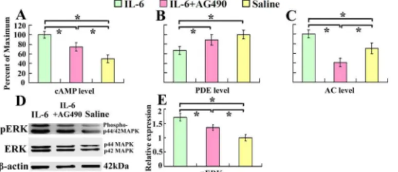
It has been well documented that cAMP is associated with intrinsic growth capability of neu-rons. cAMP degradation by PDE can be inhibited through activation of ERK [18]. We observed that intrathecal delivery of recombinant rat IL-6 for 7 days, immediately after spinal cord inju-ry, increased cAMP level (Fig 1A), and decreased PDE level (Fig 1B) respectively in the sensori-motor cortex. At the same time pERK increased 1.67 times compared with that of saline control (Fig 1D and 1E). We also demonstrated that AC level was increased after IL-6 treat-ment (Fig 1C). To explore the relevant mechanisms, we introduced AG490, an inhibitor of JAK, at the same time. As expected, AG490 partially blocked the IL-6-induced elevation of the cAMP level (Fig 1A).
Fig 1. Intrathecal delivery of IL-6 increases cAMP and AC levels and decreases PDE level in the sensorimotor cortex.Seven days after intrathecal delivery of IL-6, IL-6 and AG-490, or saline, cAMP, AC and PDE levels in the sensorimotor cortex were determined by ELISA. cAMP (A) and AC (C) levels increased while PDE (B) level decreased in IL-6 delivery group compared with that of saline group. After treatment with AG490, an inhibitor of JAK, the effects of IL-6 were partially blocked. Data were expressed as percentage of maximum (means±SEM; n = 3).*P<0.01. D and E demonstrate that IL-6 activates ERK. The expression of pERK was increased in IL-6 delivery group compared with that of saline group, which was also partially blocked by AG-490. Data were expressed as relative density of that of saline delivery after normalized to β-actin (means±SEM; n = 3).*P<0.01.
Intrathecal delivery of IL-6 increases the expression of
regeneration-associated genes in the sensorimotor cortex
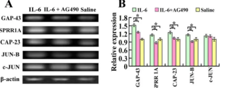
Seven days after spinal cord injury and intrathecal delivery of recombinant rat IL-6, IL-6 and AG490, or saline, total mRNA was prepared from both sides of the sensorimotor cortex for the RT-PCR assay. Quantitative analysis of the optical density observed in three independent ex-periments illustrated that the expression of mRNAs for GAP-43, SPRR1A, CAP-23 and JUN B increased significantly in the sensorimotor cortex of IL-6 group, compared to saline group (Fig 2A and 2B). AG490 treatment partially blocked IL-6-induced upregulation of GAP-43 and CAP-23 and completely blocked IL-6-induced upregulation of SPRR1A and JUN B (Fig 2A and 2B), while c-JUN did not show significant difference among these three groups (Fig 2A and 2B).
gp-130 receptor is expressed by both pyramidal and spinal cord neurons
To investigate whether IL-6 might exert its growth effect through the common signal
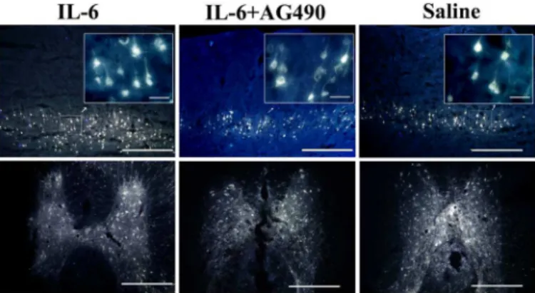
transducer, namely gp130 (glycoprotein 130, also known as CD130) of which all the IL-6 fami-ly members share [19–22], we examined the expression of gp-130 in pyramidal cells of the sen-sorimotor cortex layers V and VI and the spinal cord neurons. Firstly we injected retrograde neuronal tracer FG into the dorsal column of the spinal cord to label pyramidal cells in the sen-sorimortex cortex layers V and VI (Fig 3), combined withβⅢ-tubulin immunostaining. We showed that immunoreactivity of gp-130 was colocalized with that of FG andβⅢ-tubulin in pyramidal cells in the sensorimotor cortex (Fig 4) and in neurons of the spinal cord (Fig 4).
Intrathecal delivery of IL-6 activates JAK/STAT3 signal pathway in
pyramidal cells of the sensorimotor cortex after spinal cord injury
IL-6 activated the JAK/STAT3 signal pathway manifested by increased pSTAT3 immunoreac-tivity in pyramidal cells of the sensorimotor cortex (Fig 5A–5D) compared with that of saline group (Fig 5I–5L). Quantification of fluorescence intensity of pSTAT3 in FG andβⅢ- tubulin labeled pyramidal cells (Fig 5M) and semiquantitative analysis of pSTAT3 protein by Western blot analysis (Fig 5N) further confirmed increased pSTAT3 level in the sensorimotor cortex of IL-6 group compared with that of saline group (Fig 5M and 5N). AG490, an inhibitor of JAK, partially blocked IL-6-induced increase of pSTAT3 (Fig 5E–5H, 5M and 5N).
Fig 2. Expression of growth-associated genes in the sensorimotor cortex after intrathecal delivery of IL-6.A. Seven days after spinal cord injury and intrathecal delivery of either IL-6, IL-6 and AG490, or saline, mRNAs for GAP-43, SPRR1A, CAP-23, JUN-B and c-JUN were measured by RT-PCR. IL-6 increased the expression of mRNAs for GAP-43, SPRR1A, CAP-23 and JUN-B in comparison to that of saline delivery. While c-JUN did not show significant difference among these three groups. B. Quantification of gene expression using densitometry. Data were expressed as relative expression of saline group after normalized toβ-actin (means±SEM; n = 3).*P<0.01.
Intrathecal delivery of IL-6 activates PI3K/AKT/mTOR signal pathway in
pyramidal cells of the sensorimotor cortex after spinal cord injury
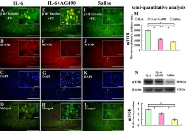
To test whether intrathecal delivery of IL-6 also activated PI3K/AKT/mTOR signal pathway in pyramidal cells in the sensorimotor cortex, we investigated mTOR expression in pyramidal cells in the sensorimotor cortex layers V and VI by immunostaining and Western blot. We showed that immunoreactivity of mTOR was upregulated in FG andβIII- tubulin labeled pyra-midal cells in the sensorimotor cortex, layers V and VI (Fig 6A–6D and 6M), which was in line with Western blot analysis (Fig 6I–6L and 6N). AG490 partially blocked IL-6-induced up-regu-lation of mTOR (Fig 6E–6H, 6M and 6N).
Discussion
We have previously shown that IL-6 promotes axonal regeneration of axotomized CST and functional recovery by enhancing synapse formation and increasing the expression of mTOR
Fig 4. Expression of gp-130 in the sensorimotor cortex and the spinal cord.Pyramidal cells were double labeled with FG andβIII- tubulin. Both pyramidal cells in the sensorimotor cortex layers V and VI (upper panel) and neurons in the spinal cord gray matter (middle and lower panel) showed gp-130 positive staining. Scale bar: 500μm for upper panel and middle panel and 200μm for lower panel. Insets of upper panel show the higher magnification of gp-130-labelled pyramidal cells in selected areas of the sensorimotor cortex layers V and VI. Scale bar: 50μm. Lower panel shows the higher magnification of neurons in the middle panel. Scale bar: 200μm. Arrowheads indicate cells are both fluorogold and tubulin labeled in the sensorimotor cortex and spinal cord, whereas arrows point to tubulin labeled cells.
doi:10.1371/journal.pone.0127772.g004
Fig 3. Expression of Fluoroglod (FG) in the sensorimotor cortex and the spinal cord.Both pyramidal cells in the V and VI layers of the sensorimotor cortex (upper panel) and neurons in the spinal cord gray matter (lower panel) contain FG (Scale bar: 500μm). Insets of upper panel show the higher magnification of FG-labelled pyramidal cells in selected areas of the sensorimotor cortex layers V and VI. Scale bar: 50μm.
in neurons around the lesion site [10]. In this study, we further delineate that intrathecal deliv-ery of IL-6 immediately after spinal cord injury induces pronounced up-regulation of cAMP level and regeneration-associated genes GAP-43, SPRR1A, CAP-23 and JUN-B and activated
Fig 5. Effect of IL-6 on the expression of pSTAT3 in the sensorimotor cortex.Pyramidal cells in the sensorimortex cortex were labelled with FG andβIII tubulin immunostaining. IL-6 delivery (A-D) increased the expression of pSTAT3 compared with that of saline treatment (I-L), and AG490 partially blocked this effect of IL-6 (E-H). These results were further confirmed by quantification of fluorescence intensity (M) and
semiquantitative analysis by Western blot (N). Lower panel of the N shows the quantification of pSTAT3 expression using densitometry. Data were expressed as relative expression of that of saline group after normalized toβ-actin (means±SEM; n = 3).*P<0.01. Scale bar: 500μm. Insets show the higher
magnification of pyramidal cells in selected areas of the sensorimotor cortex V and VI layers. Scale bar: 50μm.
doi:10.1371/journal.pone.0127772.g005
Fig 6. Effect of IL-6 on the expression of mTOR in the sensorimotor cortex.Pyramidal cells in the sensorimortex cortex were labelled with FG label andβ-III tubulin immunostaining. IL-6 delivery increased the expression of mTOR (A-D) compared with that of saline treatment (I-L), and AG490 partially blocked this effect of IL-6 (E-H). These results were further confirmed by quantification of fluorescence intensity (M) and semiquantitative analysis by Western blot (N). Lower panel of the N shows the quantification of mTOR expression using densitometry. Data were expressed as relative expression of that of saline treatment after normalized toβ-actin (means±SEM; n = 3).*P<0.01. Scale bar: 500μm. Insets show the higher magnification of the pyramidal cells in selected areas of the sensorimotor cortex layers V and VI. Scale bar: 50μm.
not only JAK/STAT3 signaling pathway but also PI3K/AKT/mTOR and ERK signaling path-ways in pyramidal cells of the sensorimotor cortex.
cortex through these IL-6R containing endosomal structures (Fig 7). Also as the coherence in the expression of gp-130 staining in both the sensorimotor cortex and in the spinal cord grey matter, it is possible that activated neurons or glia in the spinal cord grey matter may produce some unknown retrograde signals to change the biochemistry of CST neurons.Furthermore, we observed that intrathecal delivery of IL-6 immediately after spinal cord injury also elevated mTOR in sensorimotor cortex. It has been reported that mTOR pathway play an important role in determining the intrinsic axon regrowth responsiveness of injured CNS neurons[6]. Ac-tivation of the mTOR pathway in RGCs or corticospinal neurons enhanced their axonal regen-eration [5–6]. Inflammation or treatment with CNTF/LIF activated mTOR activity in retinal ganglion cells (RGCs) and facilitated long distance regeneration of RGCs after axotomy [42]. IL-6 has also been shown to activate PI3K/Akt pathway [19]. Therefore, mTOR is one possible signaling molecule that IL-6 acts through to enhance axonal regeneration of axotomized pyra-midal cells (Fig 7).
In conclusion, we further showed that intrathecal delivery of IL-6 immediately after spinal cord injury elevated cAMP level and up-regulated the expression of regeneration-associated genes including GAP-43, SPRR1A, CAP-23 and JUN-B, and the expression of pSTAT3 and mTOR in pyramidal cells of the sensorimotor cortex. It is possible that IL-6 acts through‘ sig-naling endosome’and is associated with the activation of JAK/STAT3 signaling pathway.
Acknowledgments
We appreciate Dr. Dongsheng Wu for reading and revising this manuscript. This study was supported by grants from National Natural Science Foundation of China (NSFC No: 81171719) and from Chongqing Natural Science of Foundation of China (CSTC, 2008 BB5281).
Author Contributions
Conceived and designed the experiments: PY. Performed the experiments: PY YQ CB YZ WZ. Analyzed the data: PY. Wrote the paper: PY.
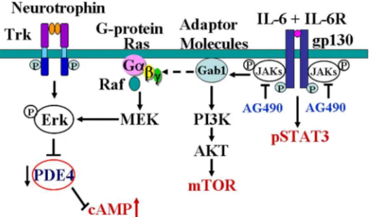
Fig 7. Diagram shows the possible mechanisms of IL-6 in regulating cAMP level in neurons.Binding of neurotrophins to the corresponding receptors (Trk) activates ERK which inhibits PDE and then inhibits the degradation of cAMP [18]. IL-6 binds to IL-6R causing dimerization of gpl30, thereby activating the intracellular tyrosine kinases of the Janus kinase family (JAKs) and subsequently activating STAT3. In the other hand, IL-6 modulates ras and raf through an adapter molecule Gab1, leading to the activation of threonine kinase of MAP kinase family, which include ERK [20–21]. IL-6 activates ERK, thus inhibits PDE and then inhibits the degradation of cAMP. IL-6 may also activate PI3K/Akt pathway [19], through which IL-6 induces mTOR expression in neurons.
References
1. Cai D, Qiu J, Cao Z, McAtee M, Bregman B, Filbin M. Neuronal cyclic AMP controls the developmental loss in ability of axons to regenerate. J Neurosci. 2001; 21(13):4731–9. PMID:11425900
2. Yang P, Yang Z. Enhancing intrinsic growth capacity promotes adult CNS regeneration. J Neurol Sci. 2012; 312(1–2):1–6. Epub 2011/09/20. S0022-510X(11)00534-X [pii] doi:10.1016/j.jns.2011.08.037 PMID:21924742.
3. Leibinger M, Müller A, Gobrecht P, Diekmann H, Andreadaki A, Fischer D. Interleukin-6 contributes to CNS axon regeneration upon inflammatory stimulation. Cell Death Dis 2013.
4. Pearse D, Pereira F, Marcillo A, Bates M, Berrocal Y, Filbin M, et al. cAMP and Schwann cells promote axonal growth and functional recovery after spinal cord injury. Nat Med. 2004; 10(6):610–6. PMID: 15156204
5. Liu K, Lu Y, Lee JK, Samara R, Willenberg R, Sears-Kraxberger I, et al. PTEN deletion enhances the regenerative ability of adult corticospinal neurons. Nat Neurosci. 2010; 13(9):1075–81. PMID: 20694004. doi:10.1038/nn.2603
6. Park KK, Liu K, Hu Y, Smith PD, Wang C, Cai B, et al. Promoting Axon Regeneration in the Adult CNS by Modulation of the PTEN/mTOR Pathway. science. 2008; 322:963–6. doi:10.1126/science.1161566 PMID:18988856
7. Qiu J, Cafferty W, McMahon S, Thompson S. Conditioning injury-induced spinal axon regeneration re-quires signal transducer and activator of transcription 3 activation. J Neurosci. 2005; 25(7):1645–53. PMID:15716400
8. Cao Z GY, Bryson JB, Hou J, Chaudhry N, Siddiq M, Martinez J, Spencer T, Carmel J, Hart RB, Filbin MT. The cytokine interleukin-6 is sufficient but not necessary to mimic the peripheral conditioning lesion effect on axonal growth. J Neurosci. 2006; 26(20):5565–73. PMID:16707807
9. Hakkoum D, Stoppini L, Muller D. Interleukin-6 promotes sprouting and functional recovery in lesioned organotypic hippocampal slice cultures. J Neurochem. 2007; 100(3):747–57. PMID:17144903 10. Yang P, Wen H, Ou S, Cui J, Fan D. IL-6 promotes regeneration and functional recovery after cortical
spinal tract injury by reactivating intrinsic growth program of neurons and enhancing synapse formation. Exp Neurol 2012; 236(1):19–27. doi:10.1016/j.expneurol.2012.03.019PMID:22504113
11. Murphy P, Grondin J, Altares M, Richardson P. Induction of interleukin-6 in axotomized sensory neu-rons. J Neurosci 1995; 15(7 Pt 2):5130–8. PMID:7623140
12. Mogi M, Harada M, Kondo T, Riederer P, Inagaki H, Minami M, et al. Interleukin-1 beta, interleukin-6, epidermal growth factor and transforming growth factor-alpha are elevated in the brain from parkinso-nian patients. Neurosci Lett. 1994; 180(2):147–50. Epub 1994/10/24. PMID:7700568.
13. Kiefer R, Lindholm D, Kreutzberg GW. Interleukin-6 and transforming growth factor-beta 1 mRNAs are induced in rat facial nucleus following motoneuron axotomy. Eur J Neurosci. 1993; 5(7):775–81. Epub 1993/07/01. PMID:8281289.
14. Kossmann T, Hans VH, Imhof HG, Stocker R, Grob P, Trentz O, et al. Intrathecal and serum interleu-kin-6 and the acute-phase response in patients with severe traumatic brain injuries. Shock. 1995; 4 (5):311–7. Epub 1995/11/01. PMID:8595516.
15. Mutlu LK, Woiciechowsky C, Bechmann I. Inflammatory response after neurosurgery. Best Pract Res Clin Anaesthesiol. 2004; 18(3):407–24. Epub 2004/06/24. PMID:15212336.
16. Kossmann T, Hans V, Imhof HG, Trentz O, Morganti-Kossmann MC. Interleukin-6 released in human cerebrospinal fluid following traumatic brain injury may trigger nerve growth factor production in astro-cytes. Brain research. 1996; 713(1–2):143–52. Epub 1996/03/25. 0006-8993(95)01501-9 [pii]. PMID: 8724985.
17. Canu M, Picquet F, Bastide B, Falempin M. Activity-dependent changes in the electrophysiological properties of regular spiking neurons in the sensorimotor cortex of the rat in vitro. Behav Brain Res. 2010; 209(2):289–94. doi:10.1016/j.bbr.2010.02.006PMID:20144900
18. Hannila SS, Filbin MT. The role of cyclic AMP signaling in promoting axonal regeneration after spinal cord injury. Exp Neurol. 2008; 209(2):321–32. PMID:17720160.
19. Spooren A, Kolmus K, Laureys G, Clinckers R, De Keyser J, Haegeman G, et al. Interleukin-6, a mental cytokine. Brain Res Rev. 2011; 67(1–2):157–83. Epub 2011/01/18. S0165-0173(11)00006-3 [pii] doi: 10.1016/j.brainresrev. 2011.01.002PMID:21238488.
20. Takahashi-Tezuka M, Yoshida Y, Fukada T, Ohtani T, Yamanaka Y, Nishida K, et al. Gab1 acts as an adapter molecule linking the cytokine receptor gp130 to ERK mitogen-activated protein kinase. Mol Cell Biol. 1998; 18(7):4109–17. PMID:9632795
22. German C, Sauer B, Howe C. The STAT3 beacon: IL-6 recurrently activates STAT 3 from endosomal structures. Exp Cell Res. 2011 317(14):1955–69. doi:10.1016/j.yexcr.2011.05.009PMID:21619877 23. Boyd J, Gordon T. Neurotrophic factors and their receptors in axonal regeneration and functional
recov-ery after peripheral nerve injury. Mol Neurobiol. 2003; 27(3):277–324. PMID:12845152
24. Chan K, Gordon T, Zochodne D, Power H. Improving Peripheral Nerve Regeneration: from Molecular Mechanisms to Potential Therapeutic Targets. Exp Neurol Epub ahead of print. 2014.
25. Benveniste EN, Tang LP, Law RM. Differential regulation of astrocyte TNF-alpha expression by the cy-tokines TGF-beta, IL-6 and IL-10. Int J Dev Neurosci. 1995; 13(3–4):341–9. Epub 1995/06/01. 0736-5748(94)00061-7 [pii]. PMID:7572286.
26. Neumann H, Schweigreiter R, Yamashita T, Rosenkranz K, Wekerle H, Barde YA. Tumor necrosis fac-tor inhibits neurite outgrowth and branching of hippocampal neurons by a rho-dependent mechanism. J Neurosci. 2002; 22(3):854–62. PMID:11826115.
27. Niederost B, Oertle T, Fritsche J, McKinney RA, Bandtlow CE. Nogo-A and myelin-associated glyco-protein mediate neurite growth inhibition by antagonistic regulation of RhoA and Rac1. J Neurosci. 2002; 22(23):10368–76. PMID:12451136.
28. Schwab JM, Brechtel K, Mueller CA, Failli V, Kaps HP, Tuli SK, et al. Experimental strategies to pro-mote spinal cord regeneration—an integrative perspective. Progress in neurobiology. 2006; 78(2):91–
116. PMID:16487649.
29. Bechmann I, Nitsch R. Identification of phagocytic glial cells after lesion-induced anterograde degener-ation using double-fluorescence labeling: combindegener-ation of axonal tracing and lectin or immunostaining. Histochem Cell Biol. 1997; 107(5):391–7. Epub 1997/05/01. PMID:9208330.
30. Schwab ME. Myelin-associated inhibitors of neurite growth. Experimental neurology. 1990; 109(1):2–5. Epub 1990/07/01. S0014-4886(05)80003-2 [pii]. PMID:2192907.
31. Batchelor PE, Liberatore GT, Wong JY, Porritt MJ, Frerichs F, Donnan GA, et al. Activated macro-phages and microglia induce dopaminergic sprouting in the injured striatum and express brain-derived neurotrophic factor and glial cell line-derived neurotrophic factor. J Neurosci. 1999; 19(5):1708–16. Epub 1999/02/19. PMID:10024357.
32. Harrington A, Ginty D. Long-distance retrograde neurotrophic factor signalling in neurons. Nat Rev Neurosci; 14(3): doi: 101038/nrn3253. 2013; 14(3):177–87. doi:10.1038/nrn3253PMID:23422909 33. He Y, Kuruvilla R, Zweifel L, Ginty D. Evidence in support of signaling endosome-based retrograde
sur-vival of sympathetic neurons. Neuron 2003; 39(1):57–68. PMID:12848932
34. Delcroix J, Valletta J, Wu C, Hunt S, Kowal A, Mobley W. NGF signaling in sensory neurons: evidence that early endosomes carry NGF retrograde signals. Neuron. 2003; 39(1):69–84. PMID:12848933 35. Heerssen H, Pazyra M, Segal R. Dynein motors transport activated Trks to promote survival of
target-dependent neurons. Nat Neurosci 2004; 7(6):596–604. PMID:15122257
36. Kurek J, Austin L, Cheema S, Bartlett P, Murphy M. Up-regulation of leukaemia inhibitory factor and in-terleukin-6 in transected sciatic nerve and muscle following denervation. Neuromuscul Disord 1996; 6 (2):105–14. PMID:8664561
37. Sun F, He Z. Neuronal intrinsic barriers for axon regeneration in the adult CNS. Current opinion in neu-robiology. 2010; 20(4):510–8. PMID:20418094. doi:10.1016/j.conb.2010.03.013
38. Kamimura D, Ishihara K, Hirano T. IL-6 signal transduction and its physiological roles: the signal or-chestration model. Rev Physiol Biochem Pharmacol. 2003; 149:1–38. PMID:12687404
39. Giordano V, Falco GD, Chiari R, Quinto I, Pelicci P, Bartholomew L, et al. Shc mediates IL-6 signaling by interacting with gp130 and Jak2 kinase. J Immunol 1997; 158(9):4097–103. PMID:9126968 40. Kim H, Baumann H. Dual signaling role of the protein tyrosine phosphatase SHP-2 in regulating
expres-sion of acute-phase plasma proteins by interleukin-6 cytokine receptors in hepatic cells. Mol Cell Biol. 1999; 19(8):5326–38. PMID:10409724
41. Beattie E, Zhou J, Grimes M, Bunnett N, Howe C, Mobley W. A signaling endosome hypothesis to ex-plain NGF actions: potential implications for neurodegeneration. Cold Spring Harb Symp Quant Biol 1996; 61:389–406. PMID:9246468