Lymphocytes Is Regulated by Mitogen-Activated Protein
Kinase Signaling
Aimee J. Marko, Rebecca A. Miller, Alina Kelman, Kenneth A. Frauwirth*
Department of Cell Biology and Molecular Genetics and Maryland Pathogen Research Institute, University of Maryland, College Park, Maryland, United States of America
Abstract
T lymphocytes play a critical role in cell-mediated immune responses. During activation, extracellular and intracellular signals alter T cell metabolism in order to meet the energetic and biosynthetic needs of a proliferating, active cell, but control of these phenomena is not well defined. Previous studies have demonstrated that signaling from the costimulatory receptor CD28 enhances glucose utilization via the phosphatidylinositol-3-kinase (PI3K) pathway. However, since CD28 ligation alone does not induce glucose metabolism in resting T cells, contributions from T cell receptor-initiated signaling pathways must also be important. We therefore investigated the role of mitogen-activated protein kinase (MAPK) signaling in the regulation of mouse T cell glucose metabolism. T cell stimulation strongly induces glucose uptake and glycolysis, both of which are severely impaired by inhibition of extracellular signal-regulated kinase (ERK), whereas p38 inhibition had a much smaller effect. Activation also induced hexokinase activity and expression in T cells, and both were similarly dependent on ERK signaling. Thus, the ERK signaling pathway cooperates with PI3K to induce glucose utilization in activated T cells, with hexokinase serving as a potential point for coordinated regulation.
Citation:Marko AJ, Miller RA, Kelman A, Frauwirth KA (2010) Induction of Glucose Metabolism in Stimulated T Lymphocytes Is Regulated by Mitogen-Activated Protein Kinase Signaling. PLoS ONE 5(11): e15425. doi:10.1371/journal.pone.0015425
Editor:Derya Unutmaz, New York University, United States of America
ReceivedJuly 26, 2010;AcceptedSeptember 20, 2010;PublishedNovember 10, 2010
Copyright:ß2010 Marko et al. This is an open-access article distributed under the terms of the Creative Commons Attribution License, which permits unrestricted use, distribution, and reproduction in any medium, provided the original author and source are credited.
Funding:This research was supported by a Howard Temin K01 Career Development Award (grant number K01 CA092156) from the National Cancer Institute (www.cancer.gov) and a New Investigator Grant from the Leukemia Research Foundation (www.leukemia-research.org) to K.A.F. The funders had no role in study design, data collection and analysis, decision to publish, or preparation of the manuscript.
Competing Interests:The authors have declared that no competing interests exist.
* E-mail: kfrauwir@umd.edu
Introduction
T cells are dependent on external supplies of glucose to maintain biosynthesis and energy metabolism during activation. Activated T cells adopt a metabolic state of ‘‘aerobic glycolysis’’, in which glucose flux through glycolysis is high, but only a small proportion of the glucose is oxidized in mitochondria [1–5]. A similar phenomenon was recognized in tumor cells more than 80 years ago [6], and was originally thought to represent a defect in mitochondrial function, perhaps as a result of mutations that occurred during oncogenic transformation. However, more recent interpretations suggest that glycolysis is a preferred metabolic pathway for highly proliferative cells, and the shift to a glycolytic phenotype is part of a larger adaptive metabolic program to support growth and proliferation [7–9]. Although there is growing appreciation for the importance of metabolic control in both immune responses and tumor development, the pathways that regulate glucose metabolism are still not well defined.
Resting lymphocytes depend upon growth signals from cy-tokines and low-level T cell receptor (TCR) stimulation in order to maintain metabolic homeostasis [10,11], whereas CD28 costim-ulation is required for induction of high level glucose uptake and glycolysis, in large part via activation of the phosphatidylinositol-3-kinase (PI3K)/Akt signaling pathway [12,13]. The inhibitory receptors cytotoxic T lymphocyte antigen-4 (CTLA-4) and pro-grammed death-1 (PD-1) both block CD28-induced Akt activa-tion, and also prevent the increase in glucose utilization [12,14],
suggesting that regulation of cellular metabolism might be a component of the inhibitory function of these receptors. Strikingly, overexpression of glucose transporter 1 (GLUT1), the major glucose transporter in hematopoietic cells [10], can partially re-place costimulation in the induction of proliferation and cytokine production, and constitutively active Akt synergizes with GLUT1 overexpression [13]. Together, these findings indicate the impor-tance of enhanced glucose utilization as a downstream effect of CD28 signaling. However, ligation of CD28 alone does not induce glucose metabolism [12]. Thus, TCR-initiated signaling pathways must cooperate with PI3K/Akt signaling to regulate glucose metabolism.
enhancing glycolysis [23–25]. We therefore investigated the role of MAPK signaling in T cell glucose metabolism. We found that the enhanced glucose uptake and glycolysis seen in activated T cells is dependent on extracellular regulated kinase (ERK) signal-ing, and that this may be due to the regulation of hexokinase expression and activity.
Results
Activation of murine T cells leads to enhanced glucose metabolism
Studies with human peripheral blood T cells have shown that stimulation via mitogenic lectins or CD3/CD28 ligation leads to an "aerobic glycolysis" phenotype, highly inducing glucose uptake and glycolysis [2,12,14,26,27]. In order to further characterize the regulation of glucose metabolism in T lymphocytes, we decided to switch to the murine system. This would allow us to take advantage of the many genetic and biochemical tools available in the murine system, as well as the lower sample-to-sample variability offered by inbred mouse strains. To confirm that glucose metabolism in murine T cells follows an induction pattern similar to that seen in human T cells, we purified splenic T cells from C57BL/6 mice and stimulated them in vitro with anti-CD3 and anti-CD28 antibodies. After 24 hours of stimulation, glucose uptake by live T cells was measured by the accumulation of radiolabeled 2-deoxyglucose, a non-metabolizable glucose analog, and glycolysis was measured by the generation of3H-labeled H2O
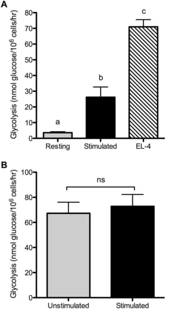
from 3H-labeled glucose, at the step catalyzed by enolase. As shown in Figure 1, activated murine T cells increased both glucose uptake and glycolysis. Thus, primary mouse T cells upregulate glucose utilization upon CD3/CD28 ligation similarly to human T cells, and comparably to previous studies using mitogenic lectins. The ‘‘aerobic glycolysis’’ phenotype was originally described in tumor cells [6]. For comparison, we therefore also examined glycolysis in the murine T lymphoma cell line EL-4. EL-4 cells showed a high rate of glycolysis without any TCR stimulation, generally 2–3 times the rate of activated T cells (Figure 2A). This is similar to what we have recently reported for glutamine utilization in EL-4 cells [22]. Thus, in the absence of TCR or CD28 ligation, EL-4 cells display elevated metabolism compared to even fully activated primary T cells. We next tested whether stimulation of EL-4 cells with anti-CD3 and anti-CD28 antibodies would further enhance glycolysis. As shown in Figure 2B, stimulation did not appreciably alter glycolysis in EL-4 cells, indicating that regulation of glucose metabolism has become unlinked from CD3/CD28 signaling.
Glucose metabolism in activated T cells is regulated by MAPK signaling
Work from several groups has established the importance of the PI3K/Akt pathway in regulating lymphocyte glucose metabolism [12,13,28], in addition to its well-known role in insulin receptor-mediated increases in glucose transport [29–34]. However, although cross-linking CD28 alone is sufficient to activate PI3K and Akt [35], it is not sufficient to increase glucose metabolism in the absence of TCR/CD3 signaling [12]. Together, these indicate that additional, TCR-induced signaling is required to cooperate with CD28-induced PI3K signals. We therefore examined other signal transduction pathways for regulation of glucose metabolism in activated T cells.
Ligation of the T cell receptor initiates numerous intracellular signaling pathways, including cascades that lead to the activation of MAPK family members. Although well established as regulators of transcription factors during T cell activation, the MAPK
pathways have also been implicated in control of amino acid [36– 38] and glucose [24,25] utilization in other cell types. We therefore tested the importance of ERK and p38 in the control of glucose metabolism during T cell activation. T cells were stimulated for 24 hours in the presence or absence of specific inhibitors for each of the MAPK pathways, and the effects on glucose uptake and glycolysis were determined. As shown in Figure 3A, the stimulation-induced increase in glucose uptake was almost completely blocked by the ERK inhibitor U0126 (82.2% inhibition), but only partially blocked by the p38 inhibitor SB203580 (51% inhibition). Inhibitors were all used at concen-trations that completely blocked T cell proliferation (data not shown) and which do not inhibit other kinases (2.6mM U0126 [39], and 20mM SB203580 [40]). As shown in Figure 3B, ERK blockade also largely prevented the induction of glycolysis (76.7% inhibition), whereas p38 inhibition had a much smaller effect on glycolysis (19.3% inhibition). Thus, glucose metabolism appears to
Figure 1. Activation induces an ‘‘aerobic glycolysis’’ phenotype in mouse T cells.Purified T cells were cultured with either control hamster IgG (resting) or anti-CD3 plus anti-CD28 antibodies (stimulat-ed). Glucose uptake (A) and glycolysis (B) rates were measured after 24 hours. ***p,0.0001, means are different; **, p,0.001, means are different. Results are representative of at least 3 independent experiments.
be regulated by MAPK signaling, with ERK playing a larger role than p38.
MAPK signaling controls hexokinase activity
Glucose uptake and subsequent metabolism are dependent on hexokinase function. Phosphorylation of glucose to glucose-6-phosphate traps glucose in the cell, and removes it from equilibrium, allowing continued diffusion through glucose transporters. Glucose-6-phosphate is also the initial substrate for glycolysis. Therefore, hexokinase is well positioned to be a common regulation point. Using an assay for total enzyme activity, we compared hexokinase in resting and activate T cells. As shown in Figure 4A, CD3/CD28 stimulation of T cells strongly increased total cellular hexokinase
activity. As with glycolysis, hexokinase activity was high in EL-4 lymphoma cells in the absence of any TCR stimulation (Figure 4B). The hexokinase activity in EL-4 cells was substantially higher than glycolysis rate, which was unlike our findings for primary T cells (compare Figure 1B to 4A for primary cells, and Figure 2 to 4B for EL-4). The disproportionately high hexokinase activity (relative to glycolysis) in EL-4 cells may reflect a large demand in tumor cells for glucose utilization via other metabolic pathways, such as the pentose phosphate shunt [41], although a deeper analysis of glucose metabolism in tumor cells would be required to draw definitive conclusions.
Figure 2. Lymphoma cells show constitutively high glycolysis.
(A) Glycolysis was measured in EL-4 lymphoma cells from continuous culture, without additional stimulation. Glycolysis in resting and stimulated T cells (as described in Figure 1) was measured for com-parison. p,0.001, means that do not share a letter differ. (B) EL-4 cells were cultured for 24 hours with either control hamster IgG (unstimu-lated) or anti-CD3 plus anti-CD28 antibodies (stimu(unstimu-lated) and glycolysis rates were measured. ns, not significantly different. Results are representative of 2 (A) or 3 (B) independent experiments.
doi:10.1371/journal.pone.0015425.g002
Figure 3. Glucose metabolism in T cells is regulated by MAPK signaling.Purified T cells were stimulated in the absence or presence of the ERK inhibitor U0126 (2.6mM) or the p38 inhibitor SB203580
(20mM) for 24 hours. Glucose uptake (A) and glycolysis (B) were
measured as in Figure 1. p,0.05, means that do not share a letter differ. Results are representative of 3 independent experiments.
Because hexokinase activity is intimately tied to both glucose uptake and glycolysis, we asked whether induction of hexokinase also required MAPK signaling. We focused on ERK, as inhibition of this pathway had much greater effects than p38 inhibition on uptake and glycolysis. In addition, ERK has been reported to positively regulate hexokinase in cultured cells [23]. Using the ERK inhibitor U0126, we found that induction of hexokinase activity downstream of TCR/CD28 ligation was strongly dependent on ERK signaling (Figure 5), similar to glucose uptake and glycolysis. Comparable results were seen using a second ERK inhibitor, PD98059 (data not shown). Thus, by regulating hexokinase activity, the ERK signaling pathway is able to control glucose metabolism in a coordinated fashion.
We recently reported that ERK positively regulates mRNA expression of the enzyme glutaminase, which catalyzes the first
step in glutamine metabolism [22]. We therefore tested whether ERK signaling in T cells also induces hexokinase gene expression,
Figure 4. T cell activation induces hexokinase activity.(A) Resting or 24-hour stimulated T cells were lysed in 0.1% Triton X-100 and enzymatic activity of hexokinase was measured. ***p,0.0001, means are different. (B) Continuously growing EL-4 cells were lysed in 0.1% Triton X-100 and hexokinase activity was compared to that in resting and 24-hour stimulated T cells. p,0.0001, means that do not share a letter differ. Results are representative of 3 (A) or 2 (B) independent experiments. doi:10.1371/journal.pone.0015425.g004
Figure 5. Hexokinase activity in activated T cells is regulated by MAPK signaling.(A) T cells were stimulated in the absence or presence of the ERK inhibitor U0126 (2.6mM), as in Figure 3, and
hexokinase activity was measured. p,0.05, means that do not share a common letter differ. (B) T cells were cultured with control (rest) or anti-CD3 and anti-CD28 antibodies for the indicated times, in the absence (stim) or presence of 40mM PD98059 (stim+PD). Hexokinase 1 (left
panel) and hexokinase 2 (right panel) mRNA levels were determined by quantitative real-time PCR. p,0.05, means that do not share a common letter differ. Results are representative of 3 (hexokinase 1) or 2 (hexokinase 2) independent experiments.
analyzing the same mRNA samples that we used previously to study glutaminase expression. T cell stimulation increased mRNA levels of hexokinase 1 and 2 within 3 hours, and hexokinase 2 consistently showed higher and longer-lived induction than hexokinase 1 (Figure 5B). Stimulation of T cells in the presence of 40mM PD98059, another highly specific ERK inhibitor [42], ablated the increase in hexokinase 1 mRNA expression, and partially blocked the increase in hexokinase 2 mRNA (Figure 5B). Thus, glucose metabolism in T cells appears to be regulated, at least partly, by ERK control of hexokinase gene expression.
Discussion
T cell activation is accompanied by a large increase in glucose uptake and glycolysis, which is necessary to support new metabolic demands. Previous studies have shown that the enhanced glucose utilization is dependent on signals from the costimulatory receptor CD28, and particularly the PI3K/Akt pathway [12,14]. However, CD28 ligation alone does not induce changes in glucose metabolism, despite strongly activating PI3K and Akt [12,35], implicating TCR-initiated signaling in this process as well. In this study, we found that the MAPK family member ERK plays an important role in the regulation of glucose metabolism during T cell activation, while p38 makes a smaller (and perhaps redundant) contribution. Blockade of ERK signaling strongly inhibited increases in glucose uptake and glycolysis. High level glucose metabolism has been shown to be required for the survival, proliferation, and cytokine production (especially IFN-c) of activated T cells [13,43]. Thus, in addition to regulating gene expression via the activation of transcription factors, ERK also controls T cell function by enhancing glucose metabolism.
Glucose metabolism is controlled by a combination of allosteric activators and inhibitors, as well as by post-translational modification of key enzymes. This provides a variety of potential targets for MAPK regulation. Hexokinase, which catalyzes the phosphorylation of glucose to glucose-6-phosphate, is a highly attractive candidate, for several reasons. Glucose uptake in lymphocytes occurs by facilitated diffusion through GLUT1, meaning that glucose can only move down a concentration gradient. Upon phosphorylation by hexokinase, glucose is removed from equilibrium and simultaneously trapped inside the cell. Thus, glucose uptake is critically dependent on hexokinase activity. In addition, phosphorylation is a required step for both glucose storage as glycogen, and glucose catabolism via glycolysis and the pentose phosphate pathway. Thus, hexokinase is likely to be a gatekeeper for cellular glucose consumption. We found that resting T cells have low hexokinase activity, which is highly induced upon activation. Strikingly, hexokinase activity levels corresponded closely to glycolysis rates, reinforcing the idea that hexokinase represents a limiting factor for glucose metabolism in T cells. As with glucose uptake and glycolysis, induction of hexokinase activity was dependent on ERK signaling, providing a point for coordinated control of glucose metabolism.
The finding that hexokinase activity in T cells is regulated by MAPK signaling also provides insight into the cooperation between the TCR and CD28 in enhancing glucose utilization. Studies of the mechanism of growth factor receptor inhibition of cell death revealed that hexokinase is a target of Akt, with evidence that Akt can both induce subcellular redistribution and increase enzymatic activity of hexokinase [44,45]. Thus, the CD28 signal (Akt) and TCR signals (ERK) intersect at hexokinase. It is unknown whether hexokinase is a direct phosphorylation target for Akt or ERK, but regulation of hexokinase activity by ERK in glomerular mesangial cells appears to require de novo gene transcription [23], consistent with our own finding that induction of hexokinase gene expression
during T cell activation is inhibited by blocking ERK signaling. However, we also found that expression of hexokinase 2 was only partially dependent on ERK, suggesting that the mechanism may involve changes in both hexokinase expression level and post-translational modifications. Further investigation will be necessary to sort out these possibilities.
A model of aerobic glycolysis, with glutamine serving as a critical Krebs cycle substrate, has been proposed to be a common feature of most or all rapidly growing and dividing cells, including tumor cells [7–9]. Indeed, we found that the EL-4 T lymphoma cell line demonstrates a pattern of glucose metabolism that is similar to that of activated primary T cells, but with the various aspects of glucose utilization further elevated. This is also similar to what we recently reported for glutamine metabolism in EL-4 cells vs. activated normal T cells [22]. Thus, the metabolic alterations that have long been observed in tumors (and which are now used diagnostically to detect cancer) may not be due to metabolic dysfunction, but rather may represent an adaptation in which increases in cellular metabolism occur without the normal requirement for receptor-mediated induction.
Materials and Methods
Ethics Statement
Animals used in this study received humane care in strict compliance with the ‘‘Guide for the Care and Use of Laboratory Animals’’ of the National Institutes of Health. All protocols were approved by the Institutional Animal Care and Use Committee of the University of Maryland (Permit Number R-09-70).
Antibodies and reagents
Anti-CD3 (mAb 145-2C11) and anti-CD28 (mAb 37.51) antibodies, control hamster IgG, and phycoerythrin-labeled anti-Thy1.2 antibodies were purchased from eBioscience (San Diego, CA). U0126, PD98059, and SB203580 were purchased from Biomol (Plymouth Meeting, PA). Hexokinase (Enzyme Commis-sion 2.7.1.1) was from MP Biomedicals (Santa Ana, CA). Glucose-6-phosphate dehydrogenase (Enzyme Commission 1.1.1.49), ATP, and NADPH were from Sigma-Aldrich (St. Louis, MO). 3-mercapto-1,2-propanediol (thioglycerol) was purchased from Fisher Scientific (Fairlawn, NJ). [3H]-2-deoxyglucose, 5-[3 H]-glucose, and [3H]-H2O were from PerkinElmer (Shelton, CT).
Animals
C57BL/6J mice (6 weeks old) were purchased from The Jackson Laboratory (Bar Harbor, ME). All mice were maintained in ventilated microisolator cages (Animal Care Systems, Centennial, CO) in the University of Maryland animal facility. Mice were euthanized by carbon dioxide inhalation, as recommended by the American Veterinary Medical Association Panel on Euthanasia, and all efforts were made to minimize suffering.
T cell purification
Murine T cells were purified from spleens using the EasySep Mouse T Cell Enrichment Kit (Stem Cell Technologies, Vancouver, BC, Canada) or the Dynal Mouse T Cell Negative Isolation Kit (Invitrogen, Carlsbad, CA) according to the manufacturer’s protocol. Purified T cells were generally .95% Thy1-positive, as determined by flow cytometry.
Cell lines and culture
supplemented with 10% FBS (Hyclone, Logan, UT), penicillin/ streptomycin, 10 mM HEPES buffer, 2 mM glutamine, and 55mM 2-mercaptoethanol at 37uC in a 5% CO2atmosphere.
T cell stimulation
Anti-CD3 and anti-CD28 antibodies were covalently attached to tosyl-activated Dynabeads (Invitrogen) following the manufac-turer’s instructions. Beads were mixed with T cells at a 3:1 ratio of beads:cells and incubated at 37uC for the desired time. For unstimulated samples, T cells were mixed with control hamster IgG-linked Dynabeads, and were then cultured in parallel with stimulated samples. For experiments with MAPK inhibitors, the inhibitors were added to culture medium at the time of stimulation, and were present throughout the incubation.
Glucose uptake, glycolysis, and hexokinase assays Glucose uptake and glycolysis were measured as described previously [12]. To measure hexokinase activity, purified T cells were lysed in 0.1% Triton X-100 at a concentration of 26108cells/mL, and 5mL of lysate was used for each reaction. The assay was carried out as described by Rathmell et al.[45], in a final volume of 100mL. Activity was determined by the change in absorbance at 340 nm, using a VersaMax spectrophotometer and SoftMax Pro software (Molecular Devices). OD readings were taken every 6–10 s for at least 30 minutes, and rate was calculated from the linear portion of the curve. A340values were converted
into substrate concentrations using Beer’s Law (A =eLc, where A is absorption,eis the extinction coefficient, L is the path length, and c is concentration) and the extinction coefficient of NAD(P)H (6.22 mM21cm21). Data points for all analyses are presented as the mean of triplicate samples6S.D.
Real-time PCR
Total RNA was extracted from unstimulated or stimulated T cells using the Nucleospin RNA II kit (Macherey-Nagel, Du¨ren, Germany) or the RNeasy Mini kit (Qiagen, Valencia, CA). cDNA
was generated using iScript reverse transcriptase reagents (Bio-Rad, Hercules, CA). Primers for real-time PCR were designed using the Primer-BLAST tool of the National Center for Biotechnology Information (http://www.ncbi.nlm.nih.gov/tools/ primer-blast/), and were as follows: hexokinase 1, forward 59 GC-CACGCTCGGTGCCATCTT 39, reverse 59 GGTCTTGTGG-AACCGCCGGG 39; hexokinase 2, forward 59 AGCTCTGTG-GCGCAGGCATG 39, reverse 59 TCGGACAGGCCACAGCA-GTG 39. Real-time PCR was performed in an iCycler iQ system (Bio-Rad) using the SensiMix SYBR & Fluorescein kit (Bioline, Taunton, MA) and analyzed with MyiQ software (Bio-Rad). Melt curve analysis was performed to confirm the presence of a single PCR product for each reaction, and agarose gel electrophoresis was used to confirm that PCR products were the expected sizes. Fold induction was calculated by theDDCt method, using 18S rRNA as the reference. Data are presented as the means of triplicate samples6SD.
Statistics
All statistical analyses were performed using Prism Version 5 software (GraphPad, San Diego, CA). The minimal level of confidence at which experimental results were considered significant was p,0.05. Statistical significance between samples was determined by one-way analysis of variance (ANOVA) with Bonferroni post-test analysis, or by two-tailed t test.
Acknowledgments
The authors thank Pau´l Clavijo for technical assistance with real-time PCR, and Drs. Jeffrey Rathmell and Brooke Humphrey for critical reading of this manuscript.
Author Contributions
Conceived and designed the experiments: AJM KAF. Performed the experiments: AJM RAM AK KAF. Analyzed the data: AJM KAF. Wrote the paper: KAF.
References
1. Cooper EH, Barkhan P, Hale AJ (1963) Observations on the Proliferation of Human Leucocytes Cultured with Phytohaemagglutinin. Brit J Haemat 9: 101–111.
2. Hedeskov CJ (1968) Early effects of phytohaemagglutinin on glucose metabolism of normal human lymphocytes. Biochem J 110: 373–380.
3. Roos D, Loos JA (1970) Changes in the carbohydrate metabolism of mitogenically stimulated human peripheral lymphocytes. I. Stimulation by phytohaemagglutinin. Biochim Biophys Acta 222: 565–582.
4. Sagone AL, Jr., LoBuglio AF, Balcerzak SP (1974) Alterations in hexose monophosphate shunt during lymphoblastic transformation. Cell Immunol 14: 443–452.
5. Culvenor JG, Weidemann MJ (1976) Phytohaemagglutinin stimulation of rat thymus lymphocytes glycolysis. Biochim Biophys Acta 437: 354–363. 6. Warburg O (1956) On respiratory impairment in cancer cells. Science 124:
269–270.
7. DeBerardinis RJ, Lum JJ, Hatzivassiliou G, Thompson CB (2008) The biology of cancer: metabolic reprogramming fuels cell growth and proliferation. Cell Metab 7: 11–20.
8. Hsu PP, Sabatini DM (2008) Cancer cell metabolism: Warburg and beyond. Cell 134: 703–707.
9. Vander Heiden MG, Cantley LC, Thompson CB (2009) Understanding the Warburg effect: the metabolic requirements of cell proliferation. Science 324: 1029–1033.
10. Rathmell JC, Vander Heiden MG, Harris MH, Frauwirth KA, Thompson CB (2000) In the absence of extrinsic signals, nutrient utilization by lymphocytes is insufficient to maintain either cell size or viability. Mol Cell 6: 683–692. 11. Rathmell JC, Farkash EA, Gao W, Thompson CB (2001) IL-7 enhances the
survival and maintains the size of naive T cells. J Immunol 167: 6869–6876. 12. Frauwirth KA, Riley JL, Harris MH, Parry RV, Rathmell JC, et al. (2002) The
CD28 Signaling Pathway Regulates Glucose Metabolism. Immunity 16: 769–777.
13. Jacobs SR, Herman CE, MacIver NJ, Wofford JA, Wieman HL, et al. (2008) Glucose Uptake Is Limiting in T Cell Activation and Requires CD28-Mediated Akt-Dependent and Independent Pathways. Journal of Immunology 180: 4476–4486.
14. Parry RV, Chemnitz JM, Frauwirth KA, Lanfranco AR, Braunstein I, et al. (2005) CTLA-4 and PD-1 receptors inhibit T-cell activation by distinct mechanisms. Mol Cell Biol 25: 9543–9553.
15. Denton RM (2009) Regulation of mitochondrial dehydrogenases by calcium ions. Biochim Biophys Acta 1787: 1309–1316.
16. Quintana A, Schwindling C, Wenning AS, Becherer U, Rettig J, et al. (2007) T cell activation requires mitochondrial translocation to the immunological synapse. Proc Natl Acad Sci U S A 104: 14418–14423.
17. Ardawi MS, Newsholme EA (1983) Glutamine metabolism in lymphocytes of the rat. Biochem J 212: 835–842.
18. Brand K, Williams JF, Weidemann MJ (1984) Glucose and glutamine metabolism in rat thymocytes. Biochem J 221: 471–475.
19. Ardawi MS (1988) Glutamine and glucose metabolism in human peripheral lymphocytes. Metabolism 37: 99–103.
20. Brand K, Fekl W, von Hintzenstern J, Langer K, Luppa P, et al. (1989) Metabolism of glutamine in lymphocytes. Metabolism 38: 29–33.
21. Koch B, Schroder MT, Schafer G, Schauder P (1990) Comparison between transport and degradation of leucine and glutamine by peripheral human lymphocytes exposed to concanavalin A. J Cell Physiol 143: 94–99. 22. Carr EL, Kelman A, Wu GS, Gopaul R, Senkevitch E, et al. (2010) Glutamine
Uptake and Metabolism Are Coordinately Regulated by ERK/MAPK During T Lymphocyte Activation. J Immunol 185: 1037–1044.
25. Zhou Q, Lam PY, Han D, Cadenas E (2008) c-Jun N-terminal kinase regulates mitochondrial bioenergetics by modulating pyruvate dehydrogenase activity in primary cortical neurons. J Neurochem 104: 325–335.
26. Roos D, Loos JA (1973) Changes in the carbohydrate metabolism of mitogenically stimulated human peripheral lymphocytes. II. Relative impor-tance of glycolysis and oxidative phosphorylation on phytohaemagglutinin stimulation. Exp Cell Res 77: 127–135.
27. Hume DA, Radik JL, Ferber E, Weidemann MJ (1978) Aerobic glycolysis and lymphocyte transformation. Biochem J 174: 703–709.
28. Doughty CA, Bleiman BF, Wagner DJ, Dufort FJ, Mataraza JM, et al. (2006) Antigen receptor-mediated changes in glucose metabolism in B lymphocytes: role of phosphatidylinositol 3-kinase signaling in the glycolytic control of growth. Blood 107: 4458–4465.
29. Kohn AD, Summers SA, Birnbaum MJ, Roth RA (1996) Expression of a constitutively active Akt Ser/Thr kinase in 3T3-L1 adipocytes stimulates glucose uptake and glucose transporter 4 translocation. J Biol Chem 271: 31372–31378. 30. Cong LN, Chen H, Li Y, Zhou L, McGibbon MA, et al. (1997) Physiological role of Akt in insulin-stimulated translocation of GLUT4 in transfected rat adipose cells. Mol Endocrinol 11: 1881–1890.
31. Barthel A, Okino ST, Liao J, Nakatani K, Li J, et al. (1999) Regulation of GLUT1 gene transcription by the serine/threonine kinase Akt1. J Biol Chem 274: 20281–20286.
32. Ueki K, Yamamoto-Honda R, Kaburagi Y, Yamauchi T, Tobe K, et al. (1998) Potential role of protein kinase B in insulin-induced glucose transport, glycogen synthesis, and protein synthesis. J Biol Chem 273: 5315–5322.
33. Taha C, Liu Z, Jin J, Al-Hasani H, Sonenberg N, et al. (1999) Opposite translational control of GLUT1 and GLUT4 glucose transporter mRNAs in response to insulin. Role of mammalian target of rapamycin, protein kinase b, and phosphatidylinositol 3-kinase in GLUT1 mRNA translation. J Biol Chem 274: 33085–33091.
34. Wang Q, Somwar R, Bilan PJ, Liu Z, Jin J, et al. (1999) Protein kinase B/Akt participates in GLUT4 translocation by insulin in L6 myoblasts. Mol Cell Biol 19: 4008–4018.
35. Parry RV, Reif K, Smith G, Sansom DM, Hemmings BA, et al. (1997) Ligation of the T cell co-stimulatory receptor CD28 activates the serine-threonine protein kinase protein kinase B. Eur J Immunol 27: 2495–2501.
36. Franchi-Gazzola R, Visigalli R, Bussolati O, Dall’Asta V, Gazzola GC (1999) Adaptive increase of amino acid transport system A requires ERK1/2 activation. J Biol Chem 274: 28922–28928.
37. Lopez-Fontanals M, Rodriguez-Mulero S, Casado FJ, Derijard B, Pastor-Anglada M (2003) The osmoregulatory and the amino acid-regulated responses of system A are mediated by different signal transduction pathways. J Gen Physiol 122: 5–16.
38. Hyde R, Cwiklinski EL, MacAulay K, Taylor PM, Hundal HS (2007) Distinct sensor pathways in the hierarchical control of SNAT2, a putative amino acid transceptor, by amino acid availability. J Biol Chem 282: 19788–19798. 39. Favata MF, Horiuchi KY, Manos EJ, Daulerio AJ, Stradley DA, et al. (1998)
Identification of a novel inhibitor of mitogen-activated protein kinase kinase. J Biol Chem 273: 18623–18632.
40. Cuenda A, Rouse J, Doza YN, Meier R, Cohen P, et al. (1995) SB 203580 is a specific inhibitor of a MAP kinase homologue which is stimulated by cellular stresses and interleukin-1. FEBS Lett 364: 229–233.
41. Vaughn AE, Deshmukh M (2008) Glucose metabolism inhibits apoptosis in neurons and cancer cells by redox inactivation of cytochrome c. Nat Cell Biol 10: 1477–1483.
42. Alessi DR, Cuenda A, Cohen P, Dudley DT, Saltiel AR (1995) PD 098059 is a specific inhibitor of the activation of mitogen-activated protein kinase kinase in vitro and in vivo. J Biol Chem 270: 27489–27494.
43. Cham CM, Gajewski TF (2005) Glucose availability regulates IFN-gamma production and p70S6 kinase activation in CD8+effector T cells. J Immunol 174: 4670–4677.
44. Gottlob K, Majewski N, Kennedy S, Kandel E, Robey RB, et al. (2001) Inhibition of early apoptotic events by Akt/PKB is dependent on the first committed step of glycolysis and mitochondrial hexokinase. Genes Dev 15: 1406–1418.