Needle Aspirates of Thyroid Nodules
Jieun Koh1, Jong Rak Choi2, Kyung Hwa Han3, Eun-Kyung Kim1, Jung Hyun Yoon1, Hee Jung Moon1, Jin
Young Kwak1*
1Department of Radiology, Research Institute of Radiological Science, Yonsei University, College of Medicine, Seoul, Korea,2Department of Laboratory Medicine, Yonsei University, College of Medicine, Seoul, Korea,3Biostatistics Collaboration Unit, Medical Research Center, Yonsei University, College of Medicine, Seoul, Korea
Abstract
Background:The aim of this study was to evaluate the proper indication of adjunctive BRAFV600Emutation analysis at the time of ultrasound-guided fine-needle aspiration in the diagnosis of thyroid nodules.
Methods:This study included 518 nodules in 479 patients who underwent ultrasound-guided fine-needle aspiration with BRAFV600Emutation. We calculated and compared the diagnostic performances of cytology and cytology with BRAFV600E mutation analysis to detect malignancy among thyroid nodules according to ultrasound features and size.
Results:Sensitivity, negative predictive value, and accuracy of cytology with BRAFV600Emutation analysis were significantly
higher than those of cytology alone in thyroid nodules with suspicious ultrasound features, regardless of size. Diagnostic performances did not show significant differences between cytology and cytology with BRAFV600E mutation analysis in
nodules without any suspicious ultrasound features, regardless of size.
Conclusion:The BRAFV600Emutation analysis was a useful adjunctive diagnostic tool in the diagnosis of thyroid nodules with suspicious ultrasound features regardless of size.
Citation:Koh J, Choi JR, Han KH, Kim E-K, Yoon JH, et al. (2013) Proper Indication of BRAFV600EMutation Testing in Fine-Needle Aspirates of Thyroid Nodules. PLoS
ONE 8(5): e64505. doi:10.1371/journal.pone.0064505
Editor:Paul J. van Diest, University Medical Centre Utrecht, The Netherlands
ReceivedJanuary 6, 2013;AcceptedApril 15, 2013;PublishedMay 24, 2013
Copyright:ß2013 Koh et al. This is an open-access article distributed under the terms of the Creative Commons Attribution License, which permits unrestricted use, distribution, and reproduction in any medium, provided the original author and source are credited.
Funding:The authors have no support or funding to report.
Competing Interests:The authors have declared that no competing interests exist. * E-mail: [email protected]
Introduction
Among the various molecular events related to thyroid cancer, BRAFV600E mutation is a highly specific somatic mutation for papillary thyroid cancer [1–3]. An activating point mutation of the T1799A point BRAF gene results in a valine-to-glutamic acid replacement at amino acid V600, resulting in the constitutive tumorogenesis [1,4]. The prevalence of the mutation in papillary thyroid cancer is highly variable especially according to region, ranging from 29% to 83% in different studies from different areas of the world [4]. Among patients diagnosed as papillary thyroid cancer in Korea, the prevalence of BRAFV600Emutation has been reported up to 84% [4–8].
Although detecting BRAFV600E mutation plays an additional role in the definitive diagnosis of thyroid nodules [8–10], performing routine BRAFV600E mutation analysis in addition to fine-needle aspiration (FNA) may be questioned, when considering its cost-effectiveness. Therefore, a proper indication for perform-ing additional BRAFV600E mutation analysis to FNA is needed. Although a few studies demonstrated proper guidelines in selecting which thyroid nodules for testing BRAFV600E mutation, these studies have mostly focused on ultrasound (US) features and the test point of analysis [4,9,11,12]. When considering that the prevalence of BRAFV600E mutation was higher in patients with papillary thyroid cancer .1 cm in size than in patients with
papillary thyroid microcarcinoma [8,13–15], size of the thyroid nodule can act as an indicative factor in deciding whether to perform additional BRAFV600E mutation analysis to FNA. Therefore, this study was to investigate the proper indication of adjunctive BRAFV600Emutation analysis at the time of ultrasound-guided fine-needle aspiration (US-FNA) in the diagnosis of thyroid nodules.
Materials and Methods
The institutional review board of severance hospital approved of this retrospective observational study and required neither patient approval nor informed consent for our review of patients’ images and records. However, written informed consent was obtained from all patients for US-FNA and BRAFV600Emutation analysis prior to each procedure as a daily practice.
Study Population
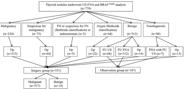
had not undergone surgery (n = 66), follow-up US showed increase in size in nodules diagnosed as benign on cytology without further cytopathologic confirmation (n = 3), and missing radiologic reports (n = 1). A total of 518 nodules in 479 patients were finally included in this study (Figure 1). Of 518 nodules, 331 nodules from 300 patients were confirmed pathologically (Surgery group), and 187 nodules from 182 patients were clinically observed by follow-up FNA (n = 112) or follow-up US (n = 75). Mean period of follow-up US was 14.7 months. Two patients had two nodules each, of which one were pathologically confirmed after surgery and the other underwent observation. One patient had three nodules of which two were pathologically confirmed and the other underwent observation.
US Analysis
For evaluation of the thyroid glands and cervical lymph nodes, a 5–12 MHz linear probe (iU22, Philips Medical Systems, Bothell, WA) or a 6–13 MHz linear probe (EUB-7500, Hitachi Medical, Tokyo, Japan) were used. Compound imaging was used in all images from the iU22 machine. Seven board-certified radiologists specialized in thyroid imaging with 1–15 years of experiences performed US and subsequent US-FNA.
US features of all thyroid nodules which had undergone US-FNA were prospectively recorded according to the internal component, echogenicity, margin, calcification, and shape at the time of US examination. Malignant US features were markedly hypoechogenicity, irregular or microlobulated margin, presence of microcalcifications, and taller than wide shape [16]. Thyroid nodules showing one or more suspicious US features described above were assessed as suspicious malignant, while those without any suspicious US features were assessed as probably benign [16]. Size of thyroid nodule was also recorded measuring the longest diameter.
US-FNA and Cytologic Analyses
US-FNA was done by the same radiologist who performed US. Fine-needle aspiration was performed on the nodules showing suspicious features, and if none showed any suspicious US features, FNA was performed targeting at the nodule with the largest size. Each nodule was aspirated at least twice using freehand technique with a 23-gauge needle attached to 2-mL disposable plastic syringe. Obtained samples were expelled on to glass slides, smeared and placed immediately into 95% alcohol for Papanico-laou staining. One of the five cytopathologists specializing in thyroid cytology interpreted the smeared samples. Cytopatholo-gists were not present during the biopsy procedures, but special staining was performed if needed. Cytology reports of June 2009 to November 2009 were based on the following 5 categories; 1) benign, 2) indeterminate cytology (follicular neoplasm or Hurthle cell neoplasm), 3) suspicious for papillary thyroid cancer, 4) malignant and 5) inadequate [9]. After December 2009 to the present, the Bethesda classifications are used in the cytology reports of thyroid aspiration studies [17].
BRAFV600Emutation analysis was performed with the remain-ing material in the syrremain-inge used in aspiration. Remainremain-ing material was rinsed in 1 mL of normal saline, and was subjected to the BRAFV600Emutation analysis.
Dual Priming Oligonucleotide-based Multiplex Polymerase Chain Reaction (DPO-PCR)
BRAFV600Emutation analysis using the DPO-PCR technology
was performed using the Seeplex BRAFACE detection system (Seegene, Seoul, Korea) as described previously [13,14].
Data and Statistical Analysis
We used cytopathological results as the ‘‘gold standard’’, pathologically confirmed malignancies classified into the positive group, and pathologically confirmed or clinical benign nodules
Figure 1. Diagram of study population.A total of 518 nodules in 479 patients were finally included in this study, and 331 nodules from 300 patients were confirmed pathologically, and 187 nodules from 182 patients were clinically observed by follow-up FNA (n = 112) or follow-up US (n = 75). Abbreviations: US-FNA, ultrasound-guided fine-needle aspiration; FN, follicular neoplasm; Op, operation; FU US, follow-up ultrasound; FU FNA, follow up fine-needle aspiration; FNA, fine-needle aspiration.
classified into the negative group. Categorical data was summa-rized using frequencies and percentages. x2 test was used in analysis of categorical variables, while independent two-sample t test was used in continuous variables.
The cytologic grouping and management was based on whether the recommendation was follow-up (clinical follow-up or repeat aspiration) or surgery, according to the Bethesda classifications [17]. Malignancy, suspicious for malignancy, indeterminate results from the study period before December 2009, suspicious for follicular neoplasm or follicular neoplasm on cytology results were included in the ‘‘positive’’ cytology group. The malignant
cytologic diagnoses were considered positive cytology when calculating diagnostic values of FNA. To calculate diagnostic performances of FNA, we compared the results to the ‘‘gold standard’’. True-positives (TP) were defined as nodules with ‘‘positive’’ cytology and a corresponding ‘‘positive’’ gold standard result. True-negatives (TN) had both ‘‘negative’’ cytology and gold standard one. False-negatives (FN) were defined as nodules with ‘‘negative’’ cytology and ‘‘positive’’ gold standard one. False-positives (FP) had positive cytology and ‘‘negative’’ gold standard one. To calculate diagnostic performances of FNA with BRAFV600Emutation analysis, a nodule was considered ‘‘positive’’ group when either FNA or BRAFV600Emutation was positive. The following statistical values were calculated as: Sensitivity = TP/ (TP+FN) 6 100; specificity = TN/(TN+FP) 6 100; positive predictive value = TP/(TP+FP) 6 100; negative predictive va-lue = TN/(TN+FN) 6 100; accuracy = (TP+TN)/ (TP+TN+FP+FN)6100. We calculated diagnostic performances of FNA and FNA with BRAFV600Emutation analysis for detecting
malignancy according to US features and size of the nodule. We also compared the diagnostic performances of FNA and FNA with BRAFV600Emutation analysis for detecting malignancy according to US features and size, using logistic regression with generalized estimating equation method.
Analysis was performed using SAS software (version 9.1.3; SAS Institute, Cary, NC). Statistical significance was assumed when the Pvalue was less than 0.05. All reportedPvalues are 2-sided.
Results
Patient and Nodule Characteristics
Of the total 518 FNA with additional BRAFV600E mutation analysis performed, 317 (61.2%) nodules were confirmed as malignancy, and the remaining 201 (38.8%) as benign (Table 1).
The mean age of patients was 48.6611.7 years. Size of the nodules ranged from 2 mm to 52 mm (mean 10.567.3 mm). Mean age of the malignant group (46.1611 years) was younger than the benign group (52.7611.6 years), showing statistical significance (P,0.001). Mean size of the benign nodules was 12.568.5 mm, which was larger than the malignant nodules (9.266.1 mm) with statistical significance (P,0.001).
Of the 317 nodules confirmed as malignant, 250 (78.9%) showed BRAFV600E mutation. Among the 201 benign nodules, three (1.5%) showed BRAFV600E mutation, two of which were pathologically confirmed as benign (adenomatous hyperplasia) by surgery, and one had undergone follow-up US for over a year with no interval change of size or characteristic.
Analyses of All 518 Thyroid Nodules
Table 2 summarizes the diagnostic performances of FNA and FNA with BRAFV600E mutation analysis. In all 518 thyroid nodules, additional BRAFV600E mutation analysis improved sensitivity of FNA alone from 67.2% to 78.9% (P,0.001), accuracy from 79.9% to 86.5% (P,0.001), and negative predictive value from 65.9% to 74.7% (P,0.001). Specificity and positive predictive value did not show statistically significant differences between FNA and FNA with BRAFV600Emutation analysis.
Of the 386 nodules with suspicious US features, sensitivity, accuracy, and negative predictive value were higher in FNA with BRAFV600Emutation analysis compared to FNA alone, 80.8% to 68.7% (P,0.001), 85% to 75.9% (P,0.001), and 60.7% to 48.9% (P,0.001), respectively, showing statistical significance. Specificity and positive predictive value did not show statistically significant differences between FNA and FNA with BRAFV600E mutation analysis. In the 132 nodules without any suspicious US features, none of diagnostic performances showed statistically significant improvement with BRAFV600Emutation analysis.
Analyses of the 175 Nodules Larger than 10 mm Sensitivity, negative predictive value, and accuracy of FNA with BRAFV600Emutation analysis were higher than FNA alone (84.1% to 75.6%, 87.5% to 82.3%, and 91.4% to 88.6%) with statistical significance in the 175 thyroid nodules larger than 10 mm. Specificity and positive predictive value did not show statistically significant differences between FNA and FNA with BRAFV600E mutation analysis.
Similar results were observed in the 99 nodules larger than 10 mm showing suspicious US features. Sensitivity, accuracy, and Table 1.Cytological diagnoses of 518 nodules according to initial fine-needle aspiration results.*
Cytological diagnoses Malignant Benign
Total BRAFV600Emutation Total BRAFV600Emutation
Positive Negative Positive Negative
Non-diagnostic (n = 20) 13/20 (65) 1/13 (7.7) 12/13 (92.3) 7/20 (35) 0/7 (0) 7/7 (100)
Benign (n = 194) 10/194 (5.2) 4/10 (40) 6/10 (60) 184/194 (94.8) 2/184 (1.1) 182/184 (98.9) Atypia (n = 22) 16/22 (72.7) 5/16 (31.3) 11/16 (68.8) 6/22 (27.3) 1/6 (16.7) 5/6 (83.3)
Follicular neoplasm or suspicious for follicular neoplasm (n = 5)
1/5 (20) 0/1 (0) 1/1 (100) 4/5 (80) 0/4 (0) 4/4 (100)
Suspicious for malignancy (n = 64) 64/64 (100) 27/64 (42.2) 37/64 (57.8) 0/64 (0)
Malignant (n = 213) 213/213 (100) 139/213 (65.3) 74/213 (34.7) 0/213 (0)
Total 317 201
negative predictive value were improved in FNA with BRAFV600E
mutation analysis compared to FNA, 89.3% to 81.3% (P =0.012), 91.9% to 85.9% (P =0.013), and 75% to 63.2% (P =0.016), respectively. Specificity and positive predictive value show same values between FNA and FNA with BRAFV600Emutation analysis. Of the 76 nodules larger than 10 mm without any suspicious US features, all values did not show statistically significant differences between FNA and FNA with BRAFV600Emutation analysis.
Analyses of the 343 Nodules Equal to or Smaller than 10 mm
Of the 343 nodules equal to or smaller than 10 mm, additional BRAFV600E mutation analysis showed improvement to FNA in sensitivity (77% to 64.3%) and negative predictive value (66.5% to 56.3%), with statistical significance. Specificity, accuracy, and positive predictive value were not improved with additional BRAFV600Emutation analysis, without statistical significance.
Diagnostic performances according to US features were analyzed among the 343 nodules. Among them, 287 were assessed Table 2.Diagnostic performances of FNA and FNA with additional BRAFV600Emutation analysis in the detection of malignancy according to US features and size of the nodules.*.
Sensitivity (%) Specificity (%) Accuracy (%) PPV (%) NPV (%) Overall (n = 518) Total (n = 518)
FNA 67.2 (213/317) 100 (201/201) 79.9 (414/518) 100 (213/213) 65.9 (201/305)
FNA with BRAFV600Emutation 78.9 (250/317) 98.5 (198/201) 86.5 (448/518) 98.8 (250/253) 74.7 (198/265)
Pvalue{
,0.001 0.081 ,0.001 0.081 ,0.001
Suspicious US feature (n = 386)
FNA 68.7 (204/297) 100 (89/89) 75.9 (293/386) 100 (204/204) 48.9 (89/182)
FNA with BRAFV600Emutation 80.8 (240/297) 98.9 (88/89) 85 (328/386) 99.6 (240/241) 60.7 (88/145)
Pvalue ,0.001 0.315 ,0.001 0.316 ,0.001
Without suspicious US feature (n = 132)
FNA 45 (9/20) 100 (112/112) 91.7 (121/132) 100 (9/9) 91.1 (112/123) FNA with BRAFV600Emutation 50 (10/20) 98.2 (110/112) 90.9 (120/132) 83.3 (10/12) 91.7 (110/120)
Pvalue 0.306 0.154 0.563 0.121 0.422
.10 mm (n = 175) Total (n = 175)
FNA 75.6 (62/82) 100 (93/93) 88.6 (155/175) 100 (62/62) 82.3 (93/113) FNA with BRAFV600Emutation 84.1 (69/82) 97.8 (91/93) 91.4 (160/175) 97.2 (69/71) 87.5 (91/104)
Pvalue 0.006 0.153 ,0.001 0.151 0.013
Suspicious US feature (n = 99)
FNA 81.3 (61/75) 100 (24/24) 85.9 (85/99) 100 (61/61) 63.2 (24/38)
FNA with BRAFV600Emutation 89.3 (67/75) 100 (24/24) 91.9 (91/99) 100 (67/67) 75 (24/32)
Pvalue 0.012 – 0.013 – 0.016
Without suspicious US feature (n = 76)
FNA 14.3 (1/7) 100 (69/69) 92.1 (70/76) 100 (1/1) 92 (69/75)
FNA with BRAFV600Emutation 28.6 (2/7) 97.1 (67/69) 90.8 (69/76) 50 (2/4) 93.1 (67/72)
Pvalue 0.295 0.151 0.563 0.046 0.405
#10 mm (n = 343) Total (n = 343)
FNA 64.3 (151/235) 100 (108/108) 75.5 (259/343) 100 (151/151) 56.3 (108/192) FNA with BRAFV600Emutation 77 (181/235) 99.1 (107/108) 84 (288/343) 99.5 (181/182) 66.5 (107/161)
Pvalue ,0.001 0.315 0.094 0.316 ,0.001
Suspicious US feature (n = 287)
FNA 64.4 (143/222) 100 (65/65) 72.5 (208/287) 100 (143/143) 45.1 (65/144)
FNA with BRAFV600Emutation 77.9 (173/222) 98.5 (64/65) 82.6 (237/287) 99.4 (173/174) 56.6 (64/113)
Pvalue ,0.001 0.314 ,0.001 0.316 ,0.001
Without suspicious US feature (n = 56)
FNA 61.5 (8/13) 100 (43/43) 91.1 (51/56) 100 (8/8) 89.6 (43/48)
FNA with BRAFV600Emutation 61.5 (8/13) 100 (43/43) 91.1 (51/56) 100 (8/8) 89.6 (43/48)
Pvalue – – – – –
Abbreviations: FNA, fine-needle aspiration; US, ultrasound; PPV, positive predictive value; NPV, negative predictive value. *Data presented in parentheses are number of nodules.
{
as suspicious malignant, and the remaining 56 as probably benign. Sensitivity, accuracy, and negative predictive value were signifi-cantly improved in FNA with BRAFV600E mutation analysis compared to FNA alone in the 287 nodules with suspicious US features, 77.9% to 64.4% (P,0.001), 82.6% to 72.5% (P,0.001), and 56.6% to 45.1% (P,0.001), respectively. Diagnostic perfor-mances showed similar values when comparing FNA to FNA with BRAFV600Emutation analysis in the 56 thyroid nodules without any suspicious US features.
Discussion
To the present date, FNA has shown acceptable diagnostic performances in the diagnosis of malignancy in thyroid nodules [18–20]. There are several limitations of FNA, however, such as false-negative and non-diagnostic results [20]. Many studies regarding molecular studies have been conducted to overcome these diagnostic limitations of FNA [11,21,22]. There have been several genetic abnormalities associated with thyroid carcinoma including point mutations such as those in the RAS and BRAF genes, and chromosomal rearrangements such as RET/PTC and PAX8/PPARc [2]. Papillary carcinoma, the most common thyroid malignancy, harbors BRAFV600E, RET/PTC rearrange-ment, or the frequently found RAS mutations [23]. RAS genes and PAX8/PPARc rearrangement are found more in follicular carcinomas [3,23]. A recent study further demonstrated that BRAFK601E was associated with the follicular variant type of papillary thyroid carcinoma [24]. Among them, BRAFV600E mutation analysis showed good performances. However, when considering its cost-effectiveness, it is unclear whether if BRAFV600E mutation analysis should routinely accompany US-FNA in the diagnosis of malignancy in patients with thyroid nodules. A proper indication for an adjunctive BRAFV600E
mutation analysis is required. Recent studies demonstrated that reflex molecular testing including the BRAFV600E mutation can offer significant improvement in the preoperative diagnosis of thyroid cancer, especially in those showing indeterminate cytologic results including follicular lesion with atypia of uncertain significance, and suspicious for papillary carcinoma [25,26]. Unfortunately, reflex molecular testing cannot always be adapted in all institutions, therefore, supporting the need for a proper guideline for the BRAFV600Emutation analysis.
Several studies regarding proper indications for the additional BRAFV600E mutation analysis demonstrated that this was more helpful when applied to nodules with suspicious features on US [4,9,11,12], and at the time of initial US-FNA [9]. Also, the size of papillary thyroid cancer may affect the diagnostic performance of BRAFV600E mutation analysis, when regarding the different prevalence of BRAFV600E mutation in papillary thyroid
micro-carcinoma and papillary thyroid cancer larger than 10 mm [8,13– 15]. In this study, we investigated the diagnostic performance of FNA and FNA with BRAFV600Emutation analysis to evaluate a
proper indication for the BRAFV600Emutation analysis, consid-ering the size and US features of the thyroid nodules. Results of our study show that the prevalence of BRAFV600Emutation was higher in papillary thyroid cancer (51/82, 62.2%) than papillary thyroid microcarcinoma (125/235, 53.2%). Sensitivity, accuracy, and negative predictive value of FNA with BRAFV600Emutation analysis were significantly higher than those of FNA alone in thyroid nodules with suspicious US features, regardless of its size. All diagnostic performances of FNA with BRAFV600E mutation analysis did not show significant differences compared to FNA alone in nodules without any suspicious US features, regardless of its size.
In the previous studies regarding the diagnostic performances of BRAFV600E mutation analysis, specificity was reported to be almost 100% [12], but several false-positive cases were reported in Korea [7,8,10]. These false-positive cases reported in literature are thought to be caused by applying highly sensitive DPO-PCR or pyrosequencing analysis. These techniques focus on improving diagnostic sensitivity, which may result in false-positive cases [7]. In this study, three cases showed false-positive results among the 201 benign nodules; two of which were pathologically confirmed as benign by surgery, and one had undergone follow-up US for over one year. To reach 100% specificity to detect BRAFV600E mutation at pyrosequencing, cut-off values were refitted to scarify sensitivity [7]. Further studies are required to evaluate the false positive results of BRAFV600Emutation and consensus also should be needed to interpret and apply the results in patients with thyroid nodules.
There are several potential limitations to this study. First, the nodules which had not undergone surgery were included, based on the cytology results. This may have affected our results in ways of false-negative or false-positive cytologic results [27,28]. Second, we divided groups based on the presence of suspicious US features. However, interobserver variability among radiologists in inter-preting US images may have affected the results [29–31], which also may not be reproducible in other institutions. Third, sample size was different in thyroid nodules when grouped into those larger or equal to or smaller than 10 mm, which may have affected the results.
Conclusion
The BRAFV600E mutation analysis was a useful adjunctive diagnostic tool in the diagnosis of thyroid nodules with suspicious US features regardless of the size.
Author Contributions
Conceived and designed the experiments: J. Kwak. Performed the experiments: J. Koh J. Kwak JRC. Analyzed the data: J. Koh J. Kwak KHH. Contributed reagents/materials/analysis tools: EKK HJM JHY. Wrote the paper: J. Koh.
References
1. Xing M (2007) BRAF mutation in papillary thyroid cancer: pathogenic role, molecular bases, and clinical implications. Endocr Rev 28: 742–762. 2. Nikiforov YE, Nikiforova MN (2011) Molecular genetics and diagnosis of
thyroid cancer. Nat Rev Endocrinol 7: 569–580.
3. Nikiforov YE, Ohori NP, Hodak SP, Carty SE, LeBeau SO, et al. (2011) Impact of mutational testing on the diagnosis and management of patients with cytologically indeterminate thyroid nodules: a prospective analysis of 1056 FNA samples. J Clin Endocrinol Metab 96: 3390–3397.
4. Hwang J, Shin JH, Han BK, Ko EY, Kang SS, et al. (2010) Papillary thyroid carcinoma with BRAFV600E mutation: sonographic prediction. AJR Am J Roentgenol 194: W425–430.
5. Lee ST, Kim SW, Ki CS, Jang JH, Shin JH, et al. (2012) Clinical implication of highly sensitive detection of the BRAF V600E mutation in fine-needle
aspirations of thyroid nodules: a comparative analysis of three molecular assays in 4585 consecutive cases in a BRAF V600E mutation-prevalent area. J Clin Endocrinol Metab 97: 2299–2306.
6. Ahn D, Park JS, Sohn JH, Kim JH, Park SK, et al. (2012) BRAFV600E mutation does not serve as a prognostic factor in Korean patients with papillary thyroid carcinoma. Auris Nasus Larynx 39: 198–203.
7. Yeo MK, Liang ZL, Oh T, Moon Y, An S, et al. (2011) Pyrosequencing cut-off value identifying BRAF(V600E) mutation in fine needle aspiration samples of thyroid nodules. Clin Endocrinol (Oxf) 75: 555–560.
9. Moon HJ, Kim EK, Chung WY, Choi JR, Yoon JH, et al. (2011) Diagnostic value of BRAF(V600E) mutation analysis of thyroid nodules according to ultrasonographic features and the time of aspiration. Ann Surg Oncol 18: 792– 799.
10. Kim SW, Lee JI, Kim JW, Ki CS, Oh YL, et al. (2010) BRAFV600E mutation analysis in fine-needle aspiration cytology specimens for evaluation of thyroid nodule: a large series in a BRAFV600E-prevalent population. J Clin Endocrinol Metab 95: 3693–3700.
11. Lee EJ, Song KH, Kim DL, Jang YM, Hwang TS, et al. (2011) The BRAF(V600E) mutation is associated with malignant ultrasonographic features in thyroid nodules. Clin Endocrinol (Oxf) 75: 844–850.
12. Nam SY, Han BK, Ko EY, Kang SS, Hahn SY, et al. (2010) BRAF V600E mutation analysis of thyroid nodules needle aspirates in relation to their ultrasongraphic classification: a potential guide for selection of samples for molecular analysis. Thyroid 20: 273–279.
13. Kwak JY, Kim EK, Chung WY, Moon HJ, Kim MJ, et al. (2009) Association of BRAFV600E mutation with poor clinical prognostic factors and US features in Korean patients with papillary thyroid microcarcinoma. Radiology 253: 854– 860.
14. Kwak JY, Kim EK, Kim JK, Han JH, Hong SW, et al. (2010) Dual priming oligonucleotide-based multiplex PCR analysis for detection of BRAFV600E mutation in FNAB samples of thyroid nodules in BRAFV600E mutation-prevalent area. Head Neck 32: 490–498.
15. Kim TY, Kim WB, Rhee YS, Song JY, Kim JM, et al. (2006) The BRAF mutation is useful for prediction of clinical recurrence in low-risk patients with conventional papillary thyroid carcinoma. Clin Endocrinol (Oxf) 65: 364–368. 16. Kim EK, Park CS, Chung WY, Oh KK, Kim DI, et al. (2002) New sonographic criteria for recommending fine-needle aspiration biopsy of nonpalpable solid nodules of the thyroid. AJR Am J Roentgenol 178: 687–691.
17. Cibas ES, Ali SZ (2009) The Bethesda System for Reporting Thyroid Cytopathology. Thyroid 19: 1159–1165.
18. Cooper DS, Doherty GM, Haugen BR, Kloos RT, Lee SL, et al. (2009) Revised American Thyroid Association management guidelines for patients with thyroid nodules and differentiated thyroid cancer. Thyroid 19: 1167–1214.
19. Lee MJ, Hong SW, Chung WY, Kwak JY, Kim MJ, et al. (2011) Cytological results of ultrasound-guided fine-needle aspiration cytology for thyroid nodules: emphasis on correlation with sonographic findings. Yonsei Med J 52: 838–844.
20. Wang CC, Friedman L, Kennedy GC, Wang H, Kebebew E, et al. (2011) A large multicenter correlation study of thyroid nodule cytopathology and histopathology. Thyroid 21: 243–251.
21. Hamfjord J, Stangeland AM, Skrede ML, Tveit KM, Ikdahl T, et al. (2011) Wobble-enhanced ARMS method for detection of KRAS and BRAF mutations. Diagn Mol Pathol 20: 158–165.
22. Tonacchera M, Agretti P, Rago T, De Marco G, Niccolai F, et al. (2012) Genetic markers to discriminate benign and malignant thyroid nodules with undetermined cytology in an area of borderline iodine deficiency. J Endocrinol Invest 35: 754–759.
23. Nikiforov YE, Steward DL, Robinson-Smith TM, Haugen BR, Klopper JP, et al. (2009) Molecular testing for mutations in improving the fine-needle aspiration diagnosis of thyroid nodules. J Clin Endocrinol Metab 94: 2092–2098. 24. Ohori NP, Singhal R, Nikiforova MN, Yip L, Schoedel KE, et al. (2012) BRAF
mutation detection in indeterminate thyroid cytology specimens: Underlying cytologic, molecular, and pathologic characteristics of papillary thyroid carcinoma. Cancer Cytopathol.
25. Adeniran AJ, Theoharis C, Hui P, Prasad ML, Hammers L, et al. (2011) Reflex BRAF testing in thyroid fine-needle aspiration biopsy with equivocal and positive interpretation: a prospective study. Thyroid 21: 717–723.
26. Hassell LA, Gillies EM, Dunn ST (2012) Cytologic and molecular diagnosis of thyroid cancers: is it time for routine reflex testing? Cancer Cytopathol 120: 7– 17.
27. Kwak JY, Koo H, Youk JH, Kim MJ, Moon HJ, et al. (2010) Value of US correlation of a thyroid nodule with initially benign cytologic results. Radiology 254: 292–300.
28. Gharib H, Goellner JR (1993) Fine-needle aspiration biopsy of the thyroid: an appraisal. Ann Intern Med 118: 282–289.
29. Park SH, Kim SJ, Kim EK, Kim MJ, Son EJ, et al. (2009) Interobserver agreement in assessing the sonographic and elastographic features of malignant thyroid nodules. AJR Am J Roentgenol 193: W416–423.
30. Kim SH, Park CS, Jung SL, Kang BJ, Kim JY, et al. (2010) Observer Variability and the Performance between Faculties and Residents: US Criteria for Benign and Malignant Thyroid Nodules. Korean J Radiol 11: 149–155.