Orthodontic management of bilateral maxillary
canine-first premolar transposition and bilateral agenesis of
maxillary lateral incisors: a case report
Elena Di Palma1, Biagio Di Giuseppe2, Michele Tepedino3, Claudio Chimenti4
Introduction: Maxillary canine-first premolar transposition (Mx.C.P1) is an uncommon dental positional anomaly that may create many orthodontic problems from both esthetic and functional points of view. Objective: In this report we show the orthodontic management of a case of Mx.C.P1 associated with bilateral maxillary lateral incisor agenesis and unilateral mandibular second premolar agenesis Methods: The patient was treated with a multibracket appliance and the extraction of the lower premolar. Results: treatment was completed without the need for any prosthetic replacement.
Keywords:Cuspid. Bicuspid. Ectopic tooth eruption. Corrective Orthodontics.
How to cite this article: Di Palma E, Di Giuseppe B, Tepedino M, Chimenti C. Orthodontic management of bilateral maxillary canine-first premolar transposi-tion and bilateral agenesis of maxillary lateral incisors: a case report. Dental Press J Orthod. 2015 Mar-Apr;20(2):100-9. DOI: http://dx.doi.org/10.1590/2176-9451.20.2.100-109.oar
Submitted: October 11, 2013 - Revised and accepted: June 03, 2014
» The authors report no commercial, proprietary or financial interest in the prod-ucts or companies described in this article.
» Patients displayed in this article previously approved the use of their facial and intraoral photographs.
Contact address: Elena Di Palma E-mail: [email protected]
1 PhD, University of L’Aquila, Department of Applied Clinical Sciences and
Biotechnology, L’Aquila, Italy.
2 Private practice, Coppito, L’Aquila, Italy.
3DDS, University of L’Aquila, Department of Applied Clinical Sciences and
Biotechnology, L’Aquila, Italy.
4 Professor, University of L’Aquila, Department of Applied Clinical Sciences and
Biotechnology, L’Aquila, Italy.
DOI: http://dx.doi.org/10.1590/2176-9451.20.2.100-109.oar
Introdução: a transposição entre canino e primeiro pré-molar superiores (Mx.C.P1) é uma anomalia de posição dentária rara, que pode causar muitos problemas ortodônticos, não só com relação à estética, mas também com relação à função do paciente.
Objetivo: no presente artigo, relatamos o manejo ortodôntico de um caso de Mx.C.P1 associada à agenesia bilateral de incisivos laterais superiores e agenesia unilateral do segundo pré-molar inferior.
Métodos: o paciente foi tratado com aparelho ortodôntico fixo e extração do pré-molar inferior.
Resultados: o tratamento foi concluído sem a necessidade de reposição protética.
INTRODUCTION
Dental transposition is an uncommon dental anomaly
involving a positional interchange of two teeth.1
Recent meta-analysis2 underlined that tooth
trans-position is a rare phenomenon (0.33%) with various, sometimes inexplicable, forms of manifestation and that its occurrence seems to have no speciic sex pre-dilection, but some maxillary predisposition is noted. Unilateral occurrence is considerably higher than the bilateral, but no let or right-side predilection in the maxilla or mandible has been evident. In contrast, other authors found that tooth transposition occurred more
frequently in the maxillary let side.1,3
The most common form of transposition is between
maxillary canine and irst premolar (Mx.C.P1).4
Dental transposition represents a multifactorial con-dition, both genetic1,5-10 and environmental1,3,11,12,13
fac-tors seem to be involved in the etiology of transposition.
A recent study conducted by Ely et al6 underlined
that large-scale population-based studies will be re-quired to further reine our understanding of the genet-ics of this anomaly.
Although in the literature there are several reports of maxillary canine and irst premolar transpositions solved
with correction of the transposition,14-17 this would not
always be advisable from a cost-beneit point of view.17
In fact, when the teeth involved in the transposition are fully erupted and completely or almost completely aligned in the transposed position, a satisfactory result can be obtained by maintaining the transposition.18-21 In this context, iatrogenic
damage to teeth and periodontal tissues can be avoided. In this report, it is shown the orthodontic manage-ment of a case of bilateral maxillary canine-irst premolar transposition (Mx.C.P1) associated with bilateral max-illary lateral incisor agenesis and unilateral mandibular second premolar agenesis.
CASE REPORT AND DIAGNOSIS
The patient came to our observation for the irst time at the age of 7 years and 6 months old (Figs 1 and 2). Ater that, she was treated for 2 years by another ortho-dontist; and later she decided to refer to us again, at the age of 10. Pre-treatment records (Figs 3-7) were taken, with previous appliances worn.
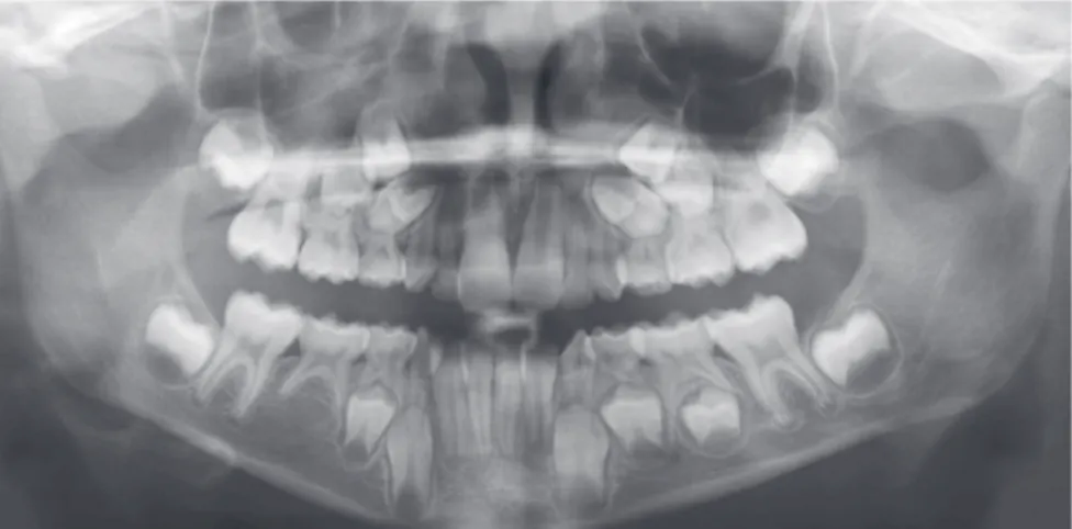
Figure 2 - Initial panoramic radiograph showing bilateral maxillary permanent lateral incisors and second right lower premolar agenesis, and initial bilateral transposition of upper canines and first premolars.
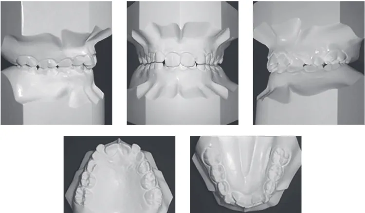
Figure 3 - Pre-treatment intraoral views.
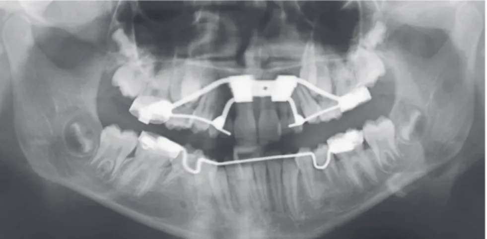
Figure 5 - Pre-treatment panoramic radiograph taken the very moment the patient came to us for the second time. The patient was wearing a lin-gual arch and a rapid palatal expander. The radio-graph shows bilateral maxillary permanent lateral incisors and second right lower premolar agene-sis, bilateral upper lateral incisors and right second molar deciduous persistence, complete bilateral transposition of upper canines and first premolars, normal periodontal support and healthy bone.
Figure 6 - Pre-treatment cephalogram. Figure 7 - Pre-treatment cephalometric tracings.
Analysis of complete diagnostic records revealed Class II division 2 malocclusion, a lat proile with bimaxillary re-trusion, the mandibular arch with moderate crowding and the retention of primary right second molar. In the max-illary arch, there was retention of primary lateral incisors and the right and let canine were erupting between irst and second premolars (Fig 3). The patient also presented regular oral hygiene and healthy periodontal tissues.
She showed a straight proile with bimaxillary retru-sion, symmetrical frontal view and normal anterior fa-cial height (Fig 4).
A panoramic radiograph showed bilateral maxillary permanent lateral incisors and mandibular second pre-molar agenesis, in addition to the bilateral transposition of canines and irst premolars (Fig 5).
Cephalometric analysis (Figs 6 and 7) did not reveal any notable deviation in the skeletal and dental patterns, as shown in Table 1: skeletal Class I relationship, hori-zontal growth tendency and lingual inclination of man-dibular incisors.
TREATMENT
Problems list
» Agenesis of left and right maxillary lateral incisors and right mandibular second premolar.
» Transposition of right and left maxillary canine. » Lingual tipping of mandibular incisors.
» Moderate crowding.
Treatment options
This case can be solved in diferent ways:
1) Considering patient’s straight proile, it would be better to maintain the spaces of lateral incisors; this treatment option requires distalization of maxillary mo-lars to correct the Class II molar relationship and to gain the spaces needed to place endosseous dental implants. Regarding the transposition:
(1a) The ideal treatment would be to correct trans-position due to functional problems related to the pres-ence of the palatal cusp of the irst premolar. The disad-vantages of this approach included a long treatment pe-riod and the risk of root resorption, loss of pulp vitality or loss of hard and sot tissues of adjacent teeth.
(1b) Leaving the transposition has some disadvan-tages related to diferences in size, shape, and tooth color between canine and premolar, which can some-times cause esthetic problems. The gingival contour of the premolar is lower in respect to the canine, and this may require a periodontal recontouring procedure. However, even if these esthetic problems are overcome, the palatal cusp of the transposed premolar might cause functional interference, despite the control of its angu-lations, torques, and even ater coronal reshaping. Pros-thetic restoration ater pulpectomy will also be neces-sary, if the size and shape of premolar are completely re-contoured, in order to make it more similar to a canine. In both cases, the space for an endosseous implant, in position 4.5 in the lower arch, must be kept.
2) The second choice, accepted by the patient and the parents, was not to correct the transposition and to
move the maxillary irst premolars into the spaces of lat-eral incisors. The disadvantages of this approach were esthetics and included the diferent color, shape and gin-gival contour of premolars in comparison to lateral inci-sors. Also, a balancing interference can occur between the palatal cusp of the premolar and the mandibular ca-nine, thus occlusal balance is oten required in order to
improve function.18
An accurate diagnostic and interdisciplinary approach is necessary to obtain improved, conservative and predictable esthetic results in an extremely estheti-cal area, such as the anterior maxillary dentition.
Treatment plan
Treatment objectives were (1) in the mandibular arch, extract the second right primary molar and the let premolar to balance the number of upper and low-er teeth and to establish a correct Class I molar rela-tionship; (2) in the maxillary arch, keep the complete bilateral transposition and replace missing maxillary lateral incisors by moving premolars mesially using a multibracket appliance; (3) establish a Class I molar and canines relationships, maintaining an ideal overjet and overbite; (4) correct lingual inclination of mandibular incisors, while maintaining the actual position of max-illary incisors; (5) maintain upper irst premolars in an ideal position to obtain good conservative and esthetic restoration; (6) maintain facial balance.
Treatment progress
In the initial phases of treatment, in the maxillary arch, 0.018-inch stainless steel sectional archwires from irst molars to irst premolars were used, and open coil springs were positioned between irst and second pre-molars to facilitate eruption of canines. Lingual arch was not removed from the lower arch (Fig 3).
When maxillary canines were completely erupted, all maxillary and mandibular teeth were bonded with a multibracket appliance ater removal of upper and low-er primary teeth and let mandibular second premolar. On mandibular irst molars, composite shims were po-sitioned to avoid interferences in occlusion. During this phase of treatment, maxillary and mandibular 0.014-inch superelastic nickel-titanium archwires were used.
In the inal phase of treatment, 0.019 x 0.025-inch stain-less steel archwires were used (Fig 8) and a panoramic ra-diograph was taken to assess correct root parallelism (Fig 9). Variables
Pre-treatment Post-treatment
(10 years and
0 months old)
(13 years and
2 months old)
SNA (degree) 84.09 84.01
SNB (degree) 82.17 83.39
ANB (degree) 1.92 0.62
GoGn/Sn (degree) 28.85 24.23
MP/FHP (degree) 19.89 15.73
PP/MP (degree) 13.58 21.26
L1 to MP (degree) 86.52 95.68
U1 to PP (degree) 108.16 108.96
Table 1 - Summary of cephalometric analysis (MP= mandibular plane;
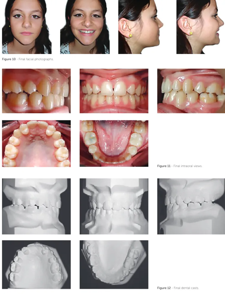
Ater 30 months of active treatment, the ixed appli-ance was removed; maxillary and mandibular removable contentions were placed for retention. Final radiograph-ic and photographradiograph-ic records were taken (Figs 10-14) and an end-treatment cephalometric analysis was performed (Fig 15) in order to check whether treatment objectives were achieved.
Treatment results
Crowding of the lower arch was corrected, a Class I molar and canine relationship was obtained as well as a good overjet and overbite (Figs 10 and 11). Lin-gual inclination of lower incisors was corrected (L1 to MP angle increased from 86.52° to 95.68°), the initial
position of upper incisors (U1 to PP angle increased from 108.16° to 108.96°) and facial balance was main-tained, as can be seen in Table 1, post-treatment ceph-alometric tracings (Fig 15) and extraoral photographs (Fig 10). Good root parallelism was achieved (Fig 13). Upper irst premolars are well positioned and with good conservative and esthetical restoration. A beauti-ful and functional result will be achieved.
DISCUSSION AND CONCLUSION
In several studies, it has been reported that trans-posed teeth are associated with dental anomalies, such as peg-shaped and congenitally missing teeth; in par-ticular, a high incidence of congenitally missing teeth
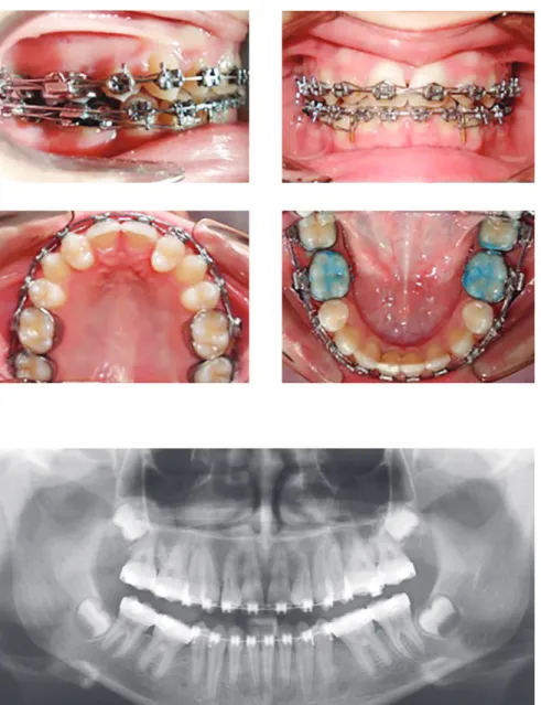
Figure 8 - In treatment intraoral views.
Figure 10 - Final facial photographs.
Figure 11 - Final intraoral views.
Figure 16 - Two-year follow-up facial photographs.
Figure 13 - Final panoramic radiograph.
Figure 17 - Two-year follow-up intraoral photo-graphs.
and peg-shaped lateral incisors are associated with Mx.C.P1.1,3,7,10,11,19
Several cases of Mx.C.P1 reported in literature are
solved with the correction of transposition;14-17,22
how-ever, this approach requires longer treatment time and stability, and esthetic and function of end results are not always granted.
In the literature, there are also many cases of trans-posed teeth that have been treated without the correc-tion of transposicorrec-tion, and cases in which congenitally missing upper lateral incisors were substituted with the
upper irst premolar.18,19,23 Nestel and Walsh18 reported a
case of bilateral Mx.C.P1 associated with agenesis of let maxillary lateral incisor, solved maintaining the trans-position in the let side and moving the premolar into the space of the missing incisor. The authors achieved
good esthetic and functional results. Parker23 reported
a very interesting case of bilateral Mx.C.P1 associated with bilateral agenesis of maxillary lateral incisors, also treated by means of maintaining the transposition and closing the spaces. Parker provided a 35-year follow-up which demonstrated that such result could be function-ally and estheticfunction-ally stable over time.
In the present case report, the chief complain for the patient and her parents was to achieve a deinitive
solution. In fact, the decision to keep the spaces of upper lateral incisors required to temporarily replace missing incisors until inal prosthesis placement was possible. There is also the probability that any ixed prosthetic device will require periodical repair or replacement throughout patient’s lifetime.
1. Peck L, Peck S, Attia Y. Maxillary canine-irst premolar transposition, associated dental anomalies and genetic basis. Angle Orthod. 1993;63(2):99-109; discussion 110.
2. Papadopoulos MA, Chatzoudi M, Kaklamanos EG. Prevalence of tooth
transposition. A meta-analysis. Angle Orthod. 2010;80(2):275-85. 3. Shapira Y, Kuftinec MM. Maxillary tooth transpositions: characteristic
features and accompanying dental anomalies. Am J Orthod Dentofacial Orthop. 2001;119(2):127-34.
4. Peck L, Peck S. Classiication of maxillary tooth transpositions. Am J Orthod Dentofacial Orthop. 1995;107(5):505-17.
5. Camilleri S. Maxillary canine anomalies and tooth agenesis. Eur J Orthod. 2005;27(5):450-6. Epub 2005 Aug 10.
6. Ely NJ, Sherrif M, Cobourne MT. Dental transposition as a disorder of genetic origin. Eur J Orthod. 2006;28(2):145-51. Epub 2005 Dec 22.
7. Peck S, Peck L, Kataja M. Concomitant occurrence of canine malposition
and tooth agenesis: evidence of orofacial genetic ields. Am J Orthod Dentofacial Orthop. 2002;122(6):657-60.
8. Chattopadhyay A, Srinivas K. Transposition of teeth and genetic etiology. Angle Orthod. 1996;66(2):147-52.
9. Shapira J, Chaushu S, Becker A. Prevalence of tooth transposition, third molar agenesis and maxillary canine impaction in individuals with Down syndrome. Angle Orthod. 2000;70(4):290-6.
10. Feichtinger CH, Rossiwall B, Wuanderrer H. Canine transposition as autosomal recessive trait in an imberg kindered, J Dent Res. 1997;56:1449-52.
11. Peck S, Peck L, Kataja M. Mandibular lateral incisor-canine transposition, concomitant dental anomalies, and genetic control. Angle Orthod. 1998;68(5):455-66.
REFERENCES
12. Dayal PK, Shodhan KH, Dave CJ. Transposition of canine with traumatic etiology. J Indian Dental Assoc. 1983;55:283-85.
13. Peck S. On the phenomenon of intraosseous migration of nonerupting teeth. Am J Orthod Dentofacial Orthop. 1998;113(5):515-7.
14. Bocchieri A, Braga G. Correction of a bilateral maxillary canine-irst premolar transposition in the late mixed dentition. Am J Orthod Dentofacial Orthop. 2002;121(2):120-8.
15. Laino A, Cacciafesta V, Martina R. Treatment of tooth impaction and transposition with a segmented-arch technique. J Clin Orthod. 2001;35(2):79-86.
16. Kuroda S, Kuroda Y. Nonextraction treatment of upper canine: premolar transposition in an adult patient. Angle Orthod. 2005;75(3):472-7. 17. Ciarlantini R, Melsen B. Maxillary tooth transposition: correct or accept?
Am J Orthod Dentofacial Orthop. 2007;132(3):385-94.
18. Nestel E, Walsh JS. Substitution of a transposed premolar for a congenitally absent lateral incisor. Am J Orthod Dentofacial Orthop. 1988;93(5):395-9. 19. Sato K, Yokozeki M, Takagi T, Moriyama K. An orthodontic case of
transposition of the upper right canine and irst premolar. Angle Orthod. 2002;72(3):275-8.
20. Shapira Y, Kuftinec MM. Tooth transpositions-a review of the literature and treatment considerations. Angle Orthod. 1989;59(4):271-6.
21. Turpin DL, Woloshyn H. Two patients with severely displaced maxillary canines respond diferently to treatment. Angle Orthod. 1995;65(1):13-22. 22. Maia FA, Maia NG. Unusual orthodontic correction of bilateral maxillary
canine: irst premolar transposition. Angle Orthod. 2005;75(2):266-76. 23. Parker WS. Transposed premolars, canines, and lateral incisors. Am J