Review Article
Versatility of Chitosan-Based Biomaterials and Their Use as
Scaffolds for Tissue Regeneration
José Carlos Viana Ribeiro,
1Rodrigo Silveira Vieira,
2Iracema Matos Melo,
1Vilana Maria Adriano Araújo,
3and Vilma Lima
31Faculty of Pharmacy, Dentistry and Nursing, Federal University of Cear´a, Rua Alexandre Bara´una, No. 949,
Rodolfo Te´oilo, 60430-160 Fortaleza, CE, Brazil
2Department of Chemical Engineering, Federal University of Cear´a, Campus do Pici, Bloco 709, Pici,
60455-760 Fortaleza, CE, Brazil
3Drug Research and Development Center, Department of Physiology and Pharmacology, Federal University of Cear´a,
Rua Coronel Nunes de Melo, No. 1000, Rodolfo Te´oilo, 60430-275 Fortaleza, CE, Brazil
Correspondence should be addressed to Vilma Lima; [email protected]
Received 23 December 2016; Revised 10 March 2017; Accepted 3 April 2017; Published 16 April 2017
Academic Editor: Ying Yang
Copyright © 2017 Jos´e Carlos Viana Ribeiro et al. his is an open access article distributed under the Creative Commons Attribution License, which permits unrestricted use, distribution, and reproduction in any medium, provided the original work is properly cited.
Chitosan is a naturally occurring polysaccharide obtained from chitin, present in abundance in the exoskeletons of crustaceans and insects. It has aroused great interest as a biomaterial for tissue engineering on account of its biocompatibility and biodegradation and its ainity for biomolecules. A signiicant number of research groups have investigated the application of chitosan as scafolds for tissue regeneration. However, there is a wide variability in terms of physicochemical characteristics of chitosan used in some studies and its combinations with other biomaterials, making it diicult to compare results and standardize its properties. he current systematic review of literature on the use of chitosan for tissue regeneration consisted of a study of 478 articles in the PubMed database, which resulted, ater applying inclusion criteria, in the selection of 61 catalogued, critically analysed works. he results demonstrated the efectiveness of chitosan-based biomaterials in 93.4% of the studies reviewed, whether or not combined with cells and growth factors, in the regeneration of various types of tissues in animals. However, the absence of clinical studies in humans, the inadequate experimental designs, and the lack of information concerning chitosan’s characteristics limit the reproducibility and relevance of studies and the clinical applicability of chitosan.
1. Introduction
Tissue engineering represents a recent area of multidisci-plinary research that applies the knowledge of materials engi-neering and biology, with the aim of reconstructing or regen-erating damaged biological tissue in clinical/pathological situations such as lesions, infections, traumas, and sequelae resulting from tumors or systemic disorders. To this end, it was sought to develop biological substitutes capable of restor-ing, maintainrestor-ing, or improving organ and tissue function. Tissue engineering approaches generally involve using a
factors, promote cell proliferation, diferentiation, and migra-tion. Biomaterials act as a scafold for the new tissue, provid-ing an environment that favors cell growth and diferentiation [1, 2].
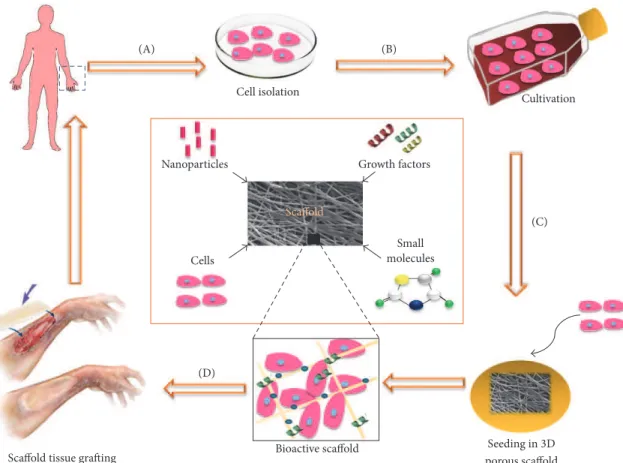
Cell isolation
Cultivation
(C)
Seeding in 3D porous scafold Growth factors
Nanoparticles
Small molecules
Scafold
Cells
(D)
Scafold tissue grafting Bioactive scafold
Figure 1: Diagram of the concept of tissue engineering. (A) Cells are isolated from humans or animals, (B) cultivated in vitro, and (C) incor-porated into a three-dimensional porous biomaterial (scafold), together with growth factors, small molecules, and/or micro/nanoparticles. (D) he bioactive scafold is then grated onto a tissue lesion, promoting its regeneration. Adapted from Dvir et al. 2011 [12] and Lanza et al. 2014 [2].
may be deined as a porous, three-dimensional, solid bioma-terial that acts as a temporary ECM during tissue regenera-tion [7]. he biomaterials used as scafolds in tissue regener-ation are of fundamental importance and may determine the success or failure of any tissue engineering approach [8, 9]. Like the ECM, scafolds must promote vascularization and provide a three-dimensional infrastructure that permits new tissue to form. Also, they must be able to act as a carrier of biologically relevant molecules and, ideally, modulate their biological responses and the regeneration process (Figure 1). hus, the use of a biomaterial as a scafold for tissue engi-neering assumes that it has certain characteristics that make it similar to the extracellular matrix [9–12].
In tissue engineering, the scafold must have a temporary structure that undergoes gradual biodegradation over a period of time consistent with the speed of new tissue forma-tion, allowing the regenerated tissue to replace it. To this end, it is necessary to possess certain essential characteristics, such as biocompatibility, biodegradability, and mechanical per-formance and surface properties such as porosity, which favor cell adhesion [7, 10]. he materials most commonly used for tissue regeneration include calcium phosphate ceramics like hydroxyapatite (HA) and beta-tricalcium phosphate
occurring biodegradable polymers such as collagen, hyalu-ronic acid, silk ibroin, gelatin, and chitosan [5, 13–15].
pos-HO
HO HO HO
OH NH2
NH2 NH2
n
O O
O NH
m
Figure 2: Chemical structure of chitin/chitosan. he index�represents the number of repeat units of glucosamine in the chain and�the
number of repeat units of acetyl-glucosamine in the chain (� + �indicating the degree of polymerization and�/� + �being the degree of
acetylation). When�is more than 50%, the polymer is called chitosan. Content of NH2increases its reactivity [16, 25].
cytokines and tissue growth factors, as a large number of these factors have an ainity with GAGs [23, 24].
he singular physicochemical and biological characteris-tics of CS have prompted numerous studies investigating its application as a scafold for tissue regeneration [26–31]. Bio-compatibility, biodegradability, mucoadhesiveness, absence of toxicity, capacity to form three-dimensional porous struc-tures, ease of handling, and low cost are just some of the beneits mentioned by the authors, which make CS a suitable biomaterial with great potential for application in tissue engineering. Moreover, the reactivity of CS also makes it possible to modify its structure through the substitution of its functional groups, which allows it to be combined with other synthetic or natural polymers, forming the so-called polymer blends, or composites with calcium and phosphate ceramics. hese modiications enable the characteristics of CS to be modulated, such as solubility, biodegradability, and mechan-ical performance, depending upon the type of tissue to be regenerated, which makes it an extremely versatile biomate-rial for application in tissue engineering [17, 32–36].
here is, however, a great diversity in the characteristics of CS used in the studies for tissue regeneration, either for the structure of the scafolds, such as membranes, sponges, ilms, and nanoibrils, or for the physicochemical parameters of chi-tosan. he degree of acetylation (DA) and molecular weight (MW), for instance, are the two major chemical properties of CS and inluence other physicochemical and biological char-acteristics, like solubility, crystallinity, mechanical perfor-mance, biocompatibility, and biodegradation [19, 37–40]. he lack of standardization in the studies, particularly with regard to the physicochemical characteristics of CS, makes it diicult to establish a consensus as to its applicability as a scafold for tissue engineering. he present study proposes a systematic review of the literature on the use of chitosan in tissue engi-neering, with a critical approach to studies on animals and humans, with the aim of establishing a relationship between the methodologies used and results obtained in tissue regen-eration, so as to make a contribution to the understanding of the application of this material and to demonstrate its poten-tial use.
2. Methodology
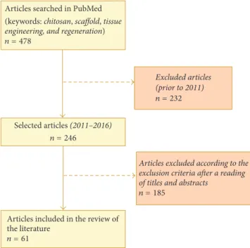
For this systematic review, a search was conducted of the
Articles searched in PubMed (keywords: chitosan, scaffold, tissue
engineering, and regeneration)
n = 478
Excluded articles (prior to 2011) n = 232
Selected articles (2011–2016) n = 246
Articles excluded according to the exclusion criteria after a reading of titles and abstracts
n = 185
Articles included in the review of the literature
n = 61
Figure 3: Flowchart of the research study and selection of articles for bibliographical review.
with the selection criteria. For the search, the following key-words were employed: chitosan, scafold, tissue engineering, and regeneration. he initial study produced 478 articles. he search was restricted to the last ive years and only preclinical studies (with animals) or clinical studies (humans) were used, resulting in 246 articles. Ater a critical reading of the titles, abstracts, and, where necessary, the articles’ methodology, only articles published in English were included. he follow-ing exclusion criteria were then applied:
(i) Literature reviews (ii) Case studies
(iii) Studies in which CS was not used to evaluate tissue regeneration
(iv) Abstracts or studies where the full content was not accessible
acteristics, main physicochemical properties of CS (when available), combinations with other polymers, incorporation of growth factors and/or cells, the in vitro and in vivo methodology evaluated, the main results in terms of tissue regeneration, and the authors’ major conclusions.
Of the 61 studies selected, 60 consisted of preclinical assays using rats (51.6%), rabbits (26.6%), mice (13.3%), dogs (5%), and guinea pigs (3.5%). Only one article demonstrated the efects of CS on human tissue [68].
As far as the analysed tissue types are concerned, 44.3% investigated the action of CS on bone tissue [8, 28, 41–52, 54– 62, 80, 85, 86], osteochondral regeneration [80, 85, 86], and bone and vascular [53] or bone and muscular tissues [56]. Moreover, 19.7% of the studies evaluated CS in the regener-ation of skin [24, 27, 29, 63–71], 14.7% in nervous tissue [30, 72–79], 11.5% in cartilage [32, 81–84, 87, 88], and 3.3% in peri-odontal [33, 90] structures. he remaining studies involved regeneration of colorectal [26], mammary [89], tympanic membrane [31], and vascular [34] tissues (1.6% each).
As for the characteristics of the scafolds, the majority of studies involved CS combined with other biomateri-als (85.25%), including HA (16%), collagen (14%), gelatin (10%), silk ibroin (10%), PLGA (8%), multiple combinations between them (20%), or forming other polymer blends (22%). Scafolds of natural CS were used in 14.75% of the studies [26, 28, 31, 33, 59, 64, 74, 75, 79]. he physicochemical characteristics of CS varied greatly. he molecular weight ranged from 22 [74] to 1,800 [73] kilodaltons (kDa), while the degree of acetylation ranged from 2% [54] to 40% [81]. In almost one-half of the studies reviewed (47.5%), the molec-ular weight and degree of acetylation of the chitosans used were not speciied. When these parameters were mentioned, 46.9% of the chitosans exhibited low molecular weight (up to 150 kDa), 37.5% were of average molecular weight (150 to 700 kDa), and 12.6% had a high molecular weight (in excess of 700 kDa). As for the macrostructure of the scafolds, these were primarily used in the form of sponges (24.6%), mem-branes (19.6%), conduits or tubules (13.1%), or even as nano-ibrils, hydrogels, microspheres, powder, and paste (11.4%) or were unspeciied (31.3%). In 63.9% of the studies, there was an incorporation into the scafolds of bioactive molecules such as growth factors, genetic factors, peptides, extracellular matrix, or cell components such as stem cells and other cells related to tissue involved in regeneration, or even drugs.
As far as the study outline is concerned, 75.4% used in vitro and in vivo assay methodologies in their investigation of scafold activities with CS, while in 24.6% of studies only in vivo assays were performed.he in vitro analyses consisted of tests on the cytocompatibility of the scafolds in cell cul-tures, evaluating cell adhesion, proliferation, viability, and diferentiation. he in vivo assays investigated the efects of scafolds on tissue regeneration and/or their biocompatibility
studies, both positive and negative controls were employed, while in 8.2% only positive controls were employed. As for sample size, 21.3% of studies used, in their in vivo assays, samples where�was less than 4 sample units; 36.1% had a value of�between 4 and 6; in 13.1%�was between 8 and 10 and 13.1% had�greater than 10. he sample size was not speciied in the methodology in 16.4% of the studies reviewed. he interrelationship between the scafolds and their efects on tissue regeneration reveals that, in 93.4% of the studies, scafolds using CS promoted in vivo or ex vivo tissue regeneration. Only 6.6% of the studies [27, 41, 71, 85] showed no evidence of the scafolds having a positive inluence on tissue regeneration.
4. Discussion
Regenerative medicine has been highlighted as one of the most promising ields of scientiic research towards which the most recent advances in biological sciences are converging, such as tissue engineering, stem-cell technology, and genetic therapy. he progress achieved in the area of tissue engineer-ing is due, for the most part, to the huge volume of studies using biomaterials that ofer potential for regeneration of almost all tissues and organs in the human body. In this con-text, chitosan has received a great attention from the scientiic research literature. Its favorable characteristics as a biomate-rial, such as biocompatibility, biodegradability, and, above all, chemical ainity with biological molecules, make it an attrac-tive material for tissue regeneration applications. Also, its chemical reactivity allows its properties to be modulated, making it an extremely versatile material [7, 19].
W
o
rld
Jo
ur
na
l
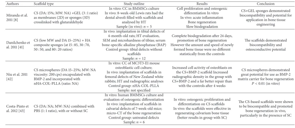
Table 1: Studies on CS-based scafolds for bone tissue regeneration.
Authors Scafold type Study outline Results Conclusion
Miranda et al. 2011 [8]
CS (DA: 15%; MW: NA) +GEL (3 : 1 ratio) as membranes (2D) or sponges (3D) crosslinked with glutaraldehyde
In vitro: CC in BMMSCs culture In vivo: 8-week-old Lewis rats had the
dental alveoli illed with scafolds and analysed by HT
Sample (in vivo):� = 5
Cell proliferation and osteogenic diferentiation In vitro In vivo: acute inlammation
Bone regeneration Slow biodegradation
CS+GEL sponges demonstrated biocompatibility and potential for
application in bone tissue engineering
Danilchenko et al. 2011 [41]
CS (low MW and DA 15–25%) + HA composite sponges (at 15 : 85, 30 : 70, 50 : 50, and 80 : 20 ratios)
In vivo: implantation in tibial defects of 4-month-old rats; HT evaluation, SEM and microhardness of tibias; serum bone-speciic alkaline phosphatase (BAP)
Control group: tibial defects without scafolds
Sample:� = 12
Complete biodegradation ater 24 days, promotion of bone regeneration However the amount and speed of newly
formed bone tissue were no diferent statistically from the controls
he scafolds demonstrated biocompatibility and osteoconductive potential
Niu et al. 2011 [42]
CS microspheres (DA 15–25%; MW: NA viscosity: 200 cps) encapsulated with BMP-2 and incorporated with nHA-COL-PLLA (ratio: NA)
In vitro: CC of MC3T3-E1 mouse osteoblastic cell culture; In vivo: implantation of scafolds in femoral defects of New Zealand white rabbits; HT and radiographic analyses
Control group: nHA-COL-PLLA Sample: not speciied
Increased cell activity of osteoblasts on the CS+BMP-2 scafold Increased radiographic density in the group with CS+BMP-2 and a far better repair than
with the controls ater 4 weeks
CS microspheres demonstrated great potential for use as BMP-2 matrix carrier for bone regeneration
� < 0.01(in vitro)
Costa-Pinto et al. 2012 [43]
CS (DA: NA; MW: NA) combined with PBS (1 : 1 ratio), with or without SC
In vitro: human BMMSCs culture and evaluation of osteogenic diferentiation In vivo: implantation of scafolds in calvarial defects of 7-week-old mice; micro-CT of the bone regeneration
Control group: untreated defects
Sample:� = 6
In vitro: osteogenic proliferation and diferentiation on CS scafolds In vivo: the scafolds were efective in
regenerating calvarium bone tissue (better results in group with SC)
he CS-based scafolds were shown to be biocompatible and promoted
he
S
cien
tiic
W
o
rld
Jo
Table 1: Continued.
Authors Scafold type Study outline Results Conclusion
Hou et al. 2012 [44]
(i) COL sponges
(ii) COL sponges with BMP2
(iii) COL sponges with CS microspheres (MW of 90 kDa; DA 5%) and BMP2 (ratio: NA)
In vitro: BMP-2 release tests In vivo: implantation of sponges in radius
defects of New Zealand white rabbits; micro-CT and HT evaluations; 3-point bending test of the regenerated bones for
mechanical evaluation Control group: normal bone
Sample:� = 23
In vitro: COL+CS+BMP2 produced a slower, more gradual release up to 35
days
In vivo: COL+CS+BMP-2 demonstrated better bone regeneration, complete closure of defects (12 weeks) and greater mechanical performance of newly formed
bone tissue
Addition of CS improved release of BMP2, promoted better bone
regeneration, and increased mechanical performance of
regenerated bone (� < 0.05)
Zhang et al. 2012 [45]
CS in gel (DA: NA; MW: NA), either pure or in a composite with nHA (ratio: NA)
In vivo: implantation in defects of the femoral condyle of New Zealand white
rabbits; CT, macroscopic, and HT analyses of the defects Control group: untreated defects
Sample:� = 10;� = 3(control)
he CS+nHA group demonstrated greater bone neoformation than the CS and control groups, and complete repair of the defects ater 12 weeks. Pure CS was
better than the control
CS+nHA has potential for use in bone regeneration of critical defects,
favoring new bone formation when
compared to CS alone (� < 0.05)
Miranda et al. 2012 [46]
CS (DA 15%; MW; NA) + GEL crosslinked with glutaraldehyde and incorporated with BMMSC (ratio: NA)
In vivo: implantation in fresh tooth sockets of Lewis rat molars; CT, HT, and
IHC analyses
Control group: contralateral untreated tooth sockets
Sample:� = 3
CS+GEL+SC group presented greater bone formation ater 21 and 35 days, with
newly formed bone tissue with a greater level of maturity. here was no control
with pure CS
here was greater alveolar bone maturation ater extraction with the
use of CS+GEL+SC (� < 0.05)
Florczyk et al. 2013 [47]
CS (DA: NA; MW: NA) +ALG sponges incorporated with BMMSC or BMP-2 (ratio: NA)
In vitro: CC in BMMSC culture of rats In vivo: implantation of scafolds in
critical calvarial defects of Sprague-Dawley rats; micro-CT, HT, and
IHC analyses
Control group: untreated defects
Sample:� = 3
CS+ALG+BMP-2 demonstrated the highest percentage of defect closure, expression of markers, and bone regeneration of all the groups, ater 16 weeks All groups showed better results
than the control
CS+ALG were biocompatible and permitted osteogenic growth and SC diferentiation In vitro and
presented osteoconductive
W
o
rld
Jo
ur
na
l
Table 1: Continued.
Authors Scafold type Study outline Results Conclusion
Jiang et al. 2013 [48]
CS+CMC (1 : 1 ratio) membranes and nHA (0, 20, 40 or 60 wt%).
DA: NA; MW: NA
In vitro: CC and osteogenic diferentiation in osteoblast cell culture;
evaluation of biodegradation In vivo: implantation in long defects in
the radius of rabbits; radiographic, micro-CT, and HT analyses Control group: untreated defects
Sample� = 3
In vitro: CS+CMC showed faster degradation; pure CS degraded more
slowly; greater cell proliferation and osteogenic diferentiation with
CS+CMC+nHA (60% wt) In vivo: there was bone regeneration of defects ater 12 weeks even in the control,
but with greater bone volume in the CS+CMC+nHA group
Cylindrical/spiral CS+CMC+nHA scafold demonstrated biomimetic behavior, promoting cell adhesion, proliferation, and diferentiation In vitro and bone regeneration in vivo
Jia et al. 2014 [28]
(i) CS sponges (MW 100–300 kDa; DA 6.63%)
(ii) CS sponges incorporated with osteogenesis and/or angiogenesis inducing genetic factors (RNA)
In vitro: RNA release tests; osteogenic proliferation and diferentiation of rat
BMSC
In vivo: Implantation of scafolds in calvarial defects of rats
Micro-CT analyses
Control groups: pure CS and CS with RNA negative control
Sample: not speciied
In vitro: CS+ both RNAs exhibited greater cell proliferation and osteogenic
diferentiation than controls In vivo: CS+ both RNAs promoted increase in area of newly formed bone
ater 3 months compared to controls
CS sponges impregnated with two RNA factors promoted greater in vitro osteogenesis and angiogenesis and bone regeneration in defects of
rat calvarias than pure CS (� < 0.05)
Cao et al. 2014 [49]
(i) GEL sponge (Gelfoam)
(ii) Gelfoam with BMP-2
(iii) Gelfoam with sulfonated CS (MW 98 kDa; DA: NA) + BMP-2 (ratio: 1 : 1) (iv) Gelfoam with sulfonated CS + BMP2 in nanoparticles
In vitro: CC, osteogenic, and angiogenic diferentiation in culture of human
umbilical vein endothelial cells In vivo: implantation in long defects in the radius of 5-mo. New Zealand rabbits;
micro-CT, HT, and micro-angiography analyses; 3-point bending test for
mechanical testing
Control groups: (Gelfoam) and normal bone
In vitro: greater cell proliferation and viability in the groups with CS+BMP
(nanoparticles) In vivo: better regeneration and angiogenesis in animals with CS+BMP (nanoparticles); mechanical performance
was similar to normal bone
CS+BMP in nanoparticles incorporated in GEL promoted greater neovascularization and bone
regeneration In vitro and in vivo than GEL alone or with BMP (� < 0.05), showing potential for
bone and vascular regeneration
Lee et al. 2014 [50]
CS (MW310 kDa; DA 10%) + HA or nano-HA composites (ratio: NA)
In vitro: CC in cell culture of MC3T3-E1 preosteoblasts
In vivo: grats in segmentary tibial defects of New Zealand rabbits; evaluation via
micro-CT and HT Control groups: NA
Sample:� = 6to 8
In vitro: CS+nHA demonstrated greater cell proliferation and viability In vivo: better histological and radiographic results with discrete ossiication in the nHA+CS group
he
S
cien
tiic
W
o
rld
Jo
Table 1: Continued.
Authors Scafold type Study outline Results Conclusion
Fernandez et al. 2014 [51]
Composite as a paste of CS (DA: NA; MW: NA) and a bioceramic of
�TCP+CaO+ZnO
(ratio: 60 : 40)
In vivo: implantation of scafolds in critical calvarial defects of 4-mo. Wistar rats; HT and histomorphometric analyses
Control groups: untreated defects
Sample:� = 4
Scafolds showed bone regeneration ater 40 days, with formation of bone marrow, vessels, and avascular cortical bone and complete closure of the defects by day 60
Control results not speciied
CS+�TCP+CaO+ZnO promoted
osteoinduction and neovascularization of the bone defects, showing potential for bone
regeneration
Fan et al. 2014 [52]
Composite sponges of CS (MW 255 kDa; DA 15–25%) + Condroitin sulfphate (ratio: 2 : 1) coated with HA;
he sponges were used with or without SC and/or BMP-2
In vitro: CC in adipose-derived SC; BMP-2 release assay
In vivo: implantation in critical defects in the jaws of rats; analyses via micro-CT,
immunoluorescence, and HT Control groups: NA
Sample:� = 4
In vivo: greater bone formation in the CS+HA+BMP-2+SC group, with greater
expression of collagen and osteocalcin, compared to blank scafolds
CS+BMP2+CS demonstrated great potential for the regeneration of
bone defects, with a synergistic
efect of the combination (� < 0.05)
Koc¸ et al. 2014 [53]
CS sponges (MW 400 kDa; DA<15%) +
HA (ratio: 9 : 1), whether or not activated with VEGF
In vitro: CC and VEGF secretion in osteoblast culture
In vivo: implantation in epigastric fasciovascular laps of Wistar rats; HT
and IHC analyses
Control groups: untreated laps and blank scafolds
Sample:� = 6
In vitro: CS+HA+VEGF: greater proliferation of osteoblasts and secretion
of VEGF
In vivo: CS+HA+VEGF +osteoblasts showed greater neovascularization and
ectopic bone formation, at 28 days compared to blank scafolds Control results not speciied
CS+HA+VEGF promoted proliferation of human osteoblasts,
induction of ectopic bone formation, and vascular
neoformation (� < 0.05)
Lai et al. 2015 [54]
Nanoibrous membranes of CS (MW: 100 kDa; DA 2%) and SF (ratio 1 : 1) + nHA (10% or 30%), either with or without stem cells.
In vitro: CC and osteogenic diferentiation of BMMSC on CS/SF with
or without nHA
In vivo: subcutaneous implantation of CS/SF/nHA30%/BMMSC in mice; HT
and IHC analyses Control group: acellular scafold
Sample: not speciied
In vitro: CS+SF+nHA30% exhibited greater osteogenic diferentiation In vivo: only CS+SF+nHA with SC induced formation of ectopic osteoid
tissue ater 8 weeks
he CS+SF+nHA scafold favored osteogenic proliferation and
diferentiation in vitro (� < 0.05)
W
o
rld
Jo
ur
na
l
Table 1: Continued.
Authors Scafold type Study outline Results Conclusion
Ghosh et al. 2015 [55]
CS (MW 710 kDa; DA<10%) crosslinked
or otherwise, with citric acid and/or carbo-di-imides
In vitro: CC and osteogenic diferentiation in culture of BMSC In vivo: implantation in tibial defects of rabbits; HT analysis of bone regeneration
Control groups not speciied Sample: not speciied
In vitro: crosslinked CS with citric acid demonstrated greater osteogenic
adhesion, proliferation and diferentiation
In vivo: dual crosslinked CS exhibited greater deposition of collagen and bone
regeneration ater 6 weeks
he dual crosslinked CS scafold demonstrated greater cytocompatibility in vitro and bone
regeneration in vivo (� < 0.05)
Caridade et al. 2015 [56]
CS membranes (MW 770 kDa; DA 22%) +ALG, crosslinked with carbo-di-imides and incorporated or otherwise with BMP-2 (ratio: NA)
In vitro: CC and myogenic and osteogenic diferentiation In vivo: implantation in subcutaneous
tissue of mice and evaluation via micro-CT
Control groups: not speciied
Sample:� = 2
In vitro: CS+BMP-2 induced osteogenic diferentiation and release of BMP-2
In vivo: only the most crosslinked membranes were capable of inducing
osteogenesis at 52 days
Crosslinked CS+BMP-2 have potential for use as a periosteum
substitute for bone regeneration
Frohbergh et al. 2015 [57]
Microibers of genipin crosslinked CS (DA 15–25%; medium MW), with or without nHA and SC (ratio: NA)
In vitro: CC and osteogenic diferentiation in murine MSC culture In vivo: implantation in calvarial defects
of 4–6-w mice; HT and micro-CT analyses
Control group: untreated defects
Sample:� = 4
In vitro: CS+nHA produced twice the osteogenic diferentiation of CS In vivo: CS+nHA+SC exhibited greater
bone neoformation than any of the others, ater 3 months
CS crosslinked with genipin has potential for use in bone regeneration; addition of nHA and
stem cells increased bone
regeneration in vivo (� < 0.01)
Dhivya et al. 2015 [58]
Hydrogels of CS-Zn (DA: NA; MW:
NA)+�-glycerophosphate + nHA (ratio:
8 : 1; 1) or without nHA
In vitro: cell proliferation and diferentiation in mouse MSC culture In vivo: insertion into tibial defects of Wistar rats; radiographic and HT
analyses
Control group: untreated defects Sample: not speciied
In vitro: scafolds favored osteoblast proliferation and diferentiation In vivo: greater mineralization and formation of collagen ater 14 days in
scafolds with nHA
CS-Zn +�-glycerophosphate
demonstrated bone regeneration potential; addition of hydroxyapatite promoted and
he
S
cien
tiic
W
o
rld
Jo
D’Mello et al. 2015 [59]
Sponges of CS (MW: 110 to 150 kDa; DA: NA), whether or not incorporated with copper sulfate (ratio: NA)
of 14-week-old Fisher rats; analyses via micro-CT and HT
Control groups: untreated defects
Sample:� = 2(control� = 3)
CS + copper exhibited greater bone neoformation than pure CS or control, both via micro-CT and via histological
analyses
CS + copper has great potential for application in bone regeneration and promoted bone regeneration in
vivo (� < 0.05)
Ji et al. 2015 [60]
3D disks of CS (low MW; DA: NA) +GEL with spherical or cylindrical nHA (ratio: 1 : 1 : 3) with or without SC.
In vitro: morphology, osteogenic proliferation, and diferentiation of
human gingival ibroblast-derived induced pluripotent SC In vivo: implantation of scafolds with or
without SC in subcutaneous tissue of mice; HT and IHC analyses of ectopic
bone-like tissue formation Control group: not speciied
Sample:� = 12
In vitro: scafolds with spherical nHA demonstrated greater osteogenic
proliferation and diferentiation (� < 0.01)
In vivo: CS+GEL+ nHA+SC showed greater bone-like tissue formation than
acellular scafolds (� < 0.01); spherical
nHA induced thicker bone-like
formation ater 12 weeks (� < 0.05)
CS+GEL+spherical nHA combined with pluripotent human cells induced ectopic bone-like tissue
formation and represent an innovative approach with the potential for application in bone
tissue engineering
Shalumon et al. 2015 [61]
Nanoibrous membranes of CS (MW 100 kDa; DA 2%) +SF+nHA+BMP2, whether or not impregnated with SC (ratio: NA)
In vitro: osteogenic proliferation and diferentiation of MSC; BMP-2 release test
In vivo: implantation of scafolds with or without MSC in subcutaneous tissue of
6–8-week-old mice; HT and IHC analyses ater 4 and 8 weeks Control groups: not speciied
Sample:� = 3
In vitro: BMP-2 increased osteogenic diferentiation of MSC on CS+SF and
CS+SF+nHA scafolds
In vivo: cellular or acellular scafolds were capable of inducing formation of ectopic
bone-like tissue, with greater intensity when SC was present
CS+SF+nHA scafolds with BMP-2 induced greater osteogenic
diferentiation In vitro (� < 0.05)
and showed great potential for application in bone regeneration in
vivo
Xie et al. 2016 [62]
Nanoibers of CS (DA<15%; MW:
NA)+HA (ratio: 7 : 3) and/or COL+SC
In vitro: CC and osteogenic diferentiation of induced pluripotent
SC+ MSC
In vivo: implantation scafolds with or without SC in critical calvarial defects of 6-week-old mice; HT analysis (4, 6, and 8
weeks) and tomographic analysis (6 weeks)
Control group: untreated defects, pure CS, and TCP
Sample:� = 2(histology);� = 6(CT)
In vitro: CS+HA+COL promoted greater osteogenic diferentiation than CS,
CS+HA, and TCP
In vivo: CS+HA+COL with SC promoted greater bone neoformation, via CT and histology, with complete regeneration of
defects
CS+COL+HA with stem cells promoted efective bone neoformation in vitro and in vivo,
with better results than controls (� < 0.01), showing potential for bone regeneration in clinical
applications
W
o
rld
Jo
ur
na
l
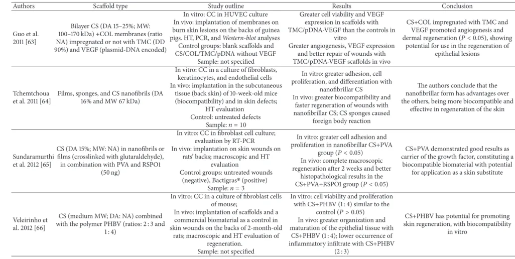
Table 2: Studies on CS-based scafolds for cutaneous tissue regeneration.
Authors Scafold type Study outline Results Conclusion
Guo et al. 2011 [63]
Bilayer CS (DA 15–25%; MW: 100–170 kDa) +COL membranes (ratio NA) impregnated or not with TMC (DD 90%) and VEGF (plasmid-DNA encoded)
In vitro: CC in HUVEC culture In vivo: implantation of membranes on burn skin lesions on the backs of guinea
pigs. HT, PCR, andWestern-blotanalyses
Control groups: blank scafolds and CS/COL/TMC/pDNA without VEGF
Sample: not speciied
Greater cell viability and VEGF expression in scafolds with TMC/pDNA-VEGF than the controls in
vitro
Greater angiogenesis, VEGF expression and better repair of wounds with TMC/pDNA-VEGF scafolds in vivo
CS+COL impregnated with TMC and VEGF promoted angiogenesis and
dermal regeneration (� < 0.05), showing
potential for use in the regeneration of epithelial lesions
Tchemtchoua et al. 2011 [64]
Films, sponges, and CS nanoibrils (DA 16% and MW 67 kDa)
In vitro: CC in a culture of ibroblasts, keratinocytes, and endothelial cells In vivo: implantation in the subcutaneous
tissue (back skin) of 10-week-old mice (biocompatibility) and in skin defects;
HT evaluation Control: untreated defects
Sample:� = 10
In vitro: greater adhesion, cell proliferation, and diferentiation with
nanoibrillar CS
In vivo: greater biocompatibility and faster regeneration of wounds with nanoibrillar CS; CS sponges caused
foreign body reaction
he authors conclude that the nanoibrillar form has advantages over the others, being more biocompatible and
efective in regeneration of the skin
Sundaramurthi et al. 2012 [65]
CS (DA 15%; MW: NA) in nanoibrils or ilms (crosslinked with glutaraldehyde), in combination with PVA and RSPO1
(50 ng)
In vitro: CC in ibroblast cell culture; evaluation by RT-PCR
In vivo: implantation on skin wounds on rats’ backs; macroscopic and HT
evaluation
Control groups: untreated wounds
(negative), Bactigras(positive)
Sample:� = 3
In vitro: greater cell adhesion and proliferation in nanoibrillar CS+PVA
group (� < 0.05)
In vivo: complete macroscopic regeneration ater 2 weeks and better
histopathological results in the
CS+PVA+RSPO1 group (� < 0.05)
CS+PVA demonstrated good results as carrier of the growth factor, constituting a
biocompatible biomaterial with potential for application as a skin substitute
Veleirinho et al. 2012 [66]
CS (medium MW; DA: NA) combined with the polymer PHBV (ratios: 2 : 3 and
1 : 4)
In vitro: CC in a culture of ibroblast cells of mouse;
In vivo: implantation of scafolds and a commercial biomaterial as a control in skin wounds on the backs of 2-month-old
rats; macroscopic and HT evaluation of regeneration.
Sample: not speciied
In vitro: cell viability and proliferation with CS+PHBV (1 : 4) similar to the
control (� > 0.05)
In vivo: greater organization and maturation of the epithelial tissue with
CS+PHBV (1 : 4); lower occurrence of inlammatory iniltrate with CS+PHBV
(2 : 3)
CS+PHBV has potential for promoting skin regeneration, with biocompatibility
he
S
cien
tiic
W
o
rld
Jo
Table 2: Continued.
Authors Scafold type Study outline Results Conclusion
Wang et al. 2013 [67]
CS membranes (MW 100 to 171 kDa; DA 15%) + COL and PLGA (ratio: NA)
In vivo: implantation of the scafolds, with or without PLGA, on skin defects in
backs of 2-month-old rats; macroscopic, HT, IHC, PCE analyses and tensile
strength tests.
Sample:� = 12
CS+COL+PLGA scafolds demonstrated better healing and greater expression of
IHC and PCR markers and higher
mechanical performance (� < 0.05)
CS+COL scafolds reinforced with PLGA demonstrated acceleration of angiogenesis and better skin regeneration
than CS+COL (� < 0.05)
Sarkar et al. 2013 [68]
Crosslinked CS membranes (MW 71 kDa;
DA<10%) whether or not combined
with COL
In vitro: CC in culture of ibroblasts and keratinocytes.
In vivo: implantation in human skin defects ex vivo; HT analyses. Control groups: not speciied
Sample:� = 3
In vitro: CS+COL demonstrated better cell adhesion, proliferation, and viability In vivo: CS+COL promoted partial reepithelialization with migration ater 14
days; pure CS did not promote regeneration
CS+COL scafold promoted better regeneration of skin wounds than pure
CS scafolds (� < 0.05)
Zeinali et al. 2014 [24]
CS membranes (medium MW; DA 15–25%) crosslinked with PHBV, with or
without SC (2 × 10⁶)
In vitro: CC in umbilical cord SC culture; In vivo: implantation in skin defects on 4–8-week-old rats’ backs; HT and IHC
analyses. Control not speciied.
Sample:� = 10
In vitro: CS+PHBV showed greater cell proliferation and viability In vivo: greater regeneration of cutaneous
tissue with CS+PHBV+SC Statistical analysis not performed
CS+PHBV added to stem cells was capable of regenerating full thickness skin
defects in rats
Guo et al. 2014 [69]
Bilayer CS (MW 100–170 kDa; DA 15–25%) +COL and silicone membranes
(ratio: NA)
In vivo: implantation of scafolds in excisional or burnt skin lesions in guinea
pigs; HT, IHC and IF evaluations; c Control group: commercial bandage;
sample:� = 2
CS+COL produced results inferior to the control in the regeneration of burn
lesions (� < 0.05) here was no
signiicant diference with excisional
lesions (� > 0.05)
CS and collagen demonstrated efectiveness similar to the commercial
W
o
rld
Jo
ur
na
l
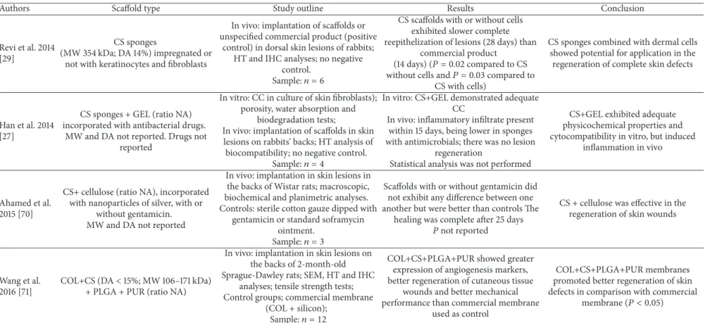
Table 2: Continued.
Authors Scafold type Study outline Results Conclusion
Revi et al. 2014 [29]
CS sponges
(MW 354 kDa; DA 14%) impregnated or not with keratinocytes and ibroblasts
In vivo: implantation of scafolds or unspeciied commercial product (positive
control) in dorsal skin lesions of rabbits; HT and IHC analyses; no negative
control.
Sample:� = 6
CS scafolds with or without cells exhibited slower complete reepithelization of lesions (28 days) than
commercial product
(14 days) (� = 0.02compared to CS
without cells and� = 0.03compared to
CS with cells)
CS sponges combined with dermal cells showed potential for application in the
regeneration of complete skin defects
Han et al. 2014 [27]
CS sponges + GEL (ratio NA) incorporated with antibacterial drugs.
MW and DA not reported. Drugs not reported
In vitro: CC in culture of skin ibroblasts); porosity, water absorption and
biodegradation tests;
In vivo: implantation of scafolds in skin lesions on rabbits’ backs; HT analysis of
biocompatibility; no negative control.
Sample:� = 4
In vitro: CS+GEL demonstrated adequate CC
In vivo: inlammatory iniltrate present within 15 days, being lower in sponges with antimicrobials; there was no lesion
regeneration
Statistical analysis was not performed
CS+GEL exhibited adequate physicochemical properties and cytocompatibility in vitro, but induced
inlammation in vivo
Ahamed et al. 2015 [70]
CS+ cellulose (ratio NA), incorporated with nanoparticles of silver, with or
without gentamicin. MW and DA not reported
In vivo: implantation in skin lesions in the backs of Wistar rats; macroscopic, biochemical and planimetric analyses. Controls: sterile cotton gauze dipped with
gentamicin or standard soframycin ointment.
Sample:� = 3
Scafolds with or without gentamicin did not exhibit any diference between one another but were better than controls he
healing was complete ater 25 days
�not reported
CS + cellulose was efective in the regeneration of skin wounds
Wang et al. 2016 [71]
COL+CS (DA<15%; MW 106–171 kDa)
+ PLGA + PUR (ratio NA)
In vivo: implantation in skin lesions on the backs of 2-month-old Sprague-Dawley rats; SEM, HT and IHC
analyses; tensile strength tests; Control groups; commercial membrane
(COL + silicon);
Sample:� = 12
COL+CS+PLGA+PUR showed greater expression of angiogenesis markers, better regeneration of cutaneous tissue
wounds and better mechanical performance than commercial membrane
used as control
COL+CS+PLGA+PUR membranes promoted better regeneration of skin defects in comparison with commercial
membrane (� < 0.05)
he
S
cien
tiic
W
o
rld
Jo
Sim˜oes et al. 2011 [72]
High MW CS membranes crosslinked with GPTMS. (DA: NA; ratio: NA)
microscopy for intracellular Ca++ In vivo: HT analysis of the subcutaneous
tissue in adult Wistar rats. No control group
Sample(in vivo):� = 4
and diferentiation in vitro In vivo: slight to intense chronic inlammation was observed in HT
analysis
Presence of ibrous capsule
biocompatibility and potential for use in the regeneration of nerve tissue However, the presence of chronic inlammation and ibrous capsules contradict the conclusion
(� < 0.05)
Wei et al. 2011 [73]
CS (MW 1800 kDa; DA 6.5%) + SF ilms (ratios: 50 : 50 or 70 : 30) impregnated
with SC
In vitro: CC in SC culture and Schwann cells
In vivo: implantation of scafolds in lesions of the sciatic nerve in adult Sprague-Dawley rats; functional and HT
evaluation
Control group: nongrated rats
Sample:� = 8
In vitro: greater adhesion and proliferation with CS and SF scafolds
when compared to pure CS In vivo: Greater functional recovery and
tissue regeneration in the groups with CS+SF impregnated with SC
CS+SF impregnated with SC promoted better regeneration in
sciatic nerve lesions and lower proliferation of ibrous scar tissue
(� < 0.05)
Chen et al. 2011 [74]
CS conduits (DA 7.7%; MW 22 kDa) whether or not impregnated with
BMMSC
In vivo: implantation of conduits in spinal cord defects in adult Sprague-Dawley
rats; functional evaluation and electromyography; HT and IHC analyses Control group: untreated defects
Sample:� = 15
Control:� = 10
Better motor and electromyographic response with CS+BMSC; better macroscopic and HT regeneration of
defects illed with scafolds with SC
CS +SC scafolds were capable of promoting axonal regeneration,
remyelination, and functional recovery ater sectioning of spinal
cord (� < 0.05)
Liao et al. 2012 [75]
CS in the form of conduits (DA 15–25%; MW: NA), either with or without SC
impregnation
In vivo: implantation of scafolds in sciatic nerve defects in adult Sprague-Dawley rats; evaluation of repair
through magnetic resonance, functional evaluation, and HT analyses. Control
group not speciied
Sample:� = 18
Nerves implanted with scafolds impregnated with MSC demonstrated
better functional recovery and better magnetic resonance results than acellular
scafolds
CS impregnated with SC promoted regeneration of nerve tissue; magnetic resonance was efective for evaluating regeneration of the
sciatic nerve (� < 0.05)
Xue et al. 2012 [76]
CS+PLGA (ratio NA) in the form of tubes, whether or not impregnated with
SC (DA: NA; MW: NA)
In vivo: grating of conduits on to sciatic nerve defects in adult Beagle dogs; functional and electroneuromyographic
evaluations and neuron count; morphometric analysis and HT analysis
of associated muscles Control groups: nongrated and
autogenous grated defects
� = 5
Better functional recovery in CS+PLGA+SC group; remyelination and recovery of nerve diameter; histologically, greater regeneration in the autogenic and
CS+PLA+SC groups
CS+PLGA scafold, either with or without stem cells, favored regeneration of extensive sciatic nerve lesion and showed viability of
carrying out a clinical study with
W
o
rld
Jo
ur
na
l
Table 3: Continued.
Authors Scafold type Study outline Results Conclusion
Xiao et al. 2013 [77]
CS+COL (ratio: 1 : 4) in the form of conduits, whether or not combined with
RGD peptide (DA: NA; MW: NA)
In vivo: implantation of scafolds in segmental defects of adult Sprague-Dawley rats sciatic nerves;
functional evaluation via electroneuromyography, neuron markers,
and histology
Control groups: untreated defects and autogenous grated defects
Sample:� = 8
CS+COL scafolds showed better functional recovery than negative control;
CS+COL+RGD showed greater management of nerve stimuli than negative control, but lower than the
autogenous control. Scafolds demonstrated greater tissue regeneration
than negative control but less than the positive control
CS+COL+RGD was capable of accelerating the regeneration of the sciatic nerve, obtaining satisfactory
results in 2 months (� < 0.05)
Biazar and Keshel 2013 [78]
CS (medium MW; DA 15–25%) in the form of conduits, whether or not
crosslinked with PHBV
In vitro: CC in Schwann cell culture In vivo: implantation of scafolds in sciatic nerve defects of 4–8-week-old Wistar rats; macroscopic and microscopic
analyses via HT and IHC Control groups: untreated defects and
autogenous grated defects
Sample:� = 5
In vitro: CS+PHBV was found to exhibit greater cell viability and proliferation
In vivo: CS (crosslinked or not with PHBV), produced regeneration results far
superior to the negative control, though inferior to the autogenous control
CS+PHBV demonstrated capacity to regenerate lesions of the sciatic
nerve in rats (� < 0.05), having
potential for application in tissue engineering and clinical studies
Gu et al. 2014 [30]
CS+SF (ratio NA) in the form of conduits impregnated with EMC (DA: NA; MW:
NA)
In vitro: isolation of Schwann cell EMC derived from rats
In vivo: implantation in sciatic nerve defects in adult Sprague-Dawley rats; HT
and IHC analyses; electrophysiological tests
Control group: acellular xenogeneic nerve grat
Sample: not speciied
In vivo: better nerve tissue regeneration and density in the CS+SF+EMC group ater 12 weeks. he electrophysiological tests got a response in all groups, though
to a lesser extent in the CS+SF group
he CS+SF+EMC scafold was efective in regenerating nerve
tissue (� < 0.05)
Wang et al. 2016 [79]
CS conduits (DD 92.3%; MW 250 kDa) or chitooligosaccharides (COS) in silicon
conduits
In vitro: CS biodegradation and CC in Schwann cell culture In vivo: implantation of scafolds in lesions of the sciatic nerves of adult Sprague-Dawley rats; HT and IHC
analyses Control: saline group Sample: not speciied
In vitro: COS promoted greater cell proliferation and diferentiation In vivo: greater expression of nerve cell
markers in the chitooligosaccharide groups
Chitooligosaccharides promote nerve cell proliferation and diferentiation, stimulating regeneration of nerve tissue
(� < 0.05/� < 0.01)
he
S
cien
tiic
W
o
rld
Jo
Chen et al. 2011 [80]
CS sponges (DA 15%; MW 400 kDa) +HA+GEL (ratio NA)
whether or not activated by growth factors (BMP-2 and TGF)
In vitro: CC and cell diferentiation in MSC culture
In vivo: implantation of sponges in osteochondral patellar defects of 4-month-old New Zealand white rabbits;
HT and IHC evaluations Control group: DNA-free composite
osteochondral grat
Sample:� = 5
Control:� = 4
Growth and osteochondral diferentiation in vitro were observed
In vivo: greater osteochondral tissue neoformation with the scafolds groups,
with or without growth factors when compared to the control
CS sponges + HA+GEL with TGF and BMP-2 promoted greater cell growth and bone and cartilage
tissue regeneration (� < 0.05)
Whu et al. 2013 [81]
CS (MW 65 kDa; DA 40%) +GEL (ratios 5 : 0, 4 : 1, 3 : 2, 1 : 1, 2 : 3, 1 : 4, or 0 : 5) in ilms and sponges, either crosslinked or
not with carbodiimide
In vitro: CC in culture of chondrocytes with scafolds
In vivo: implantation of crosslinked scafolds in cartilage defects in rabbits
feet; HT and IHC analyses Control group: untreated defects
Sample:� = 3
In vitro: greater cell proliferation and viability with crosslinked CS+GEL
scafolds
In vivo: greater regeneration of cartilage with CS+GEL impregnated with chondrocytes ater 1 month here was no comparison with pure CS or with absence
of chondrocytes
he authors conclude that carbodiimide crosslinked CS+GEL scafold demonstrated potential for
cartilage regeneration (� < 0.05)
Zhang et al. 2013 [82]
Sponges of CS (MW 40 kDa; DA: NA) +PLGA (ratio 1 : 1), either with or without
incorporation of SC
In vitro: CC in adipose-derived stem-cell culture, in chondrogenic medium In vivo: implantation of scafolds, with or
without SC, in articular defects in 4-month-old New Zealand rabbits knees;
HT, IHC, and biomechanical assays (compressive modulus and
cytonano-indentation) Control group: scafold alone
Sample:� = 5
In vitro: CS+PLGA favored chondrogenic adhesion, proliferation, and
diferentiation
In vivo: CS+PLGA+SC promoted greater regeneration of the defects and maintenance of subchondral bone, ater
12 weeks, and greater mechanical performance than scafolds without SC
CS+PLGA+SC scafolds were capable of regenerating the full thickness of the cartilage defects in
12 weeks (� < 0.05)
Deng et al. 2013 [83]
Sponges of CS (DD: NA; MW: NA) +SF (ratio 1 : 1); DA: NA; MW: NA
incorporated or not with SC
In vitro: CC in BMMSC culture In vivo: illing of defects in the cartilage of
2-3- month-old New Zealand rabbit knees with scafolds, with or without SC;
HT and IHC analyses Control group: untreated group
Sample:� = 6
In vitro: CS scafolds promoted chondrogenic diferentiation In vivo: the CS+SF+SC scafold promoted
almost complete repair of the defects and positive HT and IHC results; CS+SF scafold demonstrated better results than
the control, but not as good as in the group with SC
CS+SF scafold showed itself to be efective as SC carrier and capable of being used in the regeneration of
W
o
rld
Jo
ur
na
l
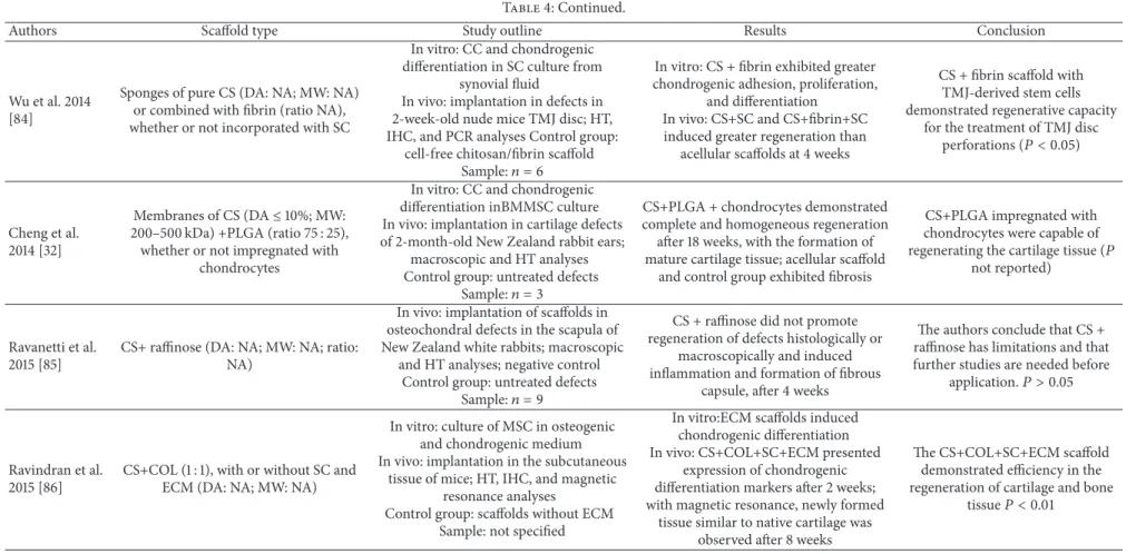
Table 4: Continued.
Authors Scafold type Study outline Results Conclusion
Wu et al. 2014 [84]
Sponges of pure CS (DA: NA; MW: NA) or combined with ibrin (ratio NA), whether or not incorporated with SC
In vitro: CC and chondrogenic diferentiation in SC culture from
synovial luid
In vivo: implantation in defects in 2-week-old nude mice TMJ disc; HT, IHC, and PCR analyses Control group:
cell-free chitosan/ibrin scafold
Sample:� = 6
In vitro: CS + ibrin exhibited greater chondrogenic adhesion, proliferation,
and diferentiation In vivo: CS+SC and CS+ibrin+SC induced greater regeneration than
acellular scafolds at 4 weeks
CS + ibrin scafold with TMJ-derived stem cells demonstrated regenerative capacity
for the treatment of TMJ disc
perforations (� < 0.05)
Cheng et al. 2014 [32]
Membranes of CS (DA≤10%; MW:
200–500 kDa) +PLGA (ratio 75 : 25), whether or not impregnated with
chondrocytes
In vitro: CC and chondrogenic diferentiation inBMMSC culture In vivo: implantation in cartilage defects of 2-month-old New Zealand rabbit ears;
macroscopic and HT analyses Control group: untreated defects
Sample:� = 3
CS+PLGA + chondrocytes demonstrated complete and homogeneous regeneration
ater 18 weeks, with the formation of mature cartilage tissue; acellular scafold
and control group exhibited ibrosis
CS+PLGA impregnated with chondrocytes were capable of
regenerating the cartilage tissue (�
not reported)
Ravanetti et al. 2015 [85]
CS+ rainose (DA: NA; MW: NA; ratio: NA)
In vivo: implantation of scafolds in osteochondral defects in the scapula of New Zealand white rabbits; macroscopic
and HT analyses; negative control Control group: untreated defects
Sample:� = 9
CS + rainose did not promote regeneration of defects histologically or
macroscopically and induced inlammation and formation of ibrous
capsule, ater 4 weeks
he authors conclude that CS + rainose has limitations and that further studies are needed before
application.� > 0.05
Ravindran et al. 2015 [86]
CS+COL (1 : 1), with or without SC and ECM (DA: NA; MW: NA)
In vitro: culture of MSC in osteogenic and chondrogenic medium In vivo: implantation in the subcutaneous
tissue of mice; HT, IHC, and magnetic resonance analyses
Control group: scafolds without ECM Sample: not speciied
In vitro:ECM scafolds induced chondrogenic diferentiation In vivo: CS+COL+SC+ECM presented
expression of chondrogenic diferentiation markers ater 2 weeks; with magnetic resonance, newly formed
tissue similar to native cartilage was observed ater 8 weeks
he CS+COL+SC+ECM scafold demonstrated eiciency in the regeneration of cartilage and bone
he
S
cien
tiic
W
o
rld
Jo
Table 4: Continued.
Authors Scafold type Study outline Results Conclusion
Meng et al. 2015 [87]
CS hydrogel, either with or without DBM particles, E7 peptide (P7), and SC (DA =
NA; MW = NA; ratio = NA)
In vitro: CC and chondrogenic diferentiation in culture of BMSC;
compression strength and elastic modulus tests
In vivo: HT and IHC analyses of subcutaneous tissue of nude mice ater 4
weeks
Control group: pure CS scafolds and composite scafolds of DBM and CS;
Sample:� = 5
In vitro: greater cell proliferation and diferentiation with CS+DBM+P7 Preparation of DBM particles might inluence the mechanical properties of
scafolds and cell proliferation In vivo: CS+DBM+P7+SC produced greater cartilage tissue formation than
pure CS or with DBM; no negative control
CS+DBM+P7 hydrogel combined with mesenchymal stem cells has
potential for regeneration of
cartilage tissue.� < 0.05
Zhang et al. 2015 [88]
CS sponges (MW 40 kDa; DA<5%)
+PLGA, with or without SC; ratio: NA; average viscosity
In vitro: CC and chondrogenic diferentiation in culture of SC In vivo: implantation in cartilage defects
in 4-month-old New Zealand rabbit knees; macroscopic, HT and IHC analyses; negative control not speciied
Control group: adherent ASC/scafold complexes
Sample:� = 5
In vitro: CS+PLGA+SC demonstrated chondrogenic diferentiation In vivo: the scafold promoted new formation of cartilage similar to hyaline, both histologically and via biomechanical
evaluation, ater 6 and 12 weeks
CS+PLGA sponges incorporated with aggregated stem cells represents a promising technique in
tissue regeneration� < 0.05
W
o
rld
Jo
ur
na
l
Table 5: Studies on CS-based scafolds for regeneration of diverse tissues.
Authors Tissue type Scafold type Study outline Results Conclusion
Gupta et al. 2011
[89] Mammary tissue
CS+SF (ratio NA) scafolds impregnated with emodin
(antitumor drug) MW and DA not reported
In vitro: breast cancer cell culture; evaluation of cell
growth and viability In vivo: implantation in breast
tumor tissue in nude rats; HT analyses
Control: lap tissue without scafolds
Sample:� = 8Control� = 7
In vitro: cell proliferation with no statistical diference in the control and CS+SF without
drugs (� > 0.05)
In vivo: there was a reduction in cancerous cells only in the CS+SF group with emodin (� < 0.05) Tissue regeneration
was similar between scafolds with or without emodin
(� > 0.05)
CS+SF scafolds were efective in absorption, release, and pharmacological activity of the therapeutic agent and in the regeneration of the tissue
defect
Seonwoo et al.
2013 [31] Tympanic membrane
CS membranes (MW 200 kDa; DA 11%), with or without EGF
In vitro: tympanic membrane cell migration and viability In vivo: implantation of CS in
tympanic perforations; endoscopic and HT analyses
Control: untreated perforations
Sample:� = 25
In vitro: CS+EGF demonstrated greater cell migration and viability than
pure CS
In vivo: CS+EGF promoted better closure of perforations and better regeneration than
the control
CS+EGF produced favorable results in vitro and in vivo
with the regeneration of tympanic perforations (� < 0.05), being a potential
alternative to surgery
Zhou et al. 2014
[34] Vascular tissue
CS+PCL (ratio NA) in the form of tubules combined
with endothelial cells MW and DA: NA
In vitro: CC in culture of endothelial cells In vivo: implantation in vascular defects in the carotids
of dogs; western-blot and RT-PCR analyses
Sample:� = 6
CS+PCL showed cytocompatibility in vitro; CS+PCL+ endothelial cells promoted normal vascular
low and endothelial regeneration in vivo
CS+PCL impregnated with endothelial cells were efective in vascular tissue regeneration
Zang et al. 2014
[33] Periodontal ligament
CS (medium MW and DA 15–25%) in the forms of
powder or solution
In vitro: CC (periodontal ligament cells) and physicochemical analyses In vivo: insertion of gel in furcation lesions in dogs’
teeth; HT analysis Negative control group
without CS
Sample:� = 8
he hydrogel obtained with autoclaved CS in the form of powder exhibited the illing of
80% of the defects. Bone and periodontal regeneration was
efective ater 12 weeks (� < 0.05)
he autoclaving of CS in the form of powder did not change its physicochemical properties; CS was efective in
he
S
cien
tiic
W
o
rld
Jo
Table 5: Continued.
Authors Tissue type Scafold type Study outline Results Conclusion
Jiang et al. 2015
[90] Periodontal ligament
CS, whether or not combined with PCE
MW and DA not reported
In vitro: CC (rat BMMSCs) and expression of periodontal
ligament markers; In vivo: insertion in periodontal defects with
BiOssin rats; HT,
immunoluorescence, and micro-CT analyses; negative
control without scafold;
sample:� = 5
CS and CS+PCE promoted periodontal regeneration, with
greater organization of ibers in the CS+PCE group ater 8
weeks (� < 0.05)
CS scafolds with PCE nanoibrils were found to have
great potential for application for regeneration of periodontal ligament
Denost et al.
2015 [26] Colorectal tissue
CS membranes with two types of hydrogel (DA 98.5% and 97%; MW 420 and 487 kDa,
resp.)
In vitro: CC and cell diferentiation (human adipose-derived stem cells)
In vivo: implantation in lesions in the intestines of rabbits; macroscopic and HT
analyses
Sample:� = 4
In vitro: CS and control (commercial membrane) were
cytocompatible In vivo: CS exhibited lower
(� > 0.05) degree of inlammation and complete colorectal regeneration ater 8
weeks
Membranes in multiple layers of CS demonstrated potential in colorectal regeneration, suggesting better results than with the commercial material
pounds of calcium and phosphate, such as HA, permits an improvement in speciic properties like solubility, biodegra-dation, and mechanical performance of CS as a function of the tissue to be regenerated [10]. In fact, some studies have clearly shown that the association of CS with collagen [42, 44, 68], silk ibroin [73], and HA [45, 57, 58] was capable of promoting greater tissue regeneration than pure CS, as in these studies pure or combined CS scafolds were implanted, making it possible to establish a comparison between them. However, in the vast majority of studies evaluating CS com-bined with other biomaterials, the favorable results obtained cannot be attributed to the associations, as pure CS was neither evaluated nor used as a control [8, 30, 32, 41, 46, 48, 49, 53, 54, 62, 63, 67, 71, 76, 77, 80, 81, 83, 86, 88].
Another aspect which merits some attention is the great variability in the chemical characteristics of CS used in the studies, such as molecular weight and degree of deacetylation. As it is a polymer which can be obtained from a variety of sources, CS may actually present a large variation in MW (ranging from 30 to 2000 kDa) and in DA (5 to 46%) resulting from the diferent processing conditions [7, 21]. However, it should be stressed that these properties drive important physical characteristics of chitosan, such as solubility, crys-tallinity, absorption of water, and mechanical performance, and also biological characteristics, such as biodegradation, antimicrobial activity, mucoadhesiveness, and biocompatibil-ity [7, 10, 21, 36, 61]. Wu et al. [38] and Zheng et al. [39], for instance, demonstrated that the in vitro bioactivity of CS on macrophages was dependant on its MW. Accordingly, it is probable that these parameters may inluence the behavior of CS as a biomaterial for tissue regeneration. hus, the use of such diverse chitosans, such as MW 22 kDa [74] and 1800 kDa [73], though both studies investigated regeneration in nerve tissue, renders any comparison of results unviable. In addition, the fact that MW and DA have not been speciied in 47.5% of the studies raises speculation that the authors have overlooked the signiicance of these parameters. In fact, the lack of information on the physicochemical characteristics and their efects on biological properties of chitosan-based scafolds is a matter of concern in the literature, since most of the useful applications of CS are determined by DA, MW, and the nature and fraction of substituents as pendant groups [19]. In the present review, only two studies accounted for the variation of MW in the methodology and interpretation of results. In the study by Cao et al. [49], chitosans of diferent molecular weights were evaluated in vitro and, based on the results, the 98 kDa CS was selected for the in vivo assay. Denost et al. [26] used layers of CS of diferent MW to fabricate the scafolds, due to the diferent characteristics they presented, corroborating the importance of this parameter on the biological activity of CS.
he use of comparison standards (control groups) is essential when investigating a particular experimental
mate-comparison. Bearing in mind that in some studies the results showed that tissue regeneration also occurred in negative control groups [31, 41, 48, 65], the absence of a negative control could cast doubt on the relevance of the results obtained with regard to the actual efectiveness of the material being investigated.
Sample size is an important consideration in the design of many studies because of its efect on statistical power. Statistical power is the probability that a statistical test will indicate a signiicant diference when there truly is one. In this review, some studies used small samples for their in vivo assays, with� = 3[32, 46–48, 61, 65, 68, 81],� = 2[27, 56, 69], and even � = 1[72]. It is important to point out that the value of�considered in this study is always the number of animals used for each treatment condition. For instance, the study by Sundaramurthi et al. [65] mentions a sample of 30 animals. However, the animals were subdivided into 5 groups and 2 evaluation periods, totaling 10 groups, resulting in three animals for each condition evaluated (� = 3). Sim˜oes et al. [72] used 4 rats that were evaluated at 4 diferent points in time, resulting in just 1 animal per period (� = 1). Small samples, such as those mentioned in these studies, may com-promise the results obtained, thus diminishing the reliability of the statistical diferences found or omitting actual difer-ences that could be observed with an increased sample size [32, 48, 69]. Given the importance of the sample size in the study outline and its inluence on the results, it is signiicant to note that 16.4% of the studies reviewed do not clearly specify this information in their methodologies. he omission of this piece of information makes it diicult to interpret the results and establish a comparison between them.
neous [64], nerve [79], colorectal [26], bone [55], periodontal ligament [33], and tympanic membrane [31] tissues.
5. Conclusion
he critical analysis of the studies reviewed conirms the potential for the application of chitosan as a biomaterial in tis-sue engineering, considering the great versatility of this poly-mer in the regeneration of various types of tissue in preclin-ical studies. Scafolds composed of CS combined with other biomaterials, cells, and growth factors were found to be efec-tive in promoting in vivo tissue regeneration. Nevertheless, the large chemical variability of the CS employed, the omis-sion of precise information about its characteristics, and the methodological limitations of the studies make it diicult to reproduce and to establish a standardization for clinical appli-cation. Further studies are required with the aim of deining chitosan’s ideal physicochemical characteristics for applica-tion with each type of tissue and, thus, propose a protocol for its use in clinical studies in order to conirm its efectiveness in tissue regeneration.
Conflicts of Interest
he authors declare that there are no conlicts of interest regarding the publication of this paper.
Acknowledgments
his work was supported by Brazilian government agencies: National Council of Scientiic and Technological Develop-ment (CNPq) (Grants: Proc. no. 157763/2013-7) and Coordi-nation for the Improvement of Higher Education Personnel (CAPES)/REUNI, Federal District, Brazil.
References
[1] R. Langer and J. P. Vacanti, “Tissue engineering,”Science, vol.
260, no. 5110, pp. 920–926, 1993.
[2] R. Lanza, R. Langer, and J. Vacanti,Principles of Tissue
Engineer-ing, Elsevier, San Diego, Calif, USA, 2014.
[3] Q. Chen and G. A. houas,Biomaterials, A Basic Introduction,
CRC Press, Boca Raton, USA, 2015.
[4] F. Schoen and R. Mitchel, “Tissues, the extracellular matrix and
cell-biomaterial interactions,” inBiomaterials Science: An
Intro-duction to Materials in Medicine, B. Ratner, A. Hofman, F. Schoen, and J. Lemons, Eds., pp. 260–281, Elsevier Academic Press, San Diego, Calif, USA, 2004.
[5] A. J. Salgado, O. P. Coutinho, and R. L. Reis, “Bone tissue
engineering: state of the art and future trends,”Macromolecular
Bioscience, vol. 4, no. 8, pp. 743–765, 2004.
[6] S. Cartmell, “Controlled release scafolds for bone tissue
engi-[8] S. C. C. C. Miranda, G. A. B. Silva, R. C. R. Hell, M. D. Martins, J. B. Alves, and A. M. Goes, “hree-dimensional culture of rat BMMSCs in a porous chitosan-gelatin scafold: a promising association for bone tissue engineering in oral reconstruction,”
Archives of Oral Biology, vol. 56, no. 1, pp. 1–15, 2011.
[9] T. J. Keane and S. F. Badylak, “Biomaterials for tissue
engineer-ing applications,”Seminars in Pediatric Surgery, vol. 23, no. 3,
pp. 112–118, 2014.
[10] I.-Y. Kim, S.-J. Seo, H.-S. Moon et al., “Chitosan and its
deriva-tives for tissue engineering applications,”Biotechnology
Advan-ces, vol. 26, no. 1, pp. 1–21, 2008.
[11] D. F. Williams, “On the nature of biomaterials,”Biomaterials,
vol. 30, no. 30, pp. 5897–5909, 2009.
[12] T. Dvir, B. P. Timko, D. S. Kohane, and R. Langer,
“Nanotech-nological strategies for engineering complex tissues,” Nature
Nanotechnology, vol. 6, no. 1, pp. 13–22, 2011.
[13] D. W. Hutmacher, “Scafolds in tissue engineering bone and
cartilage,”Biomaterials, vol. 21, no. 24, pp. 2529–2543, 2000.
[14] K. F. Leong, C. M. Cheah, and C. K. Chua, “Solid freeform fab-rication of three-dimensional scafolds for engineering
replace-ment tissues and organs,”Biomaterials, vol. 24, no. 13, pp. 2363–
2378, 2003.
[15] B. Dhandayuthapani, Y. Yoshida, T. Maekawa, and D. S. Kumar, “Polymeric scafolds in tissue engineering application: a review,”
International Journal of Polymer Science, vol. 2011, Article ID 290602, 19 pages, 2011.
[16] M. Rinaudo, “Chitin and chitosan: properties and applications,”
Progress in Polymer Science (Oxford), vol. 31, no. 7, pp. 603–632, 2006.
[17] P. Baldrick, “he safety of chitosan as a pharmaceutical
excipi-ent,”Regulatory Toxicology and Pharmacology, vol. 56, no. 3, pp.
290–299, 2010.
[18] R. A. A. Muzzarelli, “Chitins and chitosans for the repair of
wounded skin, nerve, cartilage and bone,”Carbohydrate
Poly-mers, vol. 76, no. 2, pp. 167–182, 2009.
[19] B. Bellich, I. D’Agostino, S. Semeraro, A. Gamini, and A. Ces`aro,
“he good, the bad and the ugly of chitosans,”Marine Drugs, vol.
14, no. 5, article 99, 2016.
[20] C. Peniche, M. Fern´andez, G. Rodr´ıguez et al., “Cell supports of chitosan/hyaluronic acid and chondroitin sulphate systems.
Morphology and biological behaviour,” Journal of Materials
Science: Materials in Medicine, vol. 18, no. 9, pp. 1719–1726, 2007. [21] T. W. Wong, “Chitosan and its use in design of insulin delivery
system,”Recent Patents on Drug Delivery & Formulation, vol. 3,
no. 1, pp. 8–25, 2009.
[22] I. A. Sogias, A. C. Williams, and V. V. Khutoryanskiy, “Why is
chitosan mucoadhesive?”Biomacromolecules, vol. 9, no. 7, pp.
1837–1842, 2008.
[23] S. V. Madihally and H. W. T. Matthew, “Porous chitosan
scaf-folds for tissue engineering,”Biomaterials, vol. 20, no. 12, pp.
1133–1142, 1999.