Institute of Orthepedics and Traumatology, Faculty of Medicine, University of São Paulo
Email: [email protected]
Received for publication on July 30, 2005. Accepted for publication on December 12, 2005.
ORIGINAL RESEARCH
COMPARATIVE ANALYSIS BETWEEN
OSTEOSARCOMA AND EWING’S SARCOMA:
EVALUATION OF THE TIME FROM ONSET OF SIGNS
AND SYMPTOMS UNTIL DIAGNOSIS
Raquel Bezerra Guerra, Marcelo Duarte Tostes, Leandro da Costa Miranda, Olavo Pires de Camargo, André Mathias Baptista, Marcelo Tadeu Caiero, Telma Muria dos Santos Machado, Márcia Datz Abadi, Claudia Regina G.C.M Mendes de Oliveira, and Renée Zon Filippi
Guerra RB, Tostes MD, Miranda L da C, de Camargo OP, Baptista AM, Caiero MT. Comparative analysis between Osteosarcoma and Ewing’s sarcoma: evaluation of the time from onset of signs and symptoms until diagnosis. Clinics. 2006;61(2):99-106.
OBJECTIVE: The purposes of this study were to describe the early signs and symptoms of osteosarcoma and Ewing’s sarcoma, identify symptoms that could be used to help differentiate the two types of tumors, and determine the time elapsed between the onset of signs and symptoms and the definitive diagnosis in our service, providing information and imputus for earlier diagnosis of these tumors.
METHODS: A retrospective analysis of the medical dossiers of 365 patients under 30 years of age diagnosed with osteosarcoma or Ewing’s sarcoma was performed, and the aspects of the clinical diagnosis were statistically analyzed and compared.
RESULTS: The time between the onset of signs and the symptoms was 5.25 months for osteosarcoma and 8.1 months for Ewing’s sarcoma, and the most frequent (89.5%) early symptom of osteosarcoma and Ewing’s sarcoma was local pain. Symptoms that might aid diagnosis included early local volume increase and the presence of fever.
CONCLUSION: The time until diagnosis of both neoplasias was higher than that reported for North America and Europe. Education of the lay public and medical professionals regarding suspicious early signs and symptoms might shorten the delay of diagnosis.
KEYWORDS: Ewing’s sarcoma. Osteosarcoma. Bone neoplasias. Chemotherapy. Orthopedic surgery.
INTRODUCTION
Bone tumors are uncommon in adults, but they are the sixth most frequent group of neoplasic diseases among chil-dren and the third among adolescents, less frequent only than leukemias and lymphomas.1 Although osteosarcomas
and Ewing’s sarcomas are rare in the general population, they are the most frequently occurring primary malignant
bone tumors in children.2
Until the mid-1970s, over 80% of patients affected by osteosarcoma would die within 5 years after being diag-nosed. Ewing’s sarcoma would progress, even with radiotherapic treatment, to 90% mortality.3 Amputation was
the only option.
During the last 20 years, new knowledge and studies on bone sarcomas have been presented, and great advances in the treatment of these diseases have been achieved.4
Or-thopedic surgical technology allows tumor resection while preserving the limb, because adequate resection margins reduce the probability of local tumor recurrence;5
The use of chemotherapy before and after surgery for tumor removal (adjuvant and neoadjuvant chemotherapies)6
was developed to promote local control of the tumor by reducing its size. The Huvos index, which rates chemo-therapy-induced tumor necrosis from 0% to 100%, empha-sizes the importance of the degree of necrosis in the pa-tient’s prognosis.7,8
Radiotherapy is used in those cases of Ewing’s sarco-mas where, due to their locations, resection is impractica-ble, when tumor margins are affected as well as in cases where surgery would impair the function of the limb. Im-provement in image evaluations and current chemotherapy regimens have contributed significantly to the detection and systemic control of the disease.9
Cooperation between oncologists, pathologists, radiolo-gists, and orthopedic surgeons with specialized knowledge of oncology allows more effective detection and therapy of bone tumors.1,3,4,10–15
In diagnosing a bone neoplasm, attention should be paid to early signs and symptoms in order to distinguish it from other orthopedic conditions, such as osteomyelitis and ten-donitis, and to distinguish different bone neoplasms from one another.11
An improvement in the 5-year survival rates from less than 10% to the current 50% to 60% has been reported for both osteosarcoma and Ewing’s sarcoma. Some studies show 5-year survival rates of up to 75% for patients with these 2 tumors.3,16
However, this improvement is only possible with early diagnosis of the tumor. The prognosis of osteosarcoma and Ewing’s sarcoma is mainly influenced by the local extent of the tumor and by the presence of metastases.17 Chances
of survival decrease 2 to 3 times in the presence of metastases.17,18 American and European statistics show that
diagnosis is made between 1 and 3 months after first pres-entation with symptoms. In Brazil, although no research has covered this subject, a consensus exists that definitive diagnosis of a bone neoplasm takes, on average, 6 months after the onset of symptoms.1,19 In many cases, the
treat-ment is started when the tumor has already achieved a large volume, which so that local control becomes difficult, hin-dering surgery with preservation of the limb, with a reduced chance of cure.In spite of the large number of trials on prognostic factors in oncology, many of them have little impact on the clinical practice.20 In Brazil, there have been
no clinical trials to corroborate the interval between the onset of signs and symptoms and the definitive diagnosis nor any that attribute adequate importance to these early signs and symptoms, even though the need for early diag-nosis of the tumor has been proven.
The purpose of this project is to show that it is
neces-sary to reduce the interval between the onset of signs and symptoms and the diagnosis of cases of osteosarcoma and Ewing’s sarcoma in Brazil, thus increasing the chances of survival for these patients. For this, it is essential that phy-sicians be aware of the characteristic early signs and symp-toms, such as pain, increase of local volume, fever, and in some cases, the original pathological fracture.
An analysis of the signs and symptoms presented origi-nally by patients diagnosed with osteosarcoma and Ewing’s sarcoma and a comparative evaluation of the interval be-tween the onset of signs and symptoms and the definitive diagnosis in these tumors are needed.
CASES AND METHODS
A retrospective analysis was performed on the medical dossiers of patients in the Orthopedic Oncology Group of this institution who were diagnosed with osteosarcoma and Ewing’s sarcoma between 1985 and 2001. Approximately 365 patients aged 30 years or under at the time of diagno-sis based on the histological analydiagno-sis of biopsy material were considered for this study. All of them were or con-tinued to be concon-tinued to be treated in this hospital. Of the 365 patients, 250 medical dossiers that included complete, properly referenced data were selected.
The data collected from each medical dossier was or-ganized on a standard form the purpose of which was not only to record the patient’s symptoms (such as pain, fever, and volume increase, as well as the blood count and pre-vious trauma), but also to record the main events that in-volved the patient during the treatment period (such as bi-opsy, surgery, use of neoadjuvant chemotherapy, metastases, and current orthopedic and oncologic condition).
Obtained data was organized for statistical analysis into 2 groups according to the type of tumor presented. The ab-solute and relative percentage frequency distributions of the normal (qualitative) parameters were determined, as well as were the descriptive statistics of the following ordinal (quantitative) parameters: mean (M), standard deviation (SD), standard error of the mean (SEM), maximum and minimum values, and number of cases (N).
To compare the frequency distributions of nominal pa-rameters between the groups (the osteosarcoma group and the Ewing’s sarcoma group) we used 2-sample methods, such as the chi-square, binomial, Fisher, and McNemar tests; to compare the means of the ordinal samples, we used either Student’s t test or the Mann-Whitney U test (for
para-metric and nonparapara-metric distributions, respectively). The 5% significance level (± = 0.05) was adopted.
main signs and symptoms presented in diagnosing osteosa-rcoma or Ewing’s saosteosa-rcoma. The time interval between the onset of signs and symptoms and diagnosis was correlated for each group throughout the period studied (1985 through 2001) and between groups.
RESULTS
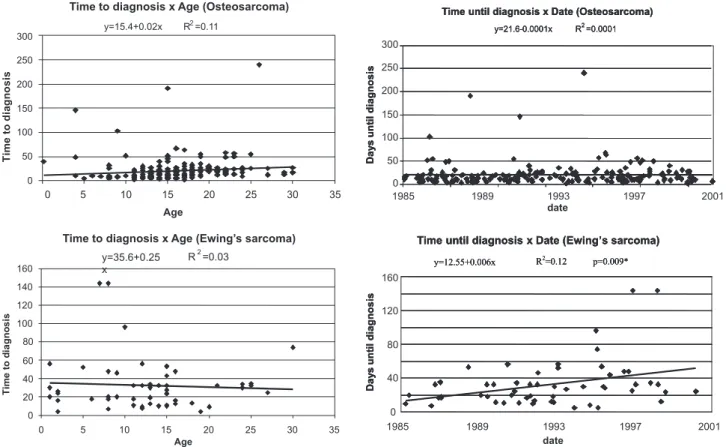
A descriptive statistical analysis of patients’ ages was performed after they were divided into 2 groups according to the type of tumor (Table 1). The evaluation showed that the mean age when an osteosarcoma occurred was 15.7 years, while for Ewing’s sarcoma the mean age of
occur-rence was 12.8 years. The comparative analysis of both groups using Mann-Whitney U test showed that the differ-ence between ages was significant, with P = 0.0005 and
percentage variation of 19%. Thus patients with Ewing’s sarcoma tended to be 2.9 years younger than patients with osteosarcoma on the dates they were diagnosed. The com-parative analysis between the patient’s age and the time of diagnosis, using the Mann-Whitney U test, did not show any correlation for either patients with osteosarcoma (P =
0.11) or for patients with Ewing’s sarcoma (P = 0.66);
therefore, the patient’s age appeared to have no influence on the time until diagnosis of these tumors (Figure 1).
The descriptive analysis of the time from onset of
symp-Table 1 - Descriptive statistics of patients’ age and time to diagnosis in the Osteosarcoma and Ewing’s Sarcoma groups. Comparison using a two-tailed Mann-Whitney u-test (±=0.05)
AGE (YEARS) WEEKS TO DIAGNOSIS
OSTEOSARCOMA Mean ± SEM: 15.8 ± 0.4 Mean ± SEM: 21.2 ± 1.8
(N = 198) minimum: 0.2 maximum: 30 minimum: 1 maximum: 240
EWING´S SARCOMA Mean ± SEM: 12.8 ± 0.9 Mean ± SEM: 32.4 ± 3.8
(N = 55) minimum:1 maximum: 30 minimum: 4 maximum: 144
P (Mann-Whitney) p=0.0005 p=0.0001
SEM: standard error of the mean; Min/Max: minimum/maximum time of occurrence
Figure 1 - Comparative analysis of patients’ age and the time to diagnosis of patients with Osteosarcoma and Ewing´s Sarcoma. Comparison using a two-tailed Mann-Whitney U-test (±=0.05)
toms until diagnosis was performed for the osteosarcoma and Ewing’s sarcoma groups. It was found that in this serv-ice, the average time until diagnosis was 21.1 weeks for osteosarcoma, while for Ewing’s sarcoma the average time until diagnosis was 32.4 weeks (Table 1).
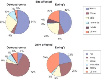
A descriptive analysis was performed on the distribu-tion of the sites affected by the tumor according to the os-teosarcoma and Ewing’s sarcoma groups. The analysis showed a difference in the distribution pattern between the tumors: osteosarcoma tended to affect the distal portion of the femur, the proximal portion of the tibia, fibula and hu-merus and, less frequently, the pelvis or other parts of the body, whereas Ewing’s sarcoma had a more homogeneous distribution in the femur and tibia portions and more com-monly affected the distal portion of the fibula, in addition to more frequently affecting the pelvis and other regions of the body (such as the spine) (Table 2).
The elevated frequency of osteosarcoma of the distal femur and of sarcoma of the proximal tibia (the knee re-gion) led us to analyze the distribution of these tumors per region of the body. The absolute and relative frequencies (Figure 3 and Table 3) were described and showed that os-teosarcoma would affect predominantly the knee, with lower frequencies in the hip, shoulder, and ankle. Ewing’s sarcoma affected the knee and hip with similar relative fre-quencies that are somewhat lower in the ankle and higher
in other joints other than the above-mentioned ones (such as the spine) and diaphyseal portions of bones.
The chi-square method was used to analyze the abso-lute and relative (%) frequency of early signs and symp-toms of patients after dividing them into 2 groups accord-ing to the type of tumor.
Since the study of fracture frequency, pain frequency, Table 3 - Absolute and relative frequency distribution (%) of sites affected by Osteosarcoma or Ewing’s Sarcoma, by joint involved, and intergroup comparison using a two-tailed chi-square test (α=0.05)
SITE OSTEOSARCOMA EWING’S
SARCOMA
N % N %
proximal femur 12 6.42 8 15.69
Pélvis 2 1.07 3 5.88
Hip 14 7.49 11 21.57
distal fêmur 83 44.39 6 11.76
proximal tibia 42 22.46 7 13.73
proximal fibula 7 3.74 0 0
Knee 132 70.59 13 25.49
distal fíbula 1 0.53 3 5.88
distal tíbia 9 4.81 3 5.88
Ankle 10 5.35 6 11.76
proximal humerus 14 7.49 1 1.96
Shoulder 14 7.49 1 1.96
distal humerus 2 1.09 0 0
Elbow 2 1.07 0 0
diaphyseal tibia 0 0 1 1.96
diaphyseal humerus 2 1.07 0 0
diaphyseal femur 6 3.21 8 15.69
diaphyseal fibula 1 0.53 1 1.96
Others 6 3.21 10 19.61
others/total 15 8.02 20 39.22
TOTAL 187 100 51 100
Table 2 - Absolute and relative frequency distribution (%) of sites affected by Osteosarcoma or Ewing’s Sarcoma in both groups, and intergroup comparison using a two-tailed chi-square test (α=0.05)
SITE OSTEOSARCOMA EWING’S
SARCOMA
n % n %
proximal femur 12 6.42 8 15.69
Diaphyseal femur 6 3.21 8 15.69
distal femur 83 44.39 6 11.76
Fêmur 101 54.01 22 43.14
proximal fibula 7 3.74 0 0
diaphyseal fibula 1 0.53 1 1.96
distal fibula 1 0.53 3 5.88
Fibula 9 4.81 4 7.84
proximal tibia 42 22.46 7 13.73
diaphyseal tibia 0 0 1 1.96
distal tibia 9 4.81 3 5.88
Tibia 51 27.27 11 21.57
proximal humerus 14 7.49 1 1.96
diaphyseal humerus 2 1.07 0 0
distal humerus 2 1.07 0 0
Humerus 18 9.63 1 1.96
Pelvis 2 1.07 3 5.88
Others 6 3.21 10 19.61
TOTAL 187 100 51 100
and pain at rest frequency did not show any difference be-tween the tumor types (P = 0.53; P = 0.49; P = 0.52,
re-spectively), these signs cannot be used as a differential in the diagnosis of these tumors (Table 4).
The study of fever frequency showed a difference be-tween the tumor types (P = 0.002); therefore, fever may
be used as a factor in the differential diagnosis of these tumors (Table 4).
The study of weight loss frequency did not show any difference between the tumor types (P = 0.93); therefore,
weight loss cannot be used as a differential in the diagno-sis of these tumors (Table 4).
The study of the local volume increase frequency showed a difference between the tumor types (P = 0.015),
with an inversion of values in the chi-square table (Table 4); therefore, early increase in local volume may be used to aid in the differential diagnosis.
A comparative analysis using Student’s t test and the
Mann-Whitney U test between the 2 groups (osteosarcoma and Ewing’s sarcoma groups) concerning the time interval between the onset of signs and symptoms and the date of diagnosis of patients showed a difference between the groups (P < 0.0001, 34.9% variation), showing that on
av-erage it took 11.3 weeks longer before for Ewing’s sarcoma was diagnosed.
Although the time from the onset of signs and symp-toms until diagnosis throughout the period analyzed (1985 though 2001) was not significantly diffenent for patients
with osteosarcoma (Figure 2), this value was significantly longer over time (P = 0.009) for patients with Ewing’s
sar-coma (Figure 2).
DISCUSSION
This study is part of an institutional effort to retrospec-tively summarize some of our most significant patient se-ries.21-23 The increased length of time until diagnosis of
tumors in Brazil as compared with that reported for the US and Europe (where it takes on average 4 to 12 weeks to reach a definitive diagnosis) is not surprising. However, this study provides statistical confirmation of a delay that should not occur in the case of malignant tumors requiring early diag-nosis in order to allow a potentially healing treatment for patients. This 11-week difference remains to be explained, although possibilities include a lack of awareness on the part of patients of early signs and symptoms that might cause a delay in seeing a physician or, more importantly, errors con-cerning differential diagnosis made by the physician who sees this patient at onset of the condition.
The finding that in recent years the delay of diagnosis of Ewing’s sarcoma has increased in our service should be discussed. Possible explanations include, among others, the following: the patient delayed his visit to a physician; the health system took too long to schedule a medical appoint-ment, both in basic health units and outsourced hospitals; errors occurred in the differential diagnosis in a primary
Table 4 - Absolute and relative frequency distribution (%) of fracture, pain, pain at rest, fever, weight loss and local volume increase as early symptoms of patients in the Osteosarcoma and Ewing’s Sarcoma groups, and intergroup comparison using a two-tailed chi-square test (±=0.05)
CHI-SQUARE(probability) OSTEOSARCOMA EWING´S SARCOMA TOTAL
N % N % N %
FRACTURE
ABSENCE 0.39 (p = 0.53) 175 70.3 50 20.1 225 90.4
PRESENCE 20 8.0 4 1.6 24 9.6
PAIN
ABSENCE 0.47 (p = 0.49) 19 7.6 7 20.1 26 10.4
PRESENCE 176 77.7 47 18.9 223 89.6
PAIN AT REST
ABSENCE 0.42 (p = 0.52) 179 71.9 51 20.5 230 92.4
PRESENCE 16 6.4 31.2 19 7.6
FEVER
ABSENCE 9.41 (p = 0.002)* 188 75.5 46 18.5 234 94.0
PRESENCE 7 2.8 8 3.2 15 6.0
WEIGHT LOSS
ABSENCE 0.07 (p = 0.93) 167 67.1 46 18.5 213 85.6
PRESENCE 28 11.2 8 3.2 36 14.4
VOLUME INCREASE
ABSENCE 5.86 (p = 0.015)* 76 30.5 31 12.4 107 43.0
PRESENCE 119 47.8 23 9.2 142 57.0
health center; there was a delay in referring the patient to a specialty service; or there was an erroneous differential diagnosis or delay in performing the biopsy due to demand that exceeded the capacity of the surgical center.
We originally proposed to study the time from onset of signs and symptoms until the diagnosis according to 3 cat-egories: (1) the time from the onset of signs and symptoms until the first visit to a physician, often in a primary health center; (2) the time from the first visit to a physician until the first visit to a bone tumor specialty service, in this case, our institution; (3) the time from the first visit to our insti-tution and the date of diagnosis, based on the biopsy re-port. The information obtained could indicate the reasons for delay or the moments when the diagnosis of osteosar-coma and Ewing’s sarosteosar-coma took more time, suggesting ac-tions for reducing this problem. It was not possible to per-form such analyses because there was not a statistically sig-nificant number of patients with medical dossiers includ-ing all information required, such as the date of the first visit; the initial symptoms and their onset dates; the date when the patient was referred to a specialty service; the date of the patient’s first visit to our service; or the evolu-tion of the patient’s clinical condievolu-tion. While this project was being performed, it was seen that basic questions of anamnesis, such as the patient’s complaint and its length, were often ignored in the patient’s medical dossier, which rarely would mention the patient’s previous history of medi-cal visits. Many explanations are possible: in some cases, the patient presented in our institution with the diagnosis either confirmed or questioned by another service; or, due to the advanced degree of evolution of the disease, the phy-sician already had in mind the diagnosis of a probable tumor. Thus, during the initial anamnesis in our service, the main questions focused on possible complications of the tumor. It is also valid to think that, due to the high vol-ume of visits, the physician in charge tried to simplify the description of the visit in the dossier, so some data, al-though elicited, was not reported.
Nevertheless, such explanations do not justify incom-plete documentation that is official in character, as is the case of the medical dossier, and suggest that the medical visit was deficient. We have focused attention on these fac-tors because as long as there is not enough information available, it will not be possible determine the source of the greatest delay for a patient in the public health serv-ice. Therefore, it was not possible to determine the cause of the difference in the time until diagnosis between this service and the American and European experiences; nor was it possible to explain why the Ewing’s sarcoma diag-nosis was increasingly delayed over time.
Formulating public intervention policies for these tumors based on this data would be risky.24 However; this
studyidentifies several early signs and symptoms that could be used to alert patients and physicians to the possibility of osteosarcoma or Ewing’s sarcoma. Additionally early indications regarding differential diagnosis between the two tumor types, were identified. Fever may be used judiciously in the differential diagnosis. Since fever is present more frequently in Ewing’s sarcoma, its presence may indicate the presence of this tumor; however, its absence does not point to osteosarcoma, given that fever alone was not very frequent in either tumor type.An increase in local volume may be used as a factor in the differential diagnosis of these tumors. Patients with osteosarcoma had a 38.6% higher probability of presenting volume increase as an early symp-tom than patients with Ewing’s sarcoma (Table 4).
Publication for the lay public in general and for medi-cal professionals in particular of the most common early signs and symptoms, the mean age at the onset, and the body regions most frequently affected in the osteosarcoma and Ewing’s sarcoma may serve as an alert for the possi-bility of bone cancer, stimulating patients to see a physi-cian without undue delay, as well as guiding physiphysi-cians to more quickly refer a patient to a specialized service for cor-rect diagnosis and treatment. This type of education could help minimize the delay in diagnosing these tumors.
CONCLUSIONS
1. Patients with Ewing’s sarcoma are younger (mean age: 12.8 years) than those with osteosarcoma (mean age: 15.7 years).
2. Patients with osteosarcoma or Ewing’s sarcoma present local pain most frequently (89.5% of the time) as an early symptom. Besides pain, local pain at rest, weight loss, and pathological fracture occur in these tumors in equal proportions.
3. Patients with osteosarcoma present volume increase more frequently, while those with Ewing’s sarcoma present fever more frequently.
4. The time interval between the onset of signs and symp-toms and diagnosis observed for osteosarcoma in our service was, on average, 21.12 weeks (5.25 months), while for Ewing’s sarcoma it was 32.4 weeks (8.1 months).
5. The time until diagnosis for either tumor was higher than that reported for North America or Europe. 6. The time until diagnosis for Ewing’s sarcoma had
RESUMO
Guerra RB, Tostes MD, Miranda L da C, de Camargo OP, Baptista AM, Caiero MT. Análise comparativa entre Osteossarcoma e Sarcoma de Ewing: avaliação do intervalo de tempo entre o início dos sinais e sintomas e o diagnóstico. Clinics. 2006;61(2):99-106.
FINALIDADE: Eeste estudo propõ-se a esclarecer as características do osteossarcoma e do sarcoma de Ewing, bem como definir o intervalo de tempo decorrido, no Brasil, entre o início dos sinais e dos sintomas e o diagnóstico definitivo, fornecendo subsídios para um diagnóstico precoce desses tumores.
MÉTODO: Alguns aspectos dos diagnósticos clínicos do prontuário de 365 pacientes com menos de 30 anos de
idade, portadores de osteossarcoma ou sarcoma de Ewing foram analisados de forma retrospectiva e comparativa, seguindo-se uma análise estatística.
RESULTADOS: O tempo entre o início de sinais e sintomas e o diagnóstico foi de 5,25 meses no osteos-sarcoma e 8,1 meses no osteos-sarcoma de Ewing; o sintoma mais freqüente (89,5) nas duas neoplasias foi a dor localizada. CONCLUSÃO: O tempo para o diagnóstico de ambas as neoplasias é maior do que o observado em estatísticas norte-americanas e européias.
UNITERMOS: Sarcoma de Ewing. Osteossarcoma. Neoplasias ósseas. Quimioterapia. Cirurgia ortopédica.
REFERENCES
1. Camargo OP, Croci AT. Diagnóstico e tratamento dos sarcomas ósseos de alta malignidade. Rev Diag Tratamento. 1999;4:20-5.
2. Yaw KM. Pediatric bone tumors. Semin Surg Oncol. 1999;16:173-83. 3. Malawer MM, Link MP, Donaldson SS. Sarcomas of bone. In: Devita VT Jr, Hellman S, Rosenberg SA, editors. Cancer: principles and practice of oncology. Philadelphia: Lippincott; 1993. p. 1509-60.
4. Vlasak R, Sim FH. Ewing’s sarcoma. Orthop Clin North Am. 1996;27:591-603.
5. Sluga M, Windhager R, Lang S, Heinzl H, Krepler P, Dkus M, et al. The role of surgery and resection margins in the treatment of Ewing’s sarcoma. Clin Orthop. 2001;392:394-9.
6. Tabone MD, Terrier P, Pacquement H, Brunat-Mentigny M, Babin-Boilletot A, Mahmoud HH, et al. Outcome of radiation-related osteosarcoma after treating childhood and adolescent cancer: a study of 23 cases. J Clin Oncol. 1999;17:2789-95.
7. Picci P, Bohling T, Bacci G, Ferrari S, Sangiorgi L, Mercuri M, Manfrini M, et al. Chemotherapy-induced tumor necrosis as a prognostic factor in localized Ewing’s sarcoma of the extremities. J Clin Oncol. 1997;15:1553-9.
8. Bacci G, Ferrari S, Bertoni F, Rimondini S, Longhi A, Bacchini F, et al. Prognostic factors in nonmetastatic Ewing’s sarcoma of bone treated with adjuvant chemotherapy: analysis of 359 patients at the Istituto Ortopedico Rizzoli. J Clin Oncol. 2000;18:4-11.
9. Hansmann HJ, Wunsch C, Darge K, Schneider B, Hess T, Gruber-Hoffmann B, et al. Diagnostic imaging for therapy control of primary bone tumors. Radiologe. 1998;38:523-9.
10. Dominkus M, Kainberger F, Lang S, Kotz, R. Primary malignant bone tumors. Clinical aspects and therapy. Vienna Bone Tumor Registry. Radiologe. 1998;38:82.
11. Widhe B, Widhe T. Initial symptoms and clinical features in osteosarcoma and Ewing sarcoma. J Bone Joint Surg Am. 2000;82:667-74.
12. Costa CML, Mendes WL, Alves L, Penna V. Tumores ósseos. In: Camargo B, Lopes LF, editors. Pediatria oncológica: noções fundamentais para o pediatra. Sao Paulo: Lemar; 2000. p. 161-73. 13. Horowitz ME, Malawer MM, Woo SY, Hicks JM. Ewing’s sarcoma
family of tumors: Ewing’s sarcoma of bone and soft tissue and the peripheral primitive neuroectodermal tumors. In: Pizzo PA, Poplack DG, editors. Principles and practice of pediatric oncology. Philadelphia: Lippincott/Raven; 1997. p. 831-63.
14. Bondar IV, Durnov LA, Lebedev VI, Goldobenko GV, Koshechkina NA, Pashkov IV, et al. Metastatic lung tumors in children. Vopr Onkol. 2000;46:446-8.
16. Exner GU, Von Hoscstetter AR, Schreiber A, Sokolovic-Dalipagic E. Osteosarcoma and Ewing’s sarcoma. Improved recovery chances in the past 25 years according to the experience of Balgrist Hospital. Schwinz Rundsch Med Prax. 1996;85:706-13.
17. Ferrari S, Bertoni F, Mercuri M, Sottili S, Versari M, Bacci G. Ewing’s sarcoma of bone: relation between clinical characteristics and staging. Oncol Rep. 2001;8:553-6.
18. Cotterill SJ, Ahrens S, Paulussen M, Jurgens HF, Voute PA, Gadner H, et al. Prognostic factors in Ewing’s sarcoma of bone: analysis of 975 patients from the European Intergroup Cooperative Ewing’s Sarcoma Study Group. J Clin Oncol. 2000;18:3108-14.
19. Lang S, Sulzbacher I. Special diagnostic problems in primary malignant bone tumors. Radiologe. 1998;38:458-66.
20. Bentzen SM. Prognostic factor studies in oncology: osteosarcoma as a clinical example. Int J Radiat Oncol Biol Phys. 2001;49:513-8.
21. Etchebehere M, Camargo OP, Croci AT Oliveira CRCM, Batista AM. Relationship between surgical procedure and outcome for patients with grade I chondrosarcomas. Clinics. 2005;60:121-126.
22. Camargo OP de, Croci AT, Oliveira CRGMC de, Baptista AM, Caiero MT. Functional and radiographic evaluation of 214 aggressive benign bone lesions treated with curettage, cauterization, and cementation: 24 years of follow-up. Clinics. 2005;60:439-444.
23. Vaz CES, Camargo OP de, Santana PJ de, Valezi AC. Accuracy of magnetic resonance in identifying traumatic intraarticular knee lesions. Clinics. 2005;60:445-450.