Int J Anat Res 2014, 2(1):249-55. ISSN 2321-4287
Original Article
M ORPHOM ETRIC ANALYSIS OF FORAM EN M AGNUM
M uralidhar P She pur *
1, M agi M
2, Nanjundappa B
3, Pavan P Havaldar
4,
Premalatha Gogi
5, Shaik Hussain Saheb
6.
ABSTRACT
Address for Correspondence: Dr M uralid har P Shepur, Assist ant professor of Anat om y, JJM M edi cal Co ll ege, Davan ger e -577004, Kar nat aka, In d i a. M ob i l e:+91-9481161036 E-M ail: [email protected]
Access this Article online
Quick Response code Web site:
* 1,4 & 6 Assistant Professor, 3 Professor in Depart ment of Anat omy, JJM M edical College, Davangere,
Karnataka, India.
2 Assistant Professor, Dept . of Anat omy, Pondicherry Inst it ute of M edical Sciences, Pondicherry,
India.
5 Taluk Heat h Officer, Nagamangal, Karnataka, India.
Background:Diam et ers of foram en m agnum are im por tant because vital st ruct ures passing t hrough it and for sex det erm inat ion of skulls. The dim ensions of t he foram en m agnum are clinically im port ant because vital st ruct ures passing t hrough it m ay endure com pression such as in cases of foram en m agnum herniat ion, foram en m agnum m eningiom as and foram en m agnum achondroplasia. The know ledge of foram en m agnum diam et ers is needed t o det erm ine som e m alform at ions such as Ar nold Chiari syndrom e, w hich show s expansion of t ransverse diam et er.
Objectives:To st udy longit udinal diam et er, t ransverse diam et er and area of foram en m agnum in relat ion t o sex.
M et hods: 150 dry skulls and 30 CT scan im ages of living subject s w ere st udied. Diam et ers of foram en m agnum w ere m easured by vernier calipers and it s area w as calculat ed by form ula. Diam et ers and area of foram en w ere m easured aut om at ically.
Results:The m ean longit udinal diam et er of foram en m agnum in m ale w as 33.4m m and fem ale w as 33.1m m and by CT Im aging m et hod in m ale w as 38.5m m and fem ale w as 35.2m m . The m ean t ransverse diam et er of foram en m agnum in m ale w as 28.5m m and fem ale w as 27.3m m and by CT Im aging m et hod in m ale w as 29.1m m and fem ale w as 27.6m m .
Conclusion:longit udinal and t ransverse diam et ers and area of foram en m agnum of m ale skulls w ere great er t han fem ales.
KEYW ORDS:Skull; Sex; Ant hropology; Foram en M agnum ; Race.
INTRODUCTION
Int J Anat Res 2014, Vol 2(1):249-55. ISSN 2321- 4287
Received: 13 Feb 2014
Peer Review : 13 Feb 2014 Published (O):30 M arch 2014 Accepted: 21 Feb 2014 Published (P):30 M arch 2014 Internat ional Journal of Anat omy and Research
ISSN 2321-4287 w w w.ijmhr.org/ ijar.ht m
The foramen magnum is a large opening in t he base of skull, it is oval, w ider behind w it h great-est diameter being antero-posterior. It contains t he low er end of t he medulla oblongat a, t he vertebral arteries and spinal accessory nerves [1]. The dimensions of t he foramen magnum are clinically im portant because t he above m en-t ioned vien-tal sen-t rucen-t ures passing en-t hrough ien-t may
Int J Anat Res 2014, 2(1):249-55. ISSN 2321-4287 m agnu m lesion s or post er ior cranial f ossa lesions, because m ore t he ant ero-post erior diameter, greater is t he cont ra lateral exposure [2]. The diam et ers and area of t he foram en magnum are greater in males t han in females, hence it s dimensions can be used t o determine sex in t he medicolegal condit ions, especially in t he follow ing circumstances, such as explosions, aircraft accident s and war fare injuries [3,4].
Foramen magnum is about 3cm w ide by 3.5cm ant eroposteriorly [5, 6]. It is locat ed m idway b et w een an d o n a l evel w i t h m ast o i d p r ocesses.22 The f or am en m agn u m i s surrounded by different part s of t he occipit al bone, squam ous part lies behind and above, basilar part in front and a condylar part on either sides [7,8]. On each side it s antero-lateral margin is encroached by occipital condyles, hence t he foram en m agnum is narrow ant er iorly. The anterior edge of the foramen magnum is slight ly t hickened and lies bet w een t he anterior ends of t he condyles. The posterior half of t he foramen magnum is t hin and semicircular. Upper ends of ant er io r an d po st er i o r at lan t o-o cci p i t al membranes are attached t o t he anterior and post er ior m argins of t he foram en m agnum respect ively, and t heir low er ends are attached t o t he superior surface of anterior and posterior arches of t he at las respect ively.[6] The foramen m agnum is a w ide com m unicat ion bet w een posterior cranial fossa and t he vertebral canal. The n ar r o w an t er i or par t o f t h e fo r am en m agnum has apical ligam ent of dens, upper f asci cul u s o f t h e cr u ci at e li gam en t an d membrana tect oria, bot h are at tached t o t he upper surface of basioccipital bone infront of the fo ram en m agnu m . It s w id e p ost er io r par t co nt ai ns t he m ed u l la o bl o n gat a an d i t s meninges. In subarachnoid space spinal rami of t he accessory nerve and vertebral arteries, w it h t heir sym pat het ic p lexu s, ascen d in t o t he cranium; t he posterior spinal arteries descend post erolat eral t o t he brain st em , w here as anterior spinal artery descends anteromedian t o t he brain st em . The cerebellar t onsi ls m ay project int o t he foramen magnum [9].
A m et rical st udy of 84 m at ure m ale Chinese skulls conduct ed at Universit y of Edinburgh, w hich show ed mean foramen magnum lengt h for Chinese t ype I skull of 35.71mm and breadt h
was 28.24mm, w hereas Chinese t ype 2 crania had mean foramen magnum lengt h of 35.21mm and breadt h was 28.00 mm[10]. The CT scan of foramen magnum of 200 children of all ages and 100 adu l t s, sho w ed an av erage f o r am en magnum lengt h of 35mm in adult s and t hat of children reached t he adult size by 3-4 years of age [11]. In a presentat ion of Brazilian skulls, t he area of foramen magnum was 673- 1195 mm2
for males and 591-961mm2 for females [12]. A
st udy conducted at Govt. M edical College, Surat for dim ensions of foram en m agnum , st udy sample was composed of 141 adult skulls (104 males, 37 females) it show ed follow ing foramen m agn u m m easu r em en t s. In m al es sku ll s anteroposterior diameter varied from 3 - 4.2 cm w it h an average of 3.55±0.28cm , t ransverse diamet er w as varied from 2.5-3.5cm w it h an average of 2.46±0.19cm, and area of foramen magnum was varied from 5.8-10.29cm2 w it h an
average of 8.19±0.94 cm2. In female skulls t he
anteroposterior diameter was varied from 2.8-3.5cm w i t h an av erage o f 3.20±0.28cm , t ransverse diameter was varied from 2.4- 3.2 cm w it h an average of 2.71±0.16cm and area of foramen magnum varied from 5.49-8.79 cm2
w it h an average of 7.71±0.90 cm2. The st udy
show ed male skulls ant eroposterior diameter, t ransverse diam eter and area of t he foramen magnum w ere significant ly higher t han female skulls [13].
In a st u d y co m pu t er ized t o m ogr aph i c dim ensions of t he foram en m agnum of 63 achondroplast ic individuals w ere compared t o st andards established for nonachondroplast ic individuals. The size of t he foramen magnum in pat ient s w it h achondroplasia was small at all ages, p ar t i cu lar l y i n t h o se w it h ser i ou s neurological problem s [ 14] . In an anat om ic met ric st udy of foramen magnum of 100 male and 100 female skulls of normal w hite people of Spain show ed a m ean sagit t al diam et er of 36.2±0.3mm and t he area of foramen magnum of 888.4±13.9mm2 in male skulls. In female skulls
m ean sagit t al d iam et er w as 34.3±0.4m m , t ransverse diam et er of 29.6±0.3m m and t he area of foram en m agnum of 801±17.1 m m2.
Foramen magnum dimension of male skulls were significant ly higher t han female skulls [14].
Int J Anat Res 2014, 2(1):249-55. ISSN 2321-4287 Bangalore on 350 skulls (175 males, 175 females) for foram en m agnum dim ensions show ed. In m al e sku l ls ant er o-p o st er io r di am et er o f foramen magnum was varied from 2.8-4.1 cm w i t h a m ean o f 3.42±0.24cm , t r an sv er se diameter was varied from 2.3-3.6cm w it h a mean o f 2.85±0.23cm an d t h e ar ea o f f o r am en magnum was varied from 5.6-11cm2 w it h a mean
of 7.69cm2. In female skulls ant ero-posterior
diameter of foramen magnum was varied form 2.2-3.3cm w it h a mean of 2.8±0.22cm and area of t he foramen magnum was varied from 5.1-10 cm2 w it h a mean of 7.8±0.98cm2[15]. The fitted
nonachondroplast ic foramen m agnum grow t h curves demonst rate t hat t he maximum grow t h o ccu r s i n t h e f i r st 18 m o n t hs and slo w s t hereafter. Indeed, the sagittal dimension almost doubles w it hin t he f irst 2 years, w hi le t he t ransverse dim ension enlarges by half t he or iginal di m ension . Grow t h of t his area is essen t i al ly com p l et e by 5 years. Th e achondroplast ic foram en magnum is small at birt h, and during t he first year it has a very severely impaired rate of grow t h essent ially in t h e t r an sv er se d i m en si o n. Thi s m ar ked l y d i m i n i sh ed gr o w t h resu l t s n ot o nl y f r o m abnorm al enchondral bone grow t h but also because of abnormal placement and fusion of t he synchondroses [16].
In a measurement of area of foramen magnum in 219 skelet ons (170 m ales, 39 fem ales) of Turkey, it was observed t hat mean area of t he foram en m agnum w as significant ly different (909.91±126.02 m m2 i n m al es,
819.01±117.,24m m2 in fem ales). Correlat ion
coefficient bet w een t he area of t he foram en magnum and sex was 0.27. The result s confirmed t hat mean area of foramen magnum is low er in females t han in males [13]. The present st udy t aken up t o st udy t he longit udinal diam eter, t ransverse diameter and t he area of the foramen magnum in relat ion t o sex.
M ATERIALS AND M ETHODS
150 dry adult human skulls (100 male, 50 female) were collected form the Depart ment of Anat omy and Forensic M edicine, J.J.M . M edical College, Davangere . Sex of each skull was determined by t he classic anat omic feat ures, t he age of t he skulls was determined by recording t he fusion of closure of t he sut ures.
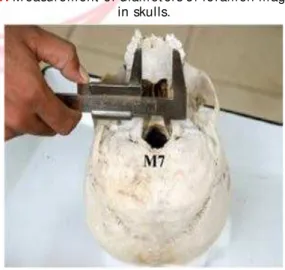
M easurements of the foramen magnum (Fig. No.1): Longitudinal diameter (LD) of t he foramen magnum - It is distance bet w een basion and opist hion. Transverse diam et er (TD) of t he foram en m agnum - It is m axim um dist ance b et w een t w o l at er al m ar gi ns [ 13] . M easurement s of t he foramen magnum w ere t aken by vernier calipers t o t he nearest of 0.1mm. M easurement s w ere taken t w ice and aver age o f t w o val u es w as t aken as f i nal measurement . Area of t he foramen magnum -It is sur face area of t he foram en m agnum calculated by t he follow ing formula [17].
AREA (A) = ¼ X p X w X h
w = W idt h, t ransverse diameter
h = Height , longit udinal diameter
= 22/7, mat hemat ical constant .CT Im age m easur em en t s o f t h e fo r am en m agnum - The best im ages of t he foram en m agn u m w as select ed , lo n git u d in al an d t ransverse diameters of t he foramen magnum w ere measured using t ools of t he soft ware. The area of t he foramen magnum was measured by planimet ry met hod (Fig. No. 2).
Fig 1:M easurem ent of diam et ers of foram en m agnum in skulls.
Int J Anat Res 2014, 2(1):249-55. ISSN 2321-4287 The Statistical methods: Result s were expressed as m ean ± st and ar d d ev iat i on and r an ge. Unpaired‘t ’ test was used t o compare bet w een males and females. P value of 0.05 or less was considered for stat ist ical significance.
RESULTS
In dry Skulls: The longit udinal diameter of t he foramen magnum in male skulls was bet w een 26.7-39.8m m , w it h a m ean of 33.4±2.6m m (mean ± SD), w hereas in females longit udinal diameter was bet w een 28-39.3mm w it h a mean o f 33.1±2.7m m (Gr ap h No . 1). Th e m ean longit udinal diam eter of m ale skulls w as not si gn i f ican t l y hi gh er t han in f em al e skul l s (p= 0.59). The t ransverse diam et er of t he foram en m agnum in m ale skulls varied from 24.3 – 37.7 mm w it h an average of 28.5±2.2mm, w hile in females t he t ransverse diameter varied from 23.6 – 33.6 mm w it h an average of 27.3± 2.0 mm (Graph No. 1). The m ean t ransverse diameter of t he foramen magnum of male skulls w as significant ly larger t han in fem ale skulls (p<0.01). The area of t he foramen magnum of male skulls w as bet w een 540.8-1002.7 mm 2, w it h an average of 748.6±97.8mm2, w hereas in female skulls t he area of t he foramen magnum w as bet w een 557.1 – 1017.7 m m2 w it h an
average of 711.1±97.7 mm2. The area of t he
foramen magnum of male skull was significant ly greater t han in female skulls (p< 0.05).
In CT Im ages: CT scan images of 30 subject s (15 male, 15 female) w it h age bet w een 6-85 year s w er e an aly sed (Tabl e. No . 1). Th e longit udinal diameter of t he foramen magnum of male subject s was varied from 32-45 mm wit h an average of 38.5±3.6mm, w hile in female it was varied from 30-39mm w it h an average of 35.2±3.1mm (Graph No. 2).
Th e lo ngit udin al d iam et er of t he f oram en magnum of male subject was significant ly large w hen co m p ar ed t o fem ale sub j ect s. Th e t ransverse diameter of t he foramen magnum in m ale subject w as bet w een 25-33m m w it h a m ean o f 29.1±2.3m m , w her eas i n f em al e subject s it was bet w een 24-33mm w it h a mean of 27.6±2.3mm(Graph No. 2). M ean t ransverse diam et er of t he foram en m agnum w as not significantly greater than in female subject s. The area of t he foramen magnum of male subject s was varied from 620- 1050mm2 w it h an average
of 862.0±119.0 m m2, w hereas in females t he
area varied from 580-940mm2 w it h an average
of 758±109m m2. The area of t he f oram en
m agnum of m ale subject s w as significant ly greater t han in female subject s.
M ALE v/ s FEM ALES
RANGE M EAN+SD RANGE M EAN+/ -SD t p
TD(mm) 24.30 - 37.70 28.50+2.20 23.50 – 33.60 27.30 + 2.00 3.39 <0.01
LD(mm) 26.70 - 39.80 33.40+2.60 28.00 – 39.3 33.10 + 2.70 0.54 0.59
A(mm2) 590.80 - 1002.70 748.60+97.80 557.1 - 1017.7 711.10 + 97.7 2.21 <0.05
TD(mm) 25.00 – 33.00 29.10+2.30 24.00 – 33.00 27.60 + 2.30 1.83 0.08
LD(mm) 32.00 – 45.00 38.5 + 3.60 30.00 – 39.00 35.20 + 3.10 2.69 <0.05
A(mm2) 620.00-1050.00 862.0 + 112.0 580.0 – 940.0 758.0 + 109.0 2.50 <0.05
CT SCAN
SAM PLES PARAM ETERS M ALES FEM ALES
SKULLS Table No. 1: Range, m ean,
st an d ar d d evi at i o n an d “ t ” values for dim ensions of foram en m agnum . Graph 1:Transverse Diam et er & Longit udinal
Diam et ers of foram en m angum : Skull
Int J Anat Res 2014, 2(1):249-55. ISSN 2321-4287
DISCUSSION
The mean longit udinal diameter of t he foramen m agnum of m ale skulls (33.4mm ) of present st udy was similar t o t he observat ions of Sayee [15] on m ale skulls of Karnat aka (34.2 m m ), how ever it was low er t han t he observat ions made by Routal [13] on Gujarat i male skulls (35.5 mm), Catalina Herrera [3] on Spain w hite male skulls and Suazo [18] on Brazilian m ale skulls (36.5 m m ). In f em al e sku l ls t he m ean longit udinal diameter of t he foramen magnum o f pr esent st ud y w as co r r el at ed w i t h t h e observat ions of Sayee [15] on Karnataka female sku l l s (33.5m m ) and W an t anab e [ 19] o n Japanese fem ale skulls (33.7m m), but it was low er t han reported by Catalina Herrera[3] on Spai n w h it e fem al e sku ll s (34.3 m m ) an d Suazo[18] on Brazilian female skulls (35.6 mm). How ever t he mean longit udinal diameter of t he foram en magnum of female skulls of present st udy was higher t han values reported by Routal [13] on Gujarat i female skulls.
In t he present st udy t h e m ean t ransv erse diameter of t he foramen magnum of male skulls was significant ly higher t han t he female skulls (P<0.01). The mean t ransverse diameter of t he foramen magnum of male skulls (28.5mm) of present st udy was similar t o t he observat ions of Sayee [15] on Karnataka male skulls (28.5mm), w hereas it was low er t han t he values reported by Catalina Herrera[3] on Spain w hite male skulls (31.1 mm), Suazo [18] on Brazilian male skulls (30.6mm) and Routal [13] on Gujarat i male skulls (30.6mm ). The m ean t ransverse diam et er of fem ale skull in present st udy (27.3m m ) w as similar t o t he observat ions made by Routal [13] on Gujarat i female skulls (27.1mm) and Sayee [15] on Karnataka female skulls (28.0mm), but it was low er t han t he values reported by Catalina Her r er a[ 3] o n Sp ai n w h i t e f em al e skul l s (29.6mm), Watanable [19] on Japan female skulls (28.6mm ) and Suazo [18] on Brazilian female skulls (29.5mm).
In t he present st udy t he area of t he foramen m agn u m o f m al e sku l ls (748.6m m2) w as
significant ly larger t han female skulls (711.1mm2
). t he mean area of t he foram en magnum of m al es in presen t st ud y w as sim i l ar t o t he observat ions made by Sayee[15] on male skulls of Karnataka (769.0mm2).
How ever it w as low er t han t he observat ions m ade by Rout al [13] on Gujarat i m ale skulls (819.0mm2), Catalina Herrera [3] on Spain w hite male skulls (888.4 mm2) and Gunay on Turkey
male skulls (909.9mm2 ). The mean area of t he
foramen magnum of female skulls (711.1 mm2)
in present st udy was similar t o t he observat ions made by Sayee[15] on Karnataka female skulls (746.0 mm2) and Routal [13] on Gujart i female
skulls (771.0mm2). It was low er in comparison
w it h observat ions of Cat alina Herrera [3] on Spain w hit e fem ale skulls (801.0m m 2) and Gunay on Turkey female skulls (819.0mm2 ).
In t he present st udy on CT scan images m ale su bj ect s sho w ed a si gn if i can t l y h i gh er longit udinal diameter and area of t he foramen magnum. The mean longit udinal diameter of the foramen magnum in present st udy on CT scan images, males was 38.5mm and in females it was 35.2m m , t hese values w ere higher t han t he values reported by M urshed [2] on Turkey people (male 37.7mm, female 34.6mm). How ever t he mean t ransverse diameter of males (29.1mm) and female (27.6mm) w ere low er t han t he values reported by M urshed[2] (male 31.6mm, female 29.6m m ). Sim ilar ly t he area of t he foram en m agn um o f m ales (862m m2) an d f em ales
(758.0mm2) w ere lower than t he values reported
b y M u rsh ed[ 2] (m ale 931.7 m m2, f em al e
795.0mm2). The conclusion was in dry skulls
-The mean t ransverse diameter and area of t he foramen m agnum of male skulls w ere higher t han fem ales, w hereas longit udinal diameter was not significant ly higher t han females. In CT scan images, t he longitudinal diameter and area of t he foramen magnum of male subject s was h i gh er t h an f em al es, w h er eas t r an sv er se diameter of t he foramen magnum of male was not significant ly higher t han females.
In a morphomet ric evaluat ion of t he foramen magnum of normal adult s by CT scan show ed. In males sagittal diameter varied from 31-45mm w it h an average of 37.2±3.43 mm, t ransverse diameter varied from 27-40mm w it h an average of 31.6±2.99 mm and area of foramen magnum was varied from 710-1266mm2 w it h an average
Int J Anat Res 2014, 2(1):249-55. ISSN 2321-4287 29.3±2.19mm and area of t he foramen magnum was varied from 671-1006mm2 w it h an average
of 795.0±99.32m m2. In t his st udy t he sagitt al
diam et er, t ransverse diam et er and area of foram en magnum of males w ere significant ly higher t han fem ales. The area of foram en magnum show ed highly significant correlat ion for bot h sagittal and t ransverse diameters [2].
The foramen magnum meningiomas are rare and comprise 0.3-3.2% of all meningiomas. It is t he most com mon neoplast ic lesion arising at t he craniocervical junction. Tw o-surgical approaches are rout inely used t o t reat foramen m agnum meningiomas; lat eral t ranscondylar approach an d i n fer i or sub o cci p i t al app r o ach w i t h m odif icat ion [20]. M et rical and nonm et rical analysis of modern female crania of nort hw est Kyushu area of Japan has report ed foramen l engt h b et w een 28-42m m w i t h m ean o f 33.7±2.13mm and foramen m agnum breadt h b et w een 24-34m m w i t h a m ean o f 28.6±1.84mm [19]. A large foram en m agnum u sual l y r esu l t s f r om chr o n ic i ncr eased int racranial pressure or from direct effect s of an expanding process w it hin foramen magnum like syringomyelia, Arnold Chiari malformat ions and also seen in children w it h Angelman syndrome or Rubinstein – Taybe syndrome. Asymmet ry of foramen magnum occurs w it h cranio-vertebral anomalies or premat ure synost oses of one or m ore of occipit al synchondroses. Key hole shaped foramen magnum has been described in hydrolit halus syndrom e [21]. According t o a st udy conducted on 211 (144 males, 71 females) Brazi li an skulls, t he m ean ant eropo st er ior diameter of t he foramen magnum for males was 36.5±2.6m m and t ran sverse diam et er w as 30.6±2.5mm. M ean antero-posterior diameter o f t h e f o ram en m agn u m f o r f em al e w as 35.6±2.5m m and t ran sverse diam et er w as 29.5±1.9mm [18].
CONCLUSION
Th e m ean l o n gi t u d i n al d i am e t e r o f t h e foram en m agnum of t he m ale and fem ale skulls was 33.4 m m and 33.1 m m respect ively, t ransverse diam et er w as 28.5m m and 27.3m m r esp ect i v el y an d ar ea w as 748.6m m2 an d
7 11 .1 m m2 r esp ect i v e l y. Hi gh si gn i f i can t
difference w as observed bet w een sexes. The dimensions of t he foramen magnum are
clinica--lly important because vital st ruct ures passing t hrough it . The know ledge of diameters of t he foram en m agnum are needed t o det erm ine radiological m alfor m at ions (Ar nold Chiar i ’s syndrome) and prior t o cutt ing off of foramen magnum or posterior cranial fossa lesions, or sex determinat ion of skulls.
Conflicts of Interests: None
REFERENCES
[1] . Standarding S. Gray’s anat omy. The anat om ical basis of clinical pract ice. 39t h ed. London : Elsevier Churchill Livingst one; 2005 .p.460.
[ 2] . M u r sh ed K A, Ci ce kei b asi A E, Tu n ce r I . M orphom et ric evaluat ion of t he foram en m agnum and variat ions in its shape. A st udy of com put erized t om ographic im ages of norm al adult s. Tur J M ed Sci 2003;33:301-306.
[3] . Sgouros S, Goldin HJ, Hockely AD, Wake M J, et al. I n t r acr an i al v o l u m e ch an ge i n ch i l d h o o d . J Neurosurg 1999;91:610-616.
[4]. Gunay Y, Alt inkok M . The value of t he size of foramen m agnum in sex det erm inat ion. J Clin foirensic M ed 2000;7(3):147-149.
[ 5] . ht t p/ / en.w i kipedia.org/ w i ki/ for am en_m agnum accessed on 18t h July 2009.
[6]. Rom anes GJ. Cunm ningham ’s t ext book of anat omy. 12t h ed. Oxford : Oxford Universit y Press; 1981 .p.114.
[ 7] . Oliveira Ed, Rhot on AL, Peace D. M icrosurgical anat omy of t he region of t he foram en m agnum . Surg Neurol 1985;24:293-352.
[8] . Rhot on AL. The foram en m agnum . Neurosurgery 2000; Suppl, 47(3):S155- S193.
[9] . Bannist er LH, Berry M M , Collins P, Dyson M , et al. Gray’s anat omy t he anat om ical basis of m edicine an d su r ge r y. 38 t h ed . Ed i n b u r gh : Ch u r ch i l l Livingst one; 1995 .p.567-568.
[10] . Singh KB. A m at er ial st udy of Chinese crania. J Anat soc India 1963;12-16.
[ 1 1] . Sch m e t ze r A. M easu r em en t o f t h e f o r am en m agnunm in children. Neuroradiology 1971;2:162. [12]. Tiexeira WRG. Sex ident if icat ion ut ilizing t he sizes of t he foram en m agnum . Am J Forensic M ed & Pat h 1982;3(3):203-206.
[ 13] . Rou t al RR, Pal GP, Bh agw at Ss, Tam ankar BP. M at er i al st u d i es w i t h sexu al d i m o r p h i sm i n foram en m agnum of hum an crania. J Anat Soc India. 1984;33(2):85-89.
[14]. Catalina – Herrera CJ. St udy of t he anat om ic m et ric values of t he foram en m agnum and it s relat ion t o sex. Acta Anat 1987;130:344-347.
[15]. Sayee R, Jankiram S, Thom as IM . Foram en m agnum m easurement s of crania from Karnataka. J Anat Soc India 1987;36(2):87-89.
Int J Anat Res 2014, 2(1):249-55. ISSN 2321-4287 [ 17] .Radinsky L. Relat ive brain size: anew m easure.
Science 1967;155:836-38.
[18]. Suazo GIL, Russo PP, Zavando M DA, Sm it h RL. Sexual dim orphism in t he foram en m agnum dim ensions. Int J M orphol 2009;27(1):21-23.
[19]. Watanabe T, Saiki K, Okam oto K, Wakebe T. M at erial and non m at er ial analysis of m odern fem ale crania
in t he nor t h w est er n Kyushu area. Ant hrop Sci 2004;112:147-159.
[20]. Pam ir M N, Kilic T, Ozdum an K, Ture V. Experience of a single inst it ut ion t reat ing foram en m agnum m eningiom as. J Clin Neuro Sci 2000;11(8):863-867. [21]. St evenson RE, Hall JG. Hum an m alform at ions and relat ed anom alies. 2nd ed. Cleveland; 2006;260-61p.