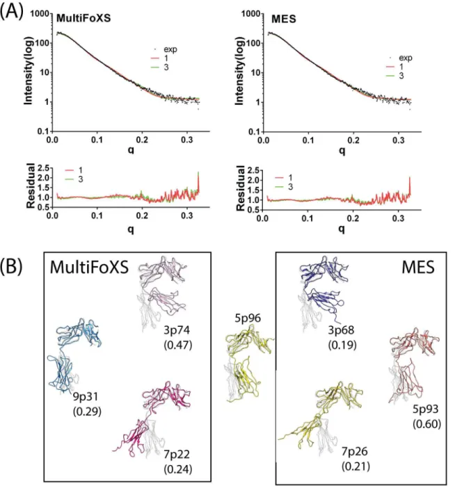
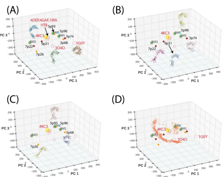
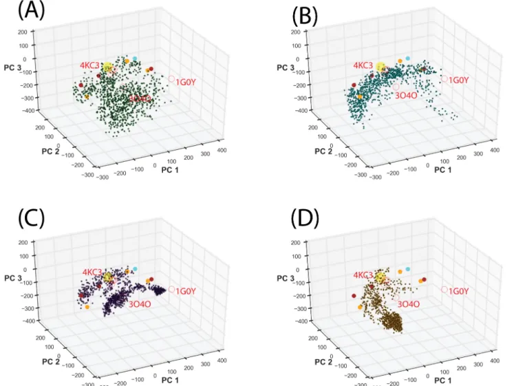
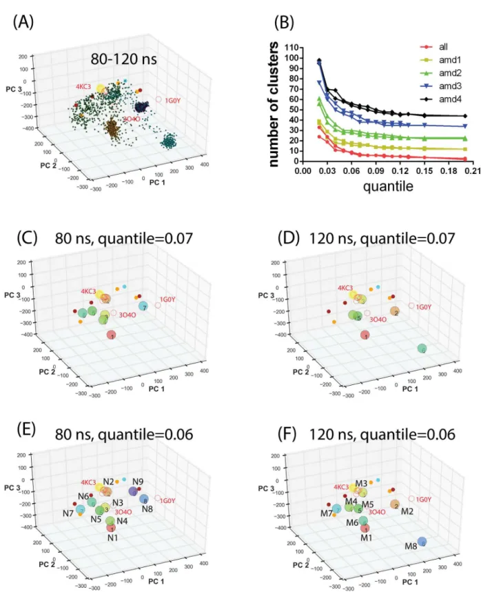
Conformational Sampling and Binding Site Assessment of Suppression of Tumorigenicity 2 Ectodomain.
Texto
Imagem
Documentos relacionados
The probability of attending school four our group of interest in this region increased by 6.5 percentage points after the expansion of the Bolsa Família program in 2007 and
Bretting, Amino acid sequence of the D-galactose binding lectin II from the sponge Axinella polypoides (Schmidt) and identification of the carbohydrate binding site in lectin II
The structure of the remelting zone of the steel C90 steel be- fore conventional tempering consitute cells, dendritic cells, sur- rounded with the cementite, inside of
Figure 3. Conformational changes of F103 during activation gate opening and SF conformations of channel states. The percentage of state was calculated from the end states at 20 ns
Since FOXO1 suppression of activin-induced Fshb transcription mapped to the 2 304/ 2 95 region of the Fshb promoter that contains multiple SMADs and FOXL2 binding sites, we
All inhibitors were subsequently docked into the binding site obtained from the receptor and conformation of the inhibitors with the lowest binding free energy was used to
The number of binding sites (n ca. 1) indicates one main binding site and molecular modeling suggests subdomain IIIA (Sudlow’s site II) as the main binding site to the
Evaluation of the number of binding sites indicate the existence of just one main binding site for the BSA:PIA association and molecular docking results suggest site I, where
