Case report
a patient with Pancoast tumor: a case report
Tomograia óssea computadorizada por emissão de fóton único (SPECT) em paciente com
tumor de Pancoast: um relato de caso
Hamid Javadi
I, Mehdi Mogharrabi
II, Isa Neshandar Asli
III, Babak Shaiei
IV, Mehrzad Bahtoee
V, Mohammad Seyedabadi
VI,
Iraj Nabipour
VII, Majid Assadi
VIIIBushehr Research Center for Nuclear Medicine, Persian Gulf Biomedical Sciences Institute, Bushehr University of Medical Sciences, Bushehr, Iran
IMD. Head of Department of Nuclear Medicine, 5th Azar Hospital, Golestan University of Medical Sciences, Gorgan, Iran.
IIMD. Assistant professor of Nuclear Medicine, Department of Nuclear Medicine, 5th Azar Hospital, Golestan University of Medical Sciences, Gorgan, Iran.
IIIMD. Head of Department of Nuclear Medicine, Taleghani Hospital, Shaheed Beheshti University of Medical Sciences, Tehran, Iran.
IVAssistant professor of Nuclear Medicine , Taleghani Hospital, Shaheed Beheshti University of Medical Sciences, Tehran, Iran.
VMD. Assistant professor of Internal Medicine, Tropical and Geographical Research Center , Persian Gulf Biomedical Sciences Institute, Bushehr University of Medical Sciences, Bushehr, Iran.
VIPhD. Pharmacologist and researcher in the Bushehr Research Center for Nuclear Medicine, Persian Gulf Biomedical Sciences Institute, Bushehr University of Medical Sciences, Bushehr, Iran. VIIMD. Professor of Internal Medicine, Tropical and Geographical Research Center, Persian Gulf Biomedical Sciences Institute, Bushehr University of Medical Sciences, Bushehr, Iran.
VIIIMD. Head of Bushehr Research Center for Nuclear Medicine, Persian Gulf Biomedical Sciences Institute, Bushehr University of Medical Sciences, Bushehr, Iran.
ABSTRACT
CONTEXT: Non-small cell lung carcinomas (NSCLCs) of the superior sulcus are considered to be the most challenging type of malignant thoracic disease. In this disease, neoplasms originating mostly from the extreme apex of the lung expand to the chest wall and thoracic inlet structures. Multiple imaging procedures have been applied to identify tumors and to stage and predict tumor resectability in surgical operations. Clinical examinations to localize pain complaints in shoulders and down the arms, and to screen for Horner’s syndrome and abnormalities seen in paraclinical assessments, have been applied extensively for differential diagnosis of superior sulcus tumors. Although several types of imaging have been utilized for diagnosing and staging Pancoast tumors, there have been almost no reports on the eficiency of whole-body bone scans (WBBS) for detecting the level of abnormality in cases of superior sulcus tumors.
CASE REPORT: We describe a case of Pancoast tumor in which technetium-99m methylene diphosphonate (Tc-99m MDP) bone single-photon emission-computed tomography (SPECT) was able to accurately detect multiple areas of abnormality in the vertebrae and ribs. In describing this case, we stress the clinical and diagnostic points, in the hope of stimulating a higher degree of suspicion and thereby facilitating appropriate diagnosis and treatment. From the results of this study, further clinical trials to evaluate the potential of SPECT as an eficient imaging tool for the work-up on cases of Pancoast tumor are recommended.
RESUMO
CONTEXTO: Carcinomas pulmonares de células não pequenas (NSCLCs) do sulco superior são considerados como o maior desaio nos tumores malignos torácicos. Nesta doença, as neoplasias se originam principalmente do ápice do pulmão e se expandem pela parede e pelas estruturas torácicas. Diversas técnicas de imagem têm sido utilizadas para identiicar e estagiar os tumores, permitindo um prognóstico para a sua ressecção em procedimentos cirúrgicos. O exame clínico tem sido bastante usado para diagnóstico diferencial de tumores do sulco superior nas queixas de dores no ombro, síndrome de Horner e anormalidades observadas em exames paraclínicos. Embora diversos tipos de exames de imagem sejam utilizados para diagnóstico e estadiamento de tumores de Pancoast, praticamente não existem relatos sobre a eicácia do exame ósseo do corpo todo (WBBS) na detecção do nível de anormalidade em casos de tumores do sulco superior.
RELATO DE CASO: Descrevemos um caso de tumor de Pancoast, em que foi realizada a tomograia computadorizada óssea por emissão de fóton único (SPECT) com metileno difosfonato de tecnécio-99 (Tc-99m MDP). Este exame foi capaz de detectar com acurácia as múltiplas áreas de anormalidade em vértebras e costelas. Ao descrever este caso, os autores ressaltam os pontos clínicos e diagnósticos, esperando estimular maior grau de suspeita, facilitando assim o diagnóstico e tratamento apropriados. A partir dos resultados deste trabalho, outros estudos clínicos podem avaliar o potencial do SPECT como uma ferramenta eiciente de imagem a ser recomendada na investigação de casos de tumor de Pancoast.
KEY WORDS:
Carcinoma, non-small-cell lung. Pancoast syndrome. Technetium Tc 99m medronate. Tomography, emission-computed, single-photon.
Magnetic resonance imaging.
PALAVRAS-CHAVE:
Carcinoma pulmonar de células não pequenas.
Síndrome de Pancoast. Medronato de tecnécio Tc 99m. Tomograia computadorizada de emissão de fóton único. Imagem por ressonância magnética.
INTRODUCTION
Pancoast tumors, irst described by Pancoast and Tobias,1-3
ac-count for less than 5% of non-small cell lung carcinomas (NSCLCs).4
hey are a rare subgroup of neoplasms that emanate mostly from the apex of the lung and expand to the chest wall and brachial plexus.5
he involvement of subclavian vessels, stellate ganglions, mediastinal
lymph nodes and adjacent vertebral bodies demands more sophisti-cated instruments for surgical treatment and may worsen the survival rate.4 he etiology of the disease includes either primary thoracic
car-cinomas, or it may be secondary to metastatic or hematological neo-plasms and a variety of infections.1
and abnormalities seen in paraclinical assessments, have been applied extensively for diferential diagnosis of superior sulcus tumors. Chest x-ray, computed tomography (CT), whole-body positron emission to-mography (PET) and magnetic resonance imaging (MRI) techniques are among the types of imaging examination used for diagnosing and staging Pancoast tumors.6 However, there have been almost no reports
on the eiciency of whole-body bone scans (WBBS) for detecting the level of abnormality in cases of superior sulcus tumors. Here, we de-scribe a case of Pancoast tumor that showed multiple areas of abnor-malities on WBBS, using planar and single-photon emission-computed tomography (SPECT) views.
CASE REPORT
A 67-year-old male subject with severe hemithorax pain and dys-pnea, along with disseminated pain and muscle weakness in the right shoulder and down the arm, was referred to our institution. Pulmonary evaluation revealed coarse as well as dull breathing sounds. Laboratory data showed normocytic normochromic anemia, whereas white blood cells, platelets, liver and renal functions, calcium, phosphorus and al-kaline phosphatase (ALP) were all within normal levels and there were no signs of organomegaly or lymphadenopathy. he chest x-ray showed collapse of the right upper lobe. he patient was further evaluated for suspected bronchogenic cancer with brachial plexus involvement. His-topathological analysis showed poorly diferentiated squamous cell car-cinoma.
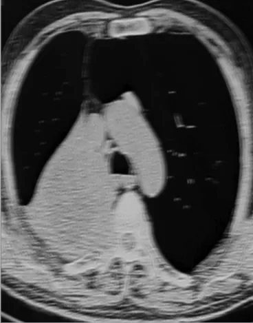
CT of the chest, upper abdomen and brain was performed with contrast and revealed some pulmonary nodules with hilar mass, resorp-tive atelectasis and obstrucresorp-tive pneumonia, along with destruction of the adjacent rib vertebra and extension to the spinal canal; there were no signs of brain involvement (Figure 1). MRI on the chest also demon-strated destruction of vertebrae and invasion into the spinal canal, there-by compressing the spinal cord (Figure 2). MRI on the spine conirmed the presence of a lytic lesion in the T4 vertebral body (Figure 3).
hree hours after administration of 750 MBq (20 mCi) technetium-99m methylene diphosphonate (Tc-technetium-99m MDP) by injection, WBBS was performed using a rotating digital gamma camera (ADAC Pegasys) equipped with a low-energy all-purpose parallel whole collimator, with a 20% window centered at 140 keV to provide energy discrimination. SPECT images were obtained in a 128 × 128 matrix, in 64 steps, with 40 s per step. he images were reconstructed and displayed on all three axes: vertical long axis, horizontal long axis and axial short axis. We found an area of diminished radiotracer uptake in the T3-T5 vertebrae and in the posterior arch of the third to ifth right ribs (Figure 4).
he patient was further evaluated for induction chemoradiotherapy and was subsequently subjected to a palliative en-bloc surgical operation with a posterior approach (Figure 5).
DISCUSSION
Assessment of patients with Pancoast tumors is mandatory not only in order to provide a prognosis, but also in order to choose the
appropri-Figure 1. Chest computed tomography with contrast: a mass is seen in the right lung apex with destruction of the adjacent rib vertebra and extension to the spinal canal. Collapse of the right upper lobe is also seen, as a result of bronchial obstruction.
Figure 3. Spine T1-magnetic resonance imaging: a lytic lesion is seen in the T4 vertebral body, suggestive of metastasis from lung carcinoma.
Figure 4. Whole body bone scan (A) in single-photon emission-computed tomography mode (B), revealing areas of decreased radiotracer uptake in the T3-T5 vertebrae and posterior arch of the third to ifth right ribs.
A
B
ate treatment approach, so that the morbidity associated with superior sulcus tumors can be minimized.7
CT scans or ultrasound-guided percutaneous transthoracic ine-needle biopsies are considered to be the most sensitive technique for identifying Pancoast tumors, whereas CT scans alone provide informa-tion about the extent of the disease and recogniinforma-tion of chest-wall in-volvement, pulmonary nodules and parenchymal diseases.7,8 Heelan et
al. investigated 32 sets of MRI and CT examinations and concluded that MRI is a procedure with greater accuracy in terms of detection of tumor invasion due to Pancoast tumors.9 MRI provides overall better
outcomes, not only in determining tumor scope and its extension to the brachial plexus, subclavian vessels, mediastinal lymph node, spinal ca-nal and vertebral bodies, but also in predicting tumor resectability and making radiosensitivity assessments.1,7,10,11
Kubota et al. reported that the C-11 methionine uptake pattern in PET showed that large-cell carcinoma with mediastinal invasion de-creased in size following radiotherapy, and this was further conirmed by biopsy. On the other hand, CT showed no noticeable change in tu-mor dimensions.12
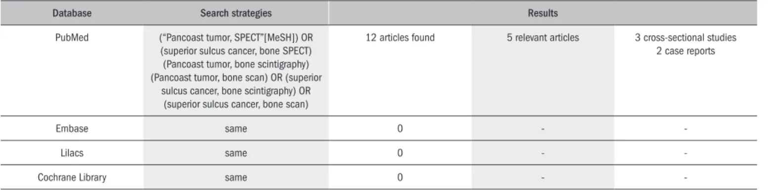
Table 1. Database search strategies for application of bone scintigraphy in cases of Pancoast tumor
Database Search strategies Results
PubMed (“Pancoast tumor, SPECT”[MeSH]) OR
(superior sulcus cancer, bone SPECT) (Pancoast tumor, bone scintigraphy) (Pancoast tumor, bone scan) OR (superior
sulcus cancer, bone scintigraphy) OR (superior sulcus cancer, bone scan)
12 articles found 5 relevant articles 3 cross-sectional studies
2 case reports
Embase same 0 -
-Lilacs same 0 -
-Cochrane Library same 0 - -
Embase = Excerpta Medica Database; Lilacs = Literatura Latino-Americana e do Caribe em Ciências da Saúde; MeSH = Medical Subject Headings.
but also in lymph node staging.13,14 Furthermore, it has been shown that
Tc-99m methoxy isobutyl isonitrile (MIBI) SPECT faithfully identi-ies not only the mediastinal lymph node metastases in patients with NSCLCs,15 but also the chemotherapy response in both small-cell lung
carcinoma (SCLC)16 and NSCLCs.17
he functional imaging mode of SPECT has a capability to visualize and measure molecular processes in vivo. However, it sufers from low
spatial resolution, in comparison with CT or MRI. One way to improve the diagnostic accuracy of this mode, despite its shortcomings, is to combine the images from this mode with images from CT and MRI.18
By integrating hybrid systems within one gamma camera, the ana-tomical inaccuracies due to image fusion would be expected to be great-ly reduced, compared with software-based image fusion, although inac-curacies in the thorax due to respiratory movements would still exist.19
A dual-headed SPECT camera with a non-diagnostic CT scanner was also recently shown to improve diagnostic speciicity with regard to diferentiating benign from malignant lesions in bone tissue.20
In searching through the main databases (PubMed, Embase, Li-lacs and Cochrane Library), we only found ive articles relating to bone scanning on Pancoast tumors (Table 1).21-25 hese studies, which
de-scribed whole-body bone scans performed on these tumors, were taken into consideration in the present study. However, none of them gave any speciic emphasis to SPECT.
Recently, some studies have shown that SPECT/spiral-CT consid-erably improves the diagnostic accuracy of radionuclide bone scanning in staging tumor patients.26,27
Bone scintigraphy is not routinely necessary in investigating Pan-coast tumors. Bone pain is the most common reason for performing WBBS,28,29 which may precede the diagnosis of metastasis. WBBS also
enables identiication of asymptomatic lesions that may produce early local and distant metastatic lesions. As shown in the present case, add-ing SPECT mode to the planar views in whole-bone scintigraphy can detect local small metastases. We have described this case of Pancoast tumor with emphasis on the clinical and diagnostic points, in the hope of stimulating a higher degree of suspicion and thereby facilitating ap-propriate diagnosis and treatment.
CONCLUSIONS
Here, we highlighted WBBS using technetium-99m methylene diphosphonate (Tc-99m MDP) in SPECT mode. We indicated that it was able to detect multiple areas of abnormality in the vertebrae and rib region that were shown using CT scans and MRI. However, addition-ally, WBBS made it possible to identify an asymptomatic lesion in these tumors, which led to changes to the management of this case.
From our observations, we recommend that a well-designed clinical trial to investigate the eicacy of Tc-99m MDP WBBS in SPECT mode should be conducted in order to investigate its potential implementa-tion for Pancoast tumor evaluaimplementa-tion.
REFERENCES
1. Arcasoy SM, Jett JR. Superior pulmonary sulcus tumors and Pancoast’s syndrome. N Engl J Med. 1997;337(19):1370-6.
2. Pancoast HK. Importance of careful roentgen-ray investigations of apical chest tumors. JAMA. 1924;83:1407-11.
3. Tobias JW. Sindrome apico-costo-vertebral-doloroso por tumor apexiano: su valor diagnos-tico en el cancer primitivo del pulmon. Rev Med Lat Am. 1932;19:1552-6.
4. Narayan S, Thomas CR. Multimodality therapy for Pancoast tumor. Nat Clin Pract Oncol. 2006;3(9):484-91.
5. Detterbeck FC. Pancoast (superior sulcus) tumors. Ann Thorac Surg. 1997;63(6):1810-8. 6. Jett JR. Superior sulcus tumors and Pancoast’s syndrome. Lung Cancer. 2003;42 Suppl
2:S17-21.
7. Rusch VW. Management of Pancoast tumours. Lancet Oncol. 2006;7(12):997-1005. 8. Urschel HC. New approaches to Pancoast and chest wall tumors. Chest. 1993;103(4
Suppl):360S-1.
9. Heelan RT, Demas BE, Caravelli JF, et al. Superior sulcus tumors: CT and MR imaging. Radio-logy. 1989;170(3 Pt 1):637-41.
10. Beale R, Slater R, Hennington M, Keagy B. Pancoast tumor: use of MRI for tumor staging. South Med J. 1992;85(12):1260-3.
11. Takasugi JE, Rapoport S, Shaw C. Superior sulcus tumors: the role of imaging. J Thorac Imaging. 1989;4(1):41-8.
12. Kubota K, Yamada S, Ishiwata K, Ito M, Ido T. Positron emission tomography for treatment evaluation and recurrence detection compared with CT in long-term follow-up cases of lung cancer. Clin Nucl Med. 1992;17(11):877-81.
13. van Tinteren H, Hoekstra OS, Smit EF, et al. Effectiveness of positron emission tomography in the preoperative assessment of patients with suspected non-small-cell lung cancer: the PLUS multicentre randomised trial. Lancet. 2002;359(9315):1388-93.
14. Vansteenkiste JF. Imaging in lung cancer: positron emission tomography scan. Eur Respir J Suppl. 2002;35:49s-60s.
15. Yamamoto Y, Nishiyama Y, Satoh K, et al. Comparative evaluation of Tc-99m MIBI and Tl-201 chloride SPECT in non-small-cell lung cancer mediastinal lymph node metastases. Clin Nucl Med. 2000;25(1):29-32.
16. Akgun A, Cok G, Karapolat I, Goksel T, Burak Z. Tc-99m MIBI SPECT in prediction of prognosis in patients with small cell lung cancer. Ann Nucl Med. 2006;20(4):269-75.
17. Nishiyama Y, Yamamoto Y, Fukunaga K, et al. Evaluation of radiotherapeutic response in non-small cell lung cancer patients by technetium-99m MIBT and thallium-201 chloride SPET. Eur J Nucl Med. 2000;27(5):536-41.
18. Schulz V, Nickel I, Nömayr A, et al. Effect of CT-based attenuation correction on uptake ratios in skeletal SPECT. Nuklearmedizin. 2007;46(1):36-42.
19. Nakamoto Y, Tatsumi M, Cohade C, et al. Accuracy of image fusion of normal upper abdomi-nal organs visualized with PET/CT. Eur J Nucl Med Mol Imaging. 2003;30(4):597-602. 20. Horger M, Eschmann SM, Pfannenberg C, et al. Evaluation of combined transmission
and emission tomography for classiication of skeletal lesions. AJR Am J Roentgenol. 2004;183(3):655-61.
21. Haniuda M, Morimoto M, Nishimura H, et al. [A case of chondrosarcoma arising from the left irst rib]. Kyobu Geka. 1990;43(10):835-8.
22. Stanford W, Barnes RP, Tucker AR. Inluence of staging in superior sulcus (Pancoast) tumors of the lung. Ann Thorac Surg. 1980;29(5):406-9.
23. Fujimura N. [Clinical study on bone metastases from bronchogenic carcinoma by bone scintigraphy with 99mTc-phosphorous compounds. III. The ability of detection by bone scin-tigraphy and roentgenography in bone destructed by Pancoast’s tumor (author’s transl)]. Nippon Igaku Hoshasen Gakkai Zasshi. 1979;39(9):984-92.
24. Sills M, Pozderac RV. Early diagnosis of Pancoast’s tumor facilitated by bone scanning. Radiology. 1974;113(2):391-2.
25. Shih WJ, DeLand FH, Domstad PA, Johnston EH. Extraosseous localization of 99mTc-MDP in ganglioneuroblastoma. Eur J Nucl Med. 1982;7(7):336-8.
26. Romer W, Nömayr A, Uder M, Bautz W, Kuwert T. SPECT-guided CT for evaluating foci of increased bone metabolism classiied as indeterminate on SPECT in cancer patients. J Nucl Med. 2006;47(7):1102-6.
27. Utsunomiya D, Shiraishi S, Imuta M, et al. Added value of SPECT/CT fusion in assessing suspected bone metastasis: comparison with scintigraphy alone and nonfused scintigraphy and CT. Radiology. 2006;238(1):264-71.
Conlict of interest: None
Sources of funding: Not declared
Date of irst submission: September 14, 2009
Last received: June 10, 2010
Accepted: June 18, 2010
Address for correspondence: Majid Assadi
Department of Nuclear Medicine, Persian Gulf Biomedical Sciences Institute, Boostan 19 Alley, Imam Khomeini Street, Bushehr, Iran