Case Report
Adenoid Cystic Carcinoma of the Buccal Mucosa with Rare Delayed
Frontal Bone Metastasis: A Case Report
Zohreh Dalirsani
1, Nooshin Mohtasham
2, Atessa Pakfetrat3, Zahra Delavarian
4,
Ala Ghazi
5, Seyedeh Asieh Rahimi
6, Zahed Anaraki
71
Associate Professor of Oral and Maxillofacial Medicine, Oral and Maxillofacial Diseases Research
Center, Faculty of Dentistry, Mashhad University of Medical Sciences, Mashhad, Iran.
2
Professor of Oral and Maxillofacial Pathology, Dental Research Center, Faculty of Dentistry,
Mashhad University of Medical Sciences, Mashhad, Iran.
3
Professor of Oral and Maxillofacial Medicine, Oral and Maxillofacial Diseases Research Center,
Faculty of Dentistry, Mashhad University of Medical Sciences, Mashhad, Iran.
4
Professor of Oral and Maxillofacial Medicine, Oral and Maxillofacial Diseases Research
Center, Faculty of Dentistry, Mashhad University of Medical Sciences, Mashhad, Iran.
5
Assistant Professor of Oral and Maxillofacial Medicine, Oral and Maxillofacial Diseases Research
Center, Faculty of Dentistry, Mashhad University of Medical Sciences, Mashhad, Iran.
6
Post-graduate Student of Oral and Maxillofacial Medicine, Faculty of Dentistry, Mashhad
University of Medical Sciences, Mashhad, Iran.
7
Cancer Research Center, Radiation Oncology Department, Omid Hospital, Mashhad University of
Medical Sciences, Mashhad, Iran
Received 22 February 2016 and Accepted 23 April 2016
Abstract
Adenoid cystic carcinoma (AdCC) is a malignant neoplasm, which accounts for 5-10% of all salivary gland tumors(1). About 50% of these tumors originate from intraoral minor salivary glands usually in the hard palate (1). Three clinically obvious characteristics of AdCC include slow growth rate, perineural invasion and high incidence of distant metastasis (1). The most commonly-affected sites of distant metastasis are bone, liver and brain, followed by lungs (2). Lymph node metastases are rare; The most common sites involved by hematogenous spread are lungs (2). This is a report about a patient with a rare form of AdCC on buccal mucosa with an unusual metastasis to the frontal region after a two-year follow up.
Key words: adenoid cystic carcinoma, buccal mucosa, frontal metastasis.
---
Dalirsani Z, Mohtasham N, Pakfetrat A, Delavarian Z, GhaziA, Rahimi SA. Multiple Adenoid Cystic Carcinoma of the Buccal Mucosa with Rare Delayed Frontal Bone Metastasis: A Case Report. J Dent Mater Tech 2016; 5(4): 208-12.Introduction
In the present case, AdCC was located in a rare region of the oral cavity, the buccal mucosa, with a subsequent metastasis in an unusual region after a two-year follow-up (i.e., the frontal area). We emphasize that the early diagnosis of malignant tumors in the oral cavity can facilitate patients’ treatment and help prevent worsening prognosis in patients with malignant tumors.
Case Report
A 47-year-old male referred to the Department of Oral Medicine, School of Dentistry, Mashhad, Iran. With the chief complaint of a mass on the right buccal mucosa (Fig. 1). The patient reported that the lesion first appeared three years before, initially observed as a small swelling in the right buccal vestibule of maxilla, interfering with proper fitting of denture. The swelling progressively increased up to its present size during the previous five months. He also reported slight pain while eating and tenderness on palpation.
Fig 1. Swelling on the right side of the face.
Clinical examination
An intraoral examination revealed a swelling on the right buccal mucosa, which was about 3 cm in the maximum dimension, extending superiorly from the tuberosity and inferiorly to the inferior border of the mandibular angle (Fig. 2a). On palpation, the swelling was firm in consistency with an irregular and well-circumscribed border (Fig. 2b). The overlying mucosa was normal in color and its surface was smooth except for a granular area of 1 cm. Moreover, leukoedema was observed on the right buccal mucosa. The color of the overlying skin was normal and no regional lymph nodes were palpable. Since the swelling was located anterior to the parotid gland, salivary secretion was not altered and there was no evidence of facial nerve dysfunction. There was also no relevant medical history. The patient underwent sonographic
examination and computed tomography (CT) scan. The sonographic image showed an infiltrative solid mass on the right buccal mucosa. The CT image revealed an infiltrative solid mass on the right buccal mucosa adjacent to the lateral wall of the right maxillary sinus and the dentoalveolar process. The tubular structure of the lesion was suggestive of hyper vascularity and vascular malformation. The adjacent bones were normal in the CT image. Based on the history and clinical examination, provisional diagnoses of salivary gland tumors and mesenchymal tumors were given.
An incisional biopsy was taken under local anesthesia. Histologically, the lesion was composed of isomorphic hyperchromatic basaloid tumor cells arranged in a cribriform pattern with pseudocystic structures. The tumor cells were small and cuboidal in shape. The tumor cells also invaded the peripheral skeletal muscle and fat tissue. In addition, superficial ulceration was observed. The histological features were suggestive of AdCC (Fig. 3). As tumoral invasion was not detected in the histological examination of the six submandibular lymph nodes, the diagnosis of stage I (T2N0M0) was established. The absence of metastases was confirmed by CT and bone scan. Surgical excision of the lesion with skin grafting was carried out (Fig. 4). The patient received 35 cycles of chemotherapy and radiotherapy. For chemotherapy regimen, cisplatin and 5-fluorouracil were applied. Two years later, the patient referred to our department with the chief complaint of a firm mass (1.5 cm) on the right side of his frontal region which had appeared since 2 months before (Fig. 5). The overlying skin was normal and the diagnosis of AdCC was made following an incisional biopsy (Fig. 6). The patient was referred to an oncologist for chemotherapy and further assessment. After performing CT-Scan, lung metastasis was also detected. Therefore stage IV was identified for the patient.
Fig 2a. Clinical picture depicting a mass of the buccal mucosa
Fig 2b. The intraoral view of lesion in buccal mucosa
Fig 3. Histopathological view of oral mass.Islands of hyperchromic cells forming cribriform and tubular
structures. (H&E)(100X).
Fig 4. Intra oral skin graft-Postoperative photograph
Fig.5. Frontal bone metastasis. The nodule was firm in palpation.
Fig 6. cribriform, trabecular and tubular structures in
Histopathologic view of frontal lision.(H&E)(100X)
Discussion
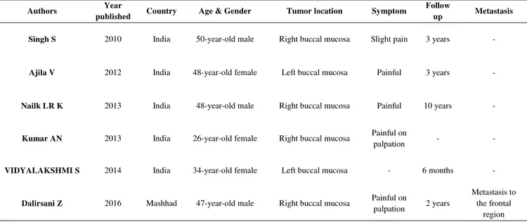
as cylandroma, accounts for 10 % of these malignancies (3).However 30-50% of primary tumors of buccal mucosa are malignant; however, it is a rare area for AdCC occurrence (2). To the best of our knowledge, our case is the sixth case with involvement of this region. Five studies performed in India have reported AdCC on the buccal mucosa (1, 2, 4, 5, 6 ) (Table 1). In the present case, the patient referred to our department two years later with a mass on his frontal region. As the distant metastasis of AdCC to the lung and bone is more frequent than metastasis to the head and neck area, the current lesion could not certainly be diagnosed as a metastatic form of AdCC in the frontal region and may simply be a second primary tumor. AdCC is the second most common malignancy of salivary glands preceded by mucoepidermoid carcinoma, occurring predominantly in the fifth and
sixth decades of life with a slight female predilection (2, 7). Nailk et al. and Sanji et al., reported AdCC in male patients, whereas other authors have mainly reported this tumor in female patients (1, 2, 4, 5, 6). Our case also occurred in a 47 year-old male. Approximately 25-50% of AdCCs metastasize distantly, especially to the lungs and bones. In addition, lymph node metastasis is relatively rare (4, 8). It should be noted that in this study, we are the first to report a case of AdCC metastasis from the buccal mucosa. AdCC is characteristically associated with small size and slow, progressive growth (2, 4, 5, 9). Our patient also complained of a slow-growing swelling for about three years. Furthermore, the tumor has a propensity for perineural invasion, which may cause pain in patients with AdCC (2, 4, 5, 9). Pain resulting from the AdCC of buccal mucosa was detected in the studies by Ajila et al., Naik et al. and Sanji et al; however,
both in our case and the case reported by Kumar et al, pain was reported on palpation (1- 3, 6).
The WHO defines AdCC as a basaloid tumor consisting of epithelial and myoepithelial cells, manifesting as three histopathological patterns (i.e., tubular, cribriform and solid). The tubular pattern has the best prognosis compared to the cribriform pattern
and the solid pattern (4, 10). AdCC is classified as grade I (with cribriform or tubular patterns), grade II (with less than 30% solid pattern) or grade III (greater than 30% solid pattern) (11). In the present case, the cribriform pattern of AdCC was dominant.
Table 1. Cases of Adenoid cystic carcinoma occurred on the buccal mucosa
Prognosis and treatment
Surgery, radiotherapy, chemotherapy and combined therapy are therapeutic modalities for AdCC. Surgery with free margins is the treatment of choice. Surgery followed by radiotherapy consists of 5 daily treatments per week, for a period of approximately 6 weeks(3). It is noted that neutron therapy can achieve more reasonable local control as the primary mode of treatment compared to photon therapy. Stereotactic
body radiation therapy such as Novalis, Cyber knife and TomoTherapy are used for destroying tumour cells (3). Chemotherapeutic regimens for AdCC include Palitaxel, Doxorubicin, Vincristin, Epirubicin, Mitoxanthrone, 5-Fluorouracil and Cisplatin. Molecular Targeted Therapy is also a promising approach in cancer treatment. It uses certain drugs such as Imatinib (against CD117), Gefitinib (against EGFR), Lapatinib and Cetoximabm in treating AdCC. The role
Metastasis Follow
up Symptom
Tumor location Age & Gender
Country Year published Authors - 3 years Slight pain Right buccal mucosa
50-year-old male India 2010 Singh S - 3 years Painful Left buccal mucosa
48-year-old female India 2012 Ajila V - 10 years Painful Right buccal mucosa
48-year-old male India
2013 Nailk LR K
- -
Painful on palpation Right buccal mucosa
26-year-old female India 2013 Kumar AN - 6 months - Left buccal mucosa
34-year-old female India 2014 VIDYALAKSHMI S Metastasis to the frontal region 2 years Painful on palpation Right buccal mucosa
47-year-old male Mashhad
of immunotherapy, gene therapy and hormonal treatment for AdCC are still undergoing clinical trials. For the present case, surgery, radiotherapy and chemotherapy were performed (3, 12, 13(.
Clinical stage, perineural invasion, observed histologic variable, surgical margin status, tumor site and cervical metastasis are the main factors determining AdCC prognosis (14). Distant metastasis is a significant characteristic of this tumor even in cases with sufficient local surgery (3). AdCC is associated with poor long-term prognosis with better prognosis in major salivary glands compared to minor types (2). In the reported case, metastasis to the frontal region was noted after two years. In other studies reporting AdCC on the buccal mucosa, no recurrence of metastasis was observed.
References
1. Kumar AN, Harish M, Alavi YA, Mallikarjuna R.
Adenoid cystic carcinoma of buccal mucosa. BMJ
case reports. 2013;2013:bcr2013009770.
2. Singh S, Gokkulakrishnan, Jain J, Pathak S, Singh
KT. Adenoid cystic carcinoma of buccal mucosa. J
Maxillofac Oral Surg.2010; Sep;9(3):273-276.
3. Vidyalakshmi S, Aravindhan R. Adenoid cystic
carcinoma of the buccal mucosa: a case report with
review of literature. Journal of Clinical and
Diagnostic Research. 2014;8(3):266-268.
4. Ajila V, Hegde S, Nair GR, Babu SG. Adenoid
cystic carcinoma of the buccal mucosa: A case
report and review of the literature. Dent Res J
(Isfahan). 2012 Sep;9(5):642-646.
5. Sa YJ, Sim SB, Kim T-J, Moon SW, Park CB.
Late-developing tongue adenoid cystic carcinoma
after pulmonary metastasectomy: a case report.
World journal of surgical oncology. 2014;12(1):1.
6. Kumaraswamy LR Naik, Pushparaja Shetty,
Padmaraj Hegde. Adenoid cystic carcinoma of
buccal mucosa with extensive hyalinization: A
unique case report. Ann Trop Med Public Health.
2013;6(5); 571-574.
7. Ellis GL, Auclair PL. Classification of salivary
gland neoplasms. Surgical Pathology of the
Salivary Glands. Philadelphia: WB Saunders &
Co. 1991; 130.
8. Giannini PJ, Shetty KV, Horan SL, Reid WD,
Litchmore LL. Adenoid cystic carcinoma of the
buccal vestibule: A case report and review of the
literature. Oral Oncol. 2006;42:1029–1032.
9. DP Vinuth, Poonam Agarwal,1 Rajesh B
Dhirawani,2 and Gunjan Dube2.Atypical case of
primary intraosseous adenoid cystic carcinoma of
mandible. J Oral Maxillofac Pathol. 2013
Sep-Dec; 17(3): 436–439.
10. Barrett AW, Speight PM. Perineural invasion in
adenoid cystic carcinoma of the salivary glands: A
valid prognostic indicator? Oral Oncol. 2009;
45:936–940.
11. Szanto PA, Luna MA, Tortoledo ME, White RA.
Histologic grading of adenoid cystic carcinoma of
the salivary glands. Cancer. 1984;54:1062–1069
12. G Papaspyrou, S Hoch, A Rinaldo, A Hominem, A
Eundem, RP Takes, CV Herpen, JA Werner, A
Ferlito. Chemotherapy and targeted therapy in
adenoid cystic carcinoma of the head and neck: A
review. Head Neck. 2011; 33:905–911.
13. Dodd RL, Slevin N. Salivary gland adenoid cystic
carcinoma: A review of chemotherapy and
molecular therapies. Oral Oncology. 2006; 42:
759– 769.
14. Huang MX, Ma DQ, Sun KH, Yu GY, Guo CB,
Gao F. Factors influencing survival rate in adenoid
cystic carcinoma of the salivary glands. Int. J. Oral
Maxillofac Surg. 1997; 26: 435-439.
Corresponding Author: Seyedeh Asieh Rahimi
Assistant of Oral and Maxillofacial Medicine, Faculty of Dentistry, Mashhad University of Medical Sciences, Mashhad, Iran.