INTRODUCTION
Article/Artigo
American tripanosomiasis: a study on the prevalence of
Trypanosoma cruzi
and
Trypanosoma
cruzi-
like
organisms in wild rodents in San Luis
province, Argentina
Tripanosomiasis americana: um estudo sobre a prevalência do
Trypanosoma cruzi
e
Trypanosoma
cruzi-like
em roedores silvestres da provincia de San Luis, Argentina
Ana María Brigada
1,2, Roberto Doña
2,3, Enrique Caviedes-Vidal
1,2,4, Edgardo Moreti
5and Beatriz Basso
5,6ABSTACT
Introduction: Chagas disease is caused by Trypanosoma cruzi. Wild and perianthropic mammals maintain the infection/transmission cycle, both in their natural habitat and in the peridomestic area. he aim of this paper was to present the results from a study on wild rodents in the central and northern regions of San Luis province, Argentina, in order to evaluate the prevalence of this infection. Methods: Sherman traps were set up in capture areas located between latitudes 32º and 33º S, and longitudes 65º and 66º W. he captured rodents were taxonomically identiied and hemolagellates were isolated. Morphological, biometric and molecular studies and in vitro cultures were performed. Infection of laboratory animals and histological examination of the cardiac muscle and inoculation area were also carried out. Parasites were detected in circulating blood in Calomys musculinus, Graomys griseolavus, Phyllotis darwini and Akodon molinae. he parasites were identiied using biological criteria. Molecular PCR studies were performed on some isolates, which conirmed the characterization of these hemolagellates as Trypanosoma cruzi. Results and Conclusions:
Forty-four percent of the 25 isolates were identiied as Trypanosomacruzi, and the remaining 56% as Trypanosomacruzi-like. hese indings provide evidence that wild rats infected with Trypanosoma cruzi and Trypanosoma cruzi-like organisms are important in areas of low endemicity.
Key-words: American trypanosomiasis. Trypanosoma cruzi. Reservoirs. Wild rodents. Chagas disease.
RESUMO
Introdução: A doença de Chagas é causada pelo Trypanosoma cruzi e os mamíferos periantrópicos e silvestres mantêm o ciclo de infecção/transmissão, tanto no ambiente natural, como no peridomicílio. O objetivo deste trabalho foi mostrar os resultados de um estudo de roedores silvestres do centro e norte da Província de San Luis, Argentina, para avaliar a prevalência da infecção. Métodos:
Estabeleceram-se lugares de caça com armadilhas tipo Sherman entre os 32º - 33º de latitude S e 65º - 66º de longitude W. Identiicou-se taxonomicamente os roedores, isolou-se os hemolagelados e izeram-se estudos morfológicos, biométricos, moleculares, cultivo in vitro, infecção a animais de laboratório, histologia de músculo cardíaco e de zona de inoculação. Observou-se parasitas em sangue circulante: Calomys musculinus, Graomys griseolavus, Phyllotis darwini e Akodon molinae. A identiicação dos parasitas foi feita utilizando critérios biológicose, em alguns, realizou estudos moleculares por PCR que conirmaram a caracterização desses hemolagelados como Trypanosoma cruzi. Resultados e Conclusões: Dos 25 isolados, 44% foram identiicados como Trypanosomacruzi e 56% como
Trypanosoma cruzi like.Este achado nosinduz a considerar a importância dos ratos do mato infectados
com Trypanosoma cruzi y Trypanosoma cruzilike, em área de baixa endemicidade.
Palavras-chaves: Tripanosomiasis americana. Tripanosoma cruzi. Reservatório. Roedores silvestres.Doença de Chagas.
1. Departamento de Bioquímica y Ciencias Biológicas, Facultad de Química, Bioquímica y Farmacia, Universidad Nacional de San Luis, San Luis, Argentina. 2. Laboratorio de Biología “Prof E. Caviedes Codelia”, Facultad de Ciencias Humanas, Universidad Nacional de San Luis, San Luis, Argentina. 3. Área de Psicobiología, Facultad de Ciencias Humanas, Universidad Nacional de San Luis, San Luis, Argentina. 4. Instituto Multidisciplinario de Investigaciones Biológicas de San Luis (IMIBIO-SL) CONICET, San Luis, Argentina. 5. Servicio Nacional del Chagas, Córdoba, Argentina. 6. Facultad e Medicina. Universidad Nacional de Córdoba, Córdoba, Argentina.
Address to: Dra. Ana Maria Brigada. Depart. de Bioquímica y Ciencias Biológicas, Facultad de Química, Bioquímica y Farmacia, Universidad Nacional de San Luis, San Luis, Argentina, Chacabuco 917, 5700 San Luis, Argentina.
Tel: 54 2652 42-4027 int 120
e-mail: [email protected];[email protected]
Received in 02/12/2008
Accepted in 16/03/2010
Chagas disease is widespread in Latin America
and is caused by the protozoan parasite Trypanosoma
cruzi. Hematophagous Triatominae(Hemiptera: Reduviidae) insects are the major vector of the disease. Wild and perianthropic mammals maintain the infection/transmission cycle in their natural
habitat as well as in the peridomesticarea1,2 .
Infection by trypanosomes depends mainly on the convergence of a given time and place, the etiological agent, the vector insect and the host animal, which are the links that comprise the wild cycle of this zoonosis. he increasing interaction between man and habitats of wild fauna favors the developmental cycle of infection, thus transmiting the parasitosis to the human environment.
Chagas disease is one of the main health problems in Latin America. However, its control is complex since the parasite can survive in the bloodstream of diferent
vertebrate hosts and Triatominae vectors3-5.
he role of wild animals as reservoirs and the low of trypanosomes across diferent habitats heighten the alert regarding the epidemic implications of this disease, and this directly afects the quality of life of the Latin
American population. Solis Franco et al6 emphasized the
importance of rodents in maintaining the wild cycle of
this disease. Trypanosoma cruzi has been reported to be
present in more than 200 mammal species7-10, including
some rodents dwelling in Argentina, such as Calomys
musculinus11, Akodon dolores12, Calomys laucha13, Phyllotis
griseolavus14 and Octodontomys gliroides15.
he aim of this work was to investigate whether wild rodents in the central and northern regions of San Luis province, Argentina (an area of low endemicity for Chagas disease) were infected with
RESULTS METHODS
Study area
Sample areas with phytogeographical characteristics representing
typical vegetation formations in San Luis province were selected16.
Lomas Blancas (32.72º S; 66.73º W); vegetation formation:
ecotone of Aspidosperma quebracho-blanco and Prosopislexuosa
forest and the Monte desert.
Mesilla del Cura (32.44º S; 65.78º W) and La Bajada (33.01º S;
66.01º W); vegetation formation: montane grasslands and forest.
La Botija (32.24º S; 66.58º S) and Daniel Donovan (33.33º S;
66.22º W); vegetation formation: Aspidosperma quebracho-blanco
and Prosopis lexuosa forest.
Beazley (33.75º S; 66.64º W); vegetation formation: low forest
of Prosopis lexuosa, Larrea divaricata and Geofroea decorticans.
Las Vizcacheras (33.56º S; 65.13º W); vegetation formation:
ecotone of Aspidosperma quebracho-blanco and Prosopis lexuosa
forest - Prosopis caldenia forest.
Capture system
In each sampling area, a capture system was established with Sherman traps bated with ground oat grains. These traps were maintained for three to six nights and were checked daily in the early morning. Samples were collected over a two-year period. Ater capture, the animals were immediately transported to the laboratory, for laboratory assays to be started.
Laboratory work
Morphological and biometric studies on the hemolagellate protozoa: blood was taken from all of the captured rodents, and
parasites were detected using the Woo technique17. Blood smears
were stained with Giemsa (1:10) after fixing with methanol. he morphology of the parasites was observed using an optical microscope (Olympus BX50). Biometric assessments was performed on microphotographs obtained using a Sony video camera and the capture X, AIMS Video Highway X-treme sotware. he parasites were measured against a standardized scale, and the following measurements were obtained: FL: lagellum length; BL: body length; TL: total length; PN: distance from the posterior end of the body to the center of the nucleus; AN: distance from the anterior end of the body to the center of the nucleus; and PN/AN: mean nuclear index
Cultures
Blood was cultured in Senekjie's culture medium18 and maintained
at 28ºC. he blood cultures were then examined over a period ofup
to 30 days,starting on the ith day ater culturing.
Infection of laboratory animals
hree to ten-day-old Balb/c albino mice (n = 300) were infected
subcutaneouslywith blood taken from wild animals, containing
between 3 and 5 x 10⁶ trypanosomes/ml. Parasitemia was controlled
starting from the seventh day ater inoculation.
Histological studies
A search for amastigotes was carried out in histological heart
sections and in the inoculation area, stainedwith hematoxylin-eosin.
Polymerase chain reaction
he polymerase chain reaction was carried out on trypanosomes
isolated from Akodon molinae was carried out. These parasites
were analyzed by amplifying the DNA of the parasite using oligonucleotides # 121 and # 122, in accordance with the technique
described by Winckler et al19. hese oligonucleotides were designed
to amplify 330 bp DNA fragments from regions of the parasite kinetoplastid minicircles (k-DNA).
Oligonucleotide Nucleotide sequence
# 121 (5'´a 3') AAA TAA TGT ACG GGT GAG ATG CAT GA # 122 (5'´a 3' GCT TCG AT GGG GT GGT GTA ATA TA
Briefly, DNA was extracted from the blood of the infected animals, then treated with a lysis bufer (SDS 1%; Tris-HCl 10mM, pH 8; EDTA 100mM) using the conventional phenol-chloroform technique, and the inal precipitate was resuspended in bidistilled sterile water and stored at -20°C. he reaction was carried out in a inal volume of 100µl, by applying 10µl of reaction bufer 10X for the Taq polymerase (Tris-HCl 100 mM, pH 8.3; KCl 500mM); 12µl of
MgCl2 25mM; 10µl of a mixture of dNTPs (10mM of each one); and
10µl of each Trypanosomacruzi-speciic primer for amplifying and
diferentiating Trypanosomacruzi kDNA. hirty-seven cycles were
performed, at between 68°C and 94°C.
Statistical analysis
he parasitemia levels of the diferent species were contrasted using the percentage of infected animals in each species, and each
sampled location, by applying the Wilcoxon matched pairs test20.
Only the species that were captured in more than ive sampling locations were used.
Parasites were detected in circulating blood in four of the seven
species analyzed: Calomys musculinus (Cm) 6.9% (percentage of
infected individuals), 8/115 (infected individuals/total captured
individuals); Graomys griseoflavus (Gg) 13.6%, 9/66; Phyllotis
darwini (Pd) 18.7%, 3/16; Akodon molinae (Am) 10.2%, 5/49. No signiicant diferences in infection prevalence according to study area were observed.
No circulating parasites were detected in Calomys venustus
(n = 7), Calomys laucha (n = 5) and Akodon sp (n = 2).
The criteria proposed by Barreto21 were used for parasite
identiication. Molecular biology techniques were also performed on some isolates.
Morphological studies
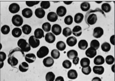
Figure 1 shows the parasites in blood smears from the infected animals. The parasites are C or S-shaped with an undulating
membrane, short lagellum,prominent subterminal kinetoplastand
well-deined nucleus.
Biometric studies
Table 1 shows the mean values from the biometric studies carried out on the parasites analyzed.
Parasite culture
FIgURE 1 - Microphotograph of isolated tripomastigotes (100X) isolated from
Calomys musculinus. Blood smear stained with giemsa.
TABLE 1 - Biometrics of the parasites isolated from the diferent rodent species.
Species FL(µ)a LC(µ)b TL(µ)c PN(µ)d AN (µ)e PN/ANf Cm
(n = 138) 5.01±0.37 20.03±2.54 25.04±2.51 9.35±0.95 10.68±1.74 0.85±0.20 Pd
(n =30) 5.10±1.0 18.38±2.00 23.60±1.50 8.82±1.40 9.57±1.50 0.94±0.20 Am
(n=36) 5.64±1.55 19.80±1.10 25.52±0.38 10.31±0.32 9.44±0.69 1.10±0.05 Gg
(n =30) 8.21±2.00 18.51±2.20 26.58±2.70 9.68±1.00 8.80±1.50 1.10±0.20 Measurements expressed in micrometers ± 1 standard deviation. alagellum length, bbody length, ctotal length, ddistance
from the posterior end of body to the center of the nucleus, edistance from the anterior end of body to the center of the
nucleus, faverage nuclear index.
Inoculation in laboratory animals
On the seventh day, circulating parasites were observed and the
morphology was similar to that of Trypanosoma cruzi. Moreover, the
biometric measurements matched Barreto’s criteria established for this species.
Histological studies
he presence ofamastigote pseudocysts was observed through
histological examination of heart tissue sections (Figure 2A).
Figure 2B shows the various stages of the parasites in Giemsa-stained inoculation areas.
ACKNOWLEDGMENTS
CONFLICT OF INTEREST
he authors declare that there is no conlict of interest.
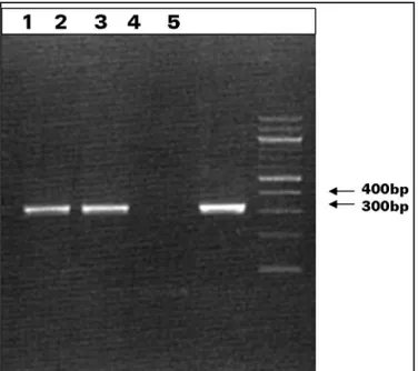
PCR analysis
DNA isolated from parasites in Akodon molinae was used for
the PCR reaction using speciic primers for Trypanosomacruzi.
Trypanosomacruzi Tulahuen DNA was used as a positive control.
DNA samples of parasites isolated from Akodon molinae showed
fragments of sizes similar to the controls (330bp) (Figure 3).
FIgURE 3 - Lane1 and 2 show the 330-bp fragment ampliied for the isolated sample of Akodon molinae. Lane 3 shows the negative control. Lane 4 shows the positive control whereas. Lane 5 shows the molecular weight markers.
DISCUSSION
Several authors have studied rodents as hosts for trypanosomes10-12,22-24. Most of these studies had the aim of describing species as trypanosome reservoirs. he present study introduces a horizontal analysis in which
various rodent species (Calomys musculinus, Graomys griseolavus,
Phyllotis darwini and Akodon molinae) were found infected with
Trypanosomacruzi or Trypanosoma cruzi-like trypanosomes. hese results support the notion that micromammals in general and rodents in particular are important as trypanosome reservoirs.
In accordance with the postulates of Barreto21, the irst factor
considered in identifying the trypanosomes was their morphological characterization. his study showed that the morphology of the
parasites isolated was similar to Trypanosoma cruzi. In fact, the
arrangement of the nuclei and the kinetoplasts of the parasites detected in the animals captured in the Lomas Blancas, Mesilla del Cura, La Bajada, Daniel Donovan and Las Vizcacheras areas matched
the morphology of the two blood typesdescribed for Trypanosoma
cruzi: a very mobile, thin shape with an elongated nucleus, a
subterminalkinetoplast and short lagellum; and a thick, slow-moving
shape with an oval nucleus and a long, free lagellum21-25.
The second parameter was the biometric measurements. In
a recent study, Martinset al26 suggested that morphometry is an
important method for biological characterization of Trypanosoma
cruzi. Biometric measurements performed on the blood forms of naturally infected animals confirmed that the parasites isolated from 11 of them were within the range of the mean nuclear index
(NA/NP) deined for Trypanosoma cruzi8. Moreover, the biometric
measurements performed on the trypanosomes obtained from the circulating blood of the Balb/c mice inoculated with blood from ield animals showed similar values to those of the respective wild strain.
One interesting observation was that the mortality rate among the laboratory Balb/c mice inoculated with wild strains was very high during the acute phase of the infection. In addition, amastigote forms found in histological sections of cardiac tissue and in the macrophages of the inoculation area of the experimentally infected Balb/c mice
were characteristic of the developmental cycle of Trypanosomacruzi
and were one of the fundamental parameters for characterizing the
strains4,8. he fact that no amastigotes were observed in the naturally
infected wild animals was in agreement with previous indings11-13.
According to Deane23, the lack of amastigotes may be due to analyzing
the wild animals during the chronic phase of the infection, when the tissue forms are scarce. In the areas where the infected wild rodents were captured, no wild triatomines (which would potentially be responsible for wild transmission) were found.
An additional xenodiagnosis performed on some of the experimentally infected wild mice revealed the characteristic metacyclic epimastigote and trypomastigote forms that were observed in the vector (data not shown).
he molecular biological studies performed on the trypanosome
isolates from Akodon molinae provided additional data that supported
the identiication of these hemolagellatesas Trypanosoma cruzi.
Together, the results from this study conirmed that 11 (44%)
of the 25 isolates were Trypanosoma cruzi. he other 14 (56%) were
classiied only as Trypanosoma cruzi-like, in terms of morphology,
biometric measurements, similar reservoir use and capture areas, since not all of the studies were performed on them. he presence of Trypanosoma lewisi was discarded because no blood spliting due to this protozoon was observed.
Finally, it is important to state that to our knowledge, this is the
irst time that the prevalence of Trypanosoma cruzi and Trypanosoma
cruzi-like organisms has beenreported in wild rodents of seven different populations from the Midwest region of Argentina, inhabiting diferent phytogeographical regions in an area where 7% of the population is serologically positive for Chagas disease. Moreover, these results conirm that this parasite is widespread in the wild environment, in spite of preventive fumigation eforts, and it maintains an independent cycle that might be inluenced by factors that are still unknown and which could inluence the human population’s health.
he authors dedicate this manuscript to Dr. Enrique Caviedes
Codelia on his 10th death anniversary and for his important
FINANCIAL SUPPORT
REFERENCES
Universidad Nacional de San Luis, Argentina.
1. Carcavallo R. Aspects of the epidemiology of Cha gas disease in Venezuela and Argentina. New Approaches in American Trypanosomiasis Research. PAHO; 1975. p.318-347.
2. Pessoa SB. Hospedeiros vertebrados (nao humanos) do Trypanosomacruzi. Goiana Medical 1958; 4: 83.
3. Chagas C. Nova tripanosomiase humana.Estudos sobre a morfolojia e o ciclo evolutivo do Schizotrypanum cruzi, ng, nSp, ajente etiológico de nova entidade morbida do homem. Mem do Inst Oswaldo Cruz 1909; 1:159-218.
4. Días JCP. Chagas disease-American trypanosomiasisits impact on transfusion and clinical medicine. In: Wendel S, Brener Z, Camargo MG, Rassi A. editor Brazil,1992.
5. Guzman-Martin E, Zavala-Castro JE, Acosta-Viana KY, Rosado-Barrera ME. Importancia de la caracterización de cepas de Trypanosoma cruzi. Rev Biomedica 1999; 10:177-184.
6. Solís-Franco R, Romo-Zapata JA, Martínez-Ibarra JA. Wild Reservoirs Infected by Trypanosoma cruzi in the Ecological Park “El Zapotal”, Tuxtla Gutiérrez, Chiapas, México. Memorias do Instituto Oswaldo Cruz 1997; 92: 163-164. 7. Brener Z. Biology of Trypanosoma cruzi. Annales Revista Microbiologia
1973;27: 347-382.
8. Ferriolli F, Barreto MP, Carvalheiro JR. Estudos sobre reservatorios e vectores silvestres do Trypanosoma cruzi. XXIV. Variação dos dados biometricos obtidos em amostras do T. cruzi isolados de casos humanos da Doenca de Chagas. Rev Soc Bras Med Trop 1968; I:2.
9. Hoare CA. Morphological and taxonomic studies on mammalian trypanosomes. X Revision of the Systematies. Journal Protozoology 1964; 11: 200-207. 10. Hoare CA. he trypanosomes of mammals. A Zoological Monograph. Blackwell
Scientiic Publications, Oxford and Edinburg; 1972.
11. Basso B, Eraso AJ, Moreti ER, Albesa I, Kra vetz F. Infección natural de Calomys musculinus (Rodentia: Cricetidae) por Trypanosoma cruzi. Asociación Argentina de Microbiología 1977; 9: 11-16.
12. Basso B, Moreti ER, Albesa I, Eraso AJ, Kravetz F, Dalessandro A. Infección natural de Akodon dolores homas 1916, (Rodentia: Cricetidae) por el Trypanosoma cruzi. Rev Inst Med Trop São Paulo1982; 24: 21-26.
13. Moreti ER, Basso B, Albesa I, Eraso AJ, Kravetz FO. Infección natural de Calomys laucha por Trypanosoma cruzi. Medicina 1980; 40 (supl 1): 182-186.
14. Rodríguez LR, Carcavallo R, Massoia EF. Estudios del Phyllotis griseolavus como reservorio de T. cruzi. Segundas Jornadas de Entomología Argentina; 1960. p.75-83. 15. Schweigmann NJ, Alberti A, Pietrokovsky S, Conti O, Riarte A, Montoya S,
Wisnivesky-Colli C. A new host of Trypanosoma cruzi from Jujuy, Argentina:
Octodontomys gliroides (Gervais & D’Orbigny, 1844) (Rodentia, Octodontidae). Mem Inst Oswaldo Cruz 1992; 87: 217.
16. Anderson DL, del Aguila J, Bernardon A. Las formaciones vegetales en la provincia de San Luis. Revista Investigaciones Agropecuarias. INTA. Serie 2, Biol Produc Vegetal 1970; VII 3:152-183.
17. Woo PT. he haematocrit centrifuge for the detec tion of trypanosomes in blood. Cana J Zoology 1969; 47:921-924.
18. Senekjie HA. Biochemical reactions, cultural characteristics and growth requirements of Trypanosoma cruzi. Ame J Trop Med Hygiene 1943; 23:523. 19. Winckler P, Brito C, Pereira JB, Cardoso MA, Oelemann W, Morel CM. Use of
a simpliied polymerase chain reaction procedure to detect Trypanosoma cruzi in blood samples from chronic chagasic patients in a rural endemic area. Am J Trop Med Hygiene 1994; 51:771-777.
20. Wilcoxon F. Individual comparisons by ranking methods. Biometrica Bulletin 1945; 1:80-83.
21. Barreto MP. Tripanossomos semelhantes ao Trypa no so ma cruzi em animais silvestres esua identiicacao com o agente etiológico doenca de Chagas. Rev Inst Med Trop Sao Paulo 1965; 7:305-315.
22. Cortez MR, Pinho A, Cuervo P, Alfaro F, SolanoM, Xavier SCC, D’Andrea PS, Fernandes O, Torrico F, Noireau F, Jansen AM. Trypanosoma cruzi
(Kinetoplastida Trypanosomatidae): Ecology of the transmission cycle in the wild enviroment of Andean vallery of Cochabamba, Bolivia. Experim Parasitol 2006; 114:305-313.
23. Deane LM. Animal reservoirs of Trypanosoma cruzi in Brazil. Rev Bras Malariol Doen Trop 1964;16: 27-48.
24. Xavier SC, Vaz VC, D’Andrea PS, Herrera L, Emperaire L, Alves R, Fernández O, Ferreira LF, Jansen AM. Mapping of the distributionof Trypanosoma cruzi
infection among small wild mammals in a conservation unit and its sorrounding (Northeast Brazil). Parasitol Internat 2007; 56:119-128.
25. Souza WA. Short Review on the morphology of Trypanosome cruzi: from 1909 to 1999. Mem Inst Oswaldo Cruz 1999; 94(Supl I):17-36.