http://dx.doi.org/10.1590/S1678-9946201759053
(1)Universidade de Fortaleza, Centro de Ciências da Saúde, Programa de Pós-Graduação em Saúde Coletiva, Fortaleza, Ceará, Brazil
(2)Universidade Federal do Rio Grande do Sul, Faculdade de Medicina, Porto Alegre, Rio Grande do Sul, Brazil
(3)Universidade Federal do Ceará, Faculdade de Medicina, Fortaleza, Ceará, Brazil
Correspondence to: Geraldo Bezerra da Silva Junior
Universidade de Fortaleza, Centro de Ciências da Saúde, Programa de Pós-Graduação em Saúde Coletiva, Av. Washington Soares, 1321, Bloco S, Sala S-01, CEP 60811-905, Fortaleza, CE, Brazil
E-mail: [email protected]
Received: 9 December 2016
Accepted: 22 February 2017
Kidney involvement in malaria: an update
Geraldo Bezerra da Silva Junior1, José Reginaldo Pinto1, Elvino José Guardão
Barros2, Geysa Maria Nogueira Farias1, Elizabeth De Francesco Daher3
ABSTRACT
Malaria is an infectious disease of great importance for Public Health, as it is the most prevalent endemic disease in the world, affecting millions of people living in tropical areas of the globe. Kidney involvement is relatively frequent in infections by P. falciparum and
P. malariae, but has also been described in the infection by P. vivax. Kidney complications in malaria mainly occur due to hemodynamic dysfunction and immune response. Liver complications leading to hepatomegaly, jaundice and hepatic dysfunction can also contribute to the occurrence of acute kidney injury. Histologic studies in malaria also evidence glomerulonephritis, acute tubular necrosis and acute interstitial nephritis. It is also possible to find chronic kidney disease associated with malaria, mainly in those patients suffering from repeated episodes of infection. Plasmodium antigens have already been detected in the glomeruli, suggesting a direct effect of the parasite in the kidney, which can trigger an inflammatory process leading to different types of glomerulonephritis. Clinical manifestations of kidney involvement in malaria include proteinuria, microalbuminuria and urinary casts, reported in 20 to 50% of cases. Nephrotic syndrome has also been described in the infection by P. falciparum, but it is rare. This paper highlights the main aspects of kidney involvement in malaria and important findings of the most recent research addressing this issue.
KEYWORDS: Malaria. Kidney disease. Acute kidney injury. Glomerulonephritis. Chronic kidney disease.
INTRODUCTION
Malaria is an infectious disease of great interest for Public Health because it remains as the most prevalent endemic disease in the world. The etiologic agents are parasitic protozoans of the genus Plasmodium. There are four species that cause disease in humans in Brazil: P. falciparum, P. vivax, P. ovale and P. malariae. P. falciparum causes falciparum malaria, which tend to be a more severe disease.
P. vivax and P. ovale causes tertian malaria, and P. malariae causes quartan malaria1.
There can be severe complications, including kidney disease, mainly in infections by P. falciparum and P. malariae2.
P. vivax is the most prevalent in tropical regions, and P. falciparum has been presenting increasing resistance to some drugs, such as chloroquine. These parasites can invade erythrocytes leading to high parasitemia levels, which correlate with disease severity and mortality.
Epidemiology
the World Health Organization, its incidence varies from 200 to 300 million new cases annually, with 200,000 to 600,000 deaths3. Brazil is responsible for the majority of
cases in the Americas, with approximately 500,000 cases each year, mainly in the Amazon region. In the last years, we have observed a decrease in its incidence, going from around 300,000 cases in 2003 to 143,000 in 20154. Among
the main strategies adopted in Brazil in the fight against Malaria, there is drug therapy, which is freely available to all patients according to guidelines periodically revised by experts and the Ministry of Health5.
Malaria outbreaks are generally influenced by multifactorial process, including environmental factors (vegetation, climate, hydrology), sociodemographic (migrations, population density, laboral activity), biological (species, Anopheles mosquito density, Plasmodium species, degree of the population immunity), and political (territory division, healthcare service organization, general infra-structure of cities)6.
Clinical manifestations
The infection begins when the infective sporozoites are inoculated in men by the vector, an insect of the genus
Anopheles. After some phases of evolutionary cycle, the schizonts appear and thousands of merozoites invade erythrocytes. The parasites multiply inside erythrocytes causing its rupture (hemolysis). The mean incubation time is 12 days for P. falciparum, 14 days for P. vivax and 30 days for P. malariae, and is usually shorter when the infection is acquired through blood transfusion2.
Malaria can be acute, fulminant or chronic. Patients present asthenia, anorexia, headache, myalgia, nausea and vomiting. The acute form is usually caused by
P. vivax, P. ovale and, less frequently, by P. malariae. Patients present fever, with intervals from 1 to 4 days, associated with chills and sweating, anemia, hepatomegaly
and splenomegaly. Diagnosis can be made through microscopy by finding the parasite inside erythrocytes in thin or thick blood films. Fulminant malaria is caused by
P. falciparum, with patient presenting anemia, adynamia, diarrhea, jaundice, acute kidney injury, hydroelectrolytic disturbances, respiratory failure, disseminated intravascular coagulation, shock and coma. Disease reactivation can be seen in the infection by P. ovale and P. vivax, due to persistence in the liver of quiescent forms, the so-called hypnozoites, and manifest with fever reappearance, anemia, malnutrition, hepatomegaly and splenomegaly. The chronic form is associated with P. malariae, which is the parasite most commonly implicated in malaria-associated glomerulonephritis, but P.vivax, despite being considered a “benign” parasite, resulting in low mortality rates, has also been implicated in severe disease7,8. Renal complications in
P.vivax infections are associated with age, hemodynamic disturbances and respiratory failure9.
Renal involvement in malaria
Malaria was the first parasitic infection to be clearly associated with glomerular diseases in tropical areas10.
Severe malaria can cause disease in glomeruli, tubules and in the interstitial region. Kidney disease in malaria is primarily due to erythrocyte abnormalities. Parasitized red cells tend to adhere to healthy erythrocytes, blood platelets and capillary endothelium, leading to formation of rosettes and clumps, which impair microcirculation1,
and these events are probable contributing factors for kidney injury, in association with hemodynamic instability, including hypovolemia and shock. Endothelial activation leads to the release of several cytokines, including thromboxane, catecholamines, endothelin and other inflammatory mediators that are also implicated in the pathogenesis of malaria-associated kidney injury. Immune system activation in malaria can go through Th1 and Th2 response. When Th2 response prevails in the infection by
P. malariae, complement activation occurs, with deposits of immune complexes leading to glomerulonephritis. Hemodynamic instability due to intense erythrocyte parasitism leads to acute tubular necrosis, as seen in the infection by P. falciparum. When Th1 response prevails, acute interstitial nephritis and acute glomerulonephritis can be seen. Cortical necrosis has also been described in malaria, characterizing a more severe kidney injury and generally associated with non-recovery of renal function and consequently development of end-stage kidney disease8.
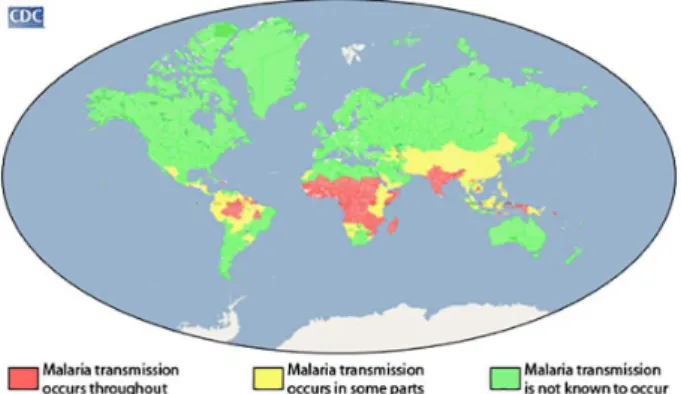
Several factors contribute to the occurrence of these complications, including hypovolemia, vasoconstriction, hemolysis (leading to hemoglobinuria), erythrocyte Figure 1 - Global distribution of malaria transmission, according
parasitemia, immune complexes deposition in glomeruli, microcirculation dysfunction (due to cytoadherence of parasite erythrocytes) and rhabdomyolysis (which is not common in malaria). Other contributing factor for kidney disease in malaria is hepatic dysfunction, with jaundice and hepatomegaly, through which hyperbilirubinemia can lead to cast nephropathy and acute kidney injury (AKI), and liver disease and its complications can also cause AKI (hepato-renal syndrome)7,9,11-13.
AKI is reported in the infection by different Plasmodium
species (P. falciparum, P. vivax, P. malariae and P. ovale), and can worsen due to low hydration and fluid loss caused by vomiting, pyrexia, sweating and dehydration. Histologic studies have showed glomerulonephritis, acute tubular necrosis and interstitial nephritis. It is also possible to find chronic kidney disease associated with malaria, mainly in those patients suffering from repeated episodes of infection1,8,14-16.
Kidney involvement by P. falciparum
AKI is a known complication of malaria and can occur in around 40% of patients with severe disease by P. falciparum in endemic regions, contributing to high mortality rate, around 75% of cases12,17,18. P. falciparum causes the most
severe form of malaria and is responsible for most cases of AKI16. In some regions of the globe, malaria is responsible
for a significant part of patients admitted with AKI (more than 10% of cases)15.
Clinical manifestations of P. falciparum malaria-associated AKI includes oligo-anuria (46-76% of cases), severe metabolic acidosis and hypercatabolic state in the majority of cases15,16. AKI in these cases occurs in the
context of severe malaria, and kidney involvement is per se one of the criteria for classifying patients as having severe falciparum malaria19. Recent studies have found
as risk factors for AKI in malaria: advanced age, referral from another hospital, hyperbilirubinemia, inotropic drugs requirement, hospital-acquired secondary infection, and factors associated with mortality: advanced age, hyperkalemia, jaundice, altered consciousness level, leukocytosis, oligo-anuria and P. falciparum infection (in comparison with P. vivax infection)16. Electrolyte
abnormalities include hyponatremia, which seem to be frequent in malaria-associated AKI, occurring in 30-50% of cases1,15, and hyperkalemia, which is associated with
hemolysis, rhabdomyolysis and acidosis1, as well as AKI
per se.
AKI pathogenesis in malaria is not yet fully understood. Renal microcirculation blockade due to parasite erythrocytes sequestration, immune-mediated glomerular injury and
volume depletion are possible mechanisms. The main kidney histopathological finding in malaria is acute tubular necrosis and, less frequently, interstitial nephritis and glomerulonephritis, suggesting a key role of hemodynamic factors in malaria-associated AKI12.
Glomerulonephritis in patients with P. falciparum
infection is not common, and it seems that children are more likely to be affected by this complication. The exact incidence of glomerulonephritis in malaria is not known, but it is estimated to be around 18%. Mild proteinuria, microalbuminuria and urinary casts are reported in 20 to 50% of cases. Nephrotic syndrome associated with P. falciparum infection is rare. This species is associated with acute tubular necrosis, cast nephropathy, inflammatory interstitial infiltrate and edema10. The mechanism that leads
to AKI in malaria is complex and includes mechanic and immune factors, cytokines release and acute phase response.
New kidney injury biomarkers have been investigated in malaria by P.falciparum, including neutrophil gelatinase-associated lipocalin (NGAL) and kidney injury molecule-1 (KIM-1), which have the advantage of detecting AKI earlier than traditional markers, such as creatinine. A recent study has found evidence that 31% of patients with malaria-associated AKI had normal levels of creatinine at presentation, illustrating the importance of new AKI biomarkers. NGAL seems to be a good biomarker for malaria-associated AKI and has already been investigated in different types of kidney injury18.
Hypergammaglobulinemia is frequently seen in malaria. The infection leads to immunosuppression secondary to cytokine production causing depletion of macrophages and T cells, which is triggered when the organism interacts with the innate immune system that in turn will activate B cells, which will produce immunoglobulins. The production of immunoglobulins leads to immune complex formation, which is related to glomerular injury. The molecular mechanisms involved in malarial nephropathy are still being discussed, and it is suggested that there is participation of TNF-α, IL-1α, IL-6, IL-10 and granulocyte-macrophage
colony-stimulating factor (GM-CSF)1,10.
There are few studies about glomerular involvement in
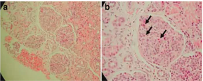
Through immunofluorescence IgM, IgG and C3 deposits have been identified in P. falciparum malaria. More recent studies have also found IgA deposits in malaria-associated AKI, with resolution after the acute phase of the infection21,22,
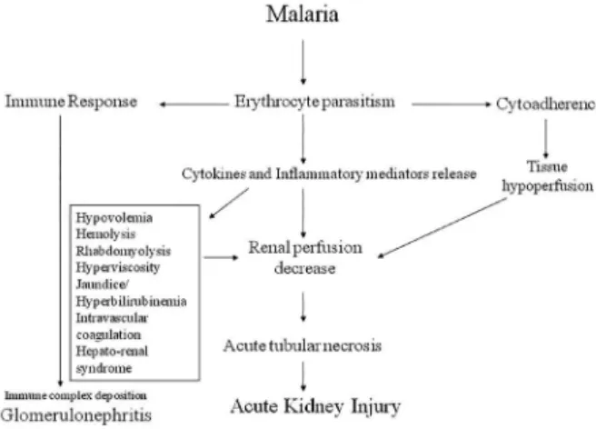
as illustrated in Figure 2. Eosinophilic glomerulonephritis has also been found in children with P. falciparum infection23
(Figure 3). Electronic microscopy findings evidence the
presence of electron-dense deposits in subendothelial and mesangial region, associated with the presence of granular, fibrilar and amorphous material. Autoantibodies have also been detected in patients with malaria-associated glomerulonephritis, but its role has yet to be determined24.
Tubular abnormalities in malaria include granular deposits of hemosiderin and the presence of urinary casts,
associated with mononuclear cells infiltrate and interstitial edema, besides acute tubular necrosis (ATN) caused by hemodynamic instability. ATN occurs in 1 to 4% of cases of P. falciparum infection. The oliguric phase can last a few days to weeks and is characterized by microcirculation abnormalities, such as peripheral vasodilation, and can be associated -with hemolysis, rhabdomyolysis and disseminated intravascular coagulation. Oxygen reactive species, TNF-α, and nitric oxide also contribute- to the
hemodynamic disturbances. Relative hypovolemia leads to an increase in catecholamine, renin, vasodilators prostaglandins and vasopressin, which contributes to AKI development. Interstitial nephritis is one of the most frequent manifestations of P. falciparum infection-associated nephropathy, which can be considered as a consequence of the host’s immune response to infection20.
Hemolytic-uremic syndrome (HUS) is also described in malaria, but its mechanisms are still poorly understood. High serum levels of cytokines are possibly related to this complication. Malaria specific treatment leads to recovery from HUS, evidencing the causal role of infection for HUS development25.
Kidney involvement by
P. malariae infection usually does not cause severe disease, but can be associated with renal complications, including AKI and glomerulopathies26. Proteinuria is found
in around 46% of cases, and is occasionally associated with microhematuria. Complement levels are normal, and there are deposits of immune complexes, leading to mesangiocapillar glomerulonephritis and, in some cases, nephrotic syndrome that develops some weeks after the beginning of infection. Clinical manifestations occur in two ways: a benign pattern, with mild and transitory proteinuria, without renal function derangement, appearing in the second or third week of infection, and a severe pattern, with persistent proteinuria or nephrotic syndrome. The most common glomerular disease associated with P. malariae
infection is the membranoproliferative glomerulonephritis. An electron microscopy study has shown the thickening of glomerular capillary walls caused by subendothelial deposits, with “tram track” appearance and mesangial proliferation23. Immunofluorescence has shown granular
deposits through the endothelium with IgG, IgM, C3 and parasite’s antigens deposits. Proliferative lesions, involving mainly the mesangium, are frequent, with crescent formation in rare cases. The initial lesion is focal and can evolve to glomerular sclerosis. As in other parasitic diseases, glomerular lesion progression in malaria depends on many factors and requires the association between immune Figure 3 - Kidney biopsy from a patient with P. falcifarum
-associated glomerulonephritis: A) Eosinophilic diffuse proliferative glomerulonephritis (hematoxylin and eosin staining); B) One glomerulus showing some eosinophils (arrows). Reproduced by Walker et al.23, with permission. ©2007
Elsevier – The International Society of Nephrology
Figure 2 - Kidney biopsy from a patient with P. falcifarum -associated immunoglobulin A (IgA) nephropathy: A) The renal biopsy specimen showed mild mesangial proliferation and expansion (original magnification × 400); B) Acute and chronic inflammatory cell infiltration in the tubulointerstitium with multifocal hemosiderin casts (original magnification × 200); C) Direct immunofluorescence showed mesangial staining for IgA (2+); D) Electron microscopy showed multifocal electron-dense deposits within the mesangium and irregularly thickened glomerular basement membrane ranging from 800 nm to 1,200 nm in thickness. Diffusely effaced foot processes were also observed. Reproduced by Yoo et al.21, with permission. © 2012
complexes deposits and other aspects such as genetic factors of the host and pathogenic mechanism associated with the infection.
Rapidly progressive glomerulonephritis associated with P. malariae infection is not common, but has been observed in endemic areas. There is no specific clinical presentation for this condition in malaria. Micro-hematuria is occasionally observed in older patients, nephritic syndrome occurs with variable incidence, and hypertension can be seen as a late manifestation. The disease can evolve despite parasite elimination, causing chronic kidney disease, detectable after 3 to 5 years of primary infection10.
AKI due to P. vivax infection is not common, although recent studies in endemic areas have evidenced
P. vivax infection in 16% of malaria-associated AKI, and histologic findings (among 15 patients who have undergone biopsies) included acute tubular necrosis, cortical necrosis, tubulointerstitial nephritis and crescentic glomerulonephritis8.
The pathophysiology of malarial nephropathy is illustrated in Figure 4.
Treatment
Treatment of malaria-associated kidney disease includes appropriated antimalarial drugs, besides all supportive measures that AKI requires (hydroelectrolytic disturbances corrections, fluid replacement and dialysis)27,28.
A recent study by Luz et al.5 has evidenced that in
Brazil there are a variety of therapeutic specific schemes for malaria, including drugs that can be used during pregnancy, such as chloroquine (as monotherapy). This scheme is also preconized in our area due to the high prevalence of P. vivax
infection, which usually causes a milder disease than that observed in infections by other Plasmodium species.
Chloroquine is the drug of choice for treatment of uncomplicated malaria, although there is already resistance, mainly for P. falciparum, against which therapy should include primaquine, quinine or mefloquine. To guarantee a good efficacy and low toxicity, it is necessary to adjust antimalarial drugs doses to the patients’ weight. In malaria by P. vivax or P. ovale it is also necessary to include primaquine, to eradicate hypnozoites. All antimalarial drugs can induce hemolysis in patients with G-6-PD deficiency, mainly when the intravenous route is used, leading to unknown origin fever. Quinine and chloroquine interacts with cyclosporine so that renal transplant patients require higher doses of these antimalarials to maintain adequate serum concentrations of the drugs19.
For severe malaria treatment, the World Health Organization recommends the use of intravenous or intramuscular antimalarial drugs, and the most effective drug for this purpose is artesunate, which should be administered for at least 24 h and until patients can tolerate oral medication. The available formulation is the artesunic acid in powder, which should be dissolved in sodium bicarbonate (5%) to form sodium artesunate. This solution in then diluted in 5 mL of 5% dextrose and given intravenously or intramuscularly19.
Renal replacement therapy (dialysis) should be considered in AKI treatment, with hemodialysis being more effective. Dialysis is required in 46 to 76% of cases, and complete renal function recovery is reported to occur in approximately 64% of cases in both P. falciparum and
P. vivax malaria-associated AKI8,15. Antimalarial drugs
are not adequately depurated in hemofiltration dialysis, so dialysis does not interfere with the specific treatment of malaria. Early initiation of dialysis, for any AKI cause, has been associated with better outcomes, and a recent meta-analysis found a 25% reduction in all-cause mortality and 30% increase in renal recovery among patients with AKI receiving early renal replacement therapy29, so we
recommend early dialysis initiation for AKI associated with severe malaria.
Vaccines for malaria are being developed and tested, so it is possible that in a near future the burden of the disease decreases, as well as renal complications30.
REFERENCES
1. Barsoum RS. Malarial acute renal failure. J Am Soc Nephrol. 2000;11:2147-54.
2. Fairhurst RM, Wellems TE. Plasmodium species (Malaria). In: Mandell GL, Bennett JE, Dolin R, editors. Mandell, Douglas, and Bennett’s Principles and practice of infectious diseases. 7th ed. Philadelphia: Churchill Livingstone; 2010. p.3437-62.
3. World Health Organization. World Malaria Report 2015. Geneva: WHO; 2015. [cited 2017 Jan 20]. Available from: http://www. who.int/malaria/publications/world-malaria-report-2015/ wmr2015-without-profiles.pdf?ua=1
4. Brasil. Ministério da Saúde. Secretaria de Vigilância em Saúde. Situação epidemiológica da malária no Brasil, 2012 e 2013. Bol Epidemiol. 2015;46(43):1-17.
5. Luz TC, Miranda ES, Freitas LF, Osório-de-Castro CG. Prescrições para tratamento de malária não complicada em gestantes na Amazônia Legal: evidências do Projeto Mafalda. Rev Bras Epidemiol. 2013;16:409-19.
6. Braz RM, Guimarães RF, Carvalho Júnior OA, Tauil PL. Spatial dependence of malaria epidemics in municipalities of the Brazilian Amazon. Rev Bras Epidemiol. 2014;17:615-28.
7. Kute VB, Trivedi HL, Vanikar AV, Shah PR, Gumber MR, Patel HV, et al. Plasmodium vivax malaria–associated acute kidney injury, India, 2010–2011. Emerg Infect Dis. 2012;18:842-5.
8. Naqvi R. Plasmodium vivax causing acute kidney injury: a foe less addressed. Pak J Med Sci. 2015;31:1472-5.
9. Saravu K, Rishikesh K, Parikh CR. Risk factors and outcomes stratified by severity of acute kidney injury in Malaria. PLoS One. 2014;9:e90419.
10. Elsheikha HM, Sheashaa HA. Epidemiology, pathophysiology, management and outcome of renal dysfunction associated with plasmodia infection. Parasitol Res. 2007;101:1183-90.
11. Gomes AP, Vitorino RR, Costa AP, Mendonça EG, Oliveira MG, Siqueira-Batista R. Malária grave por Plasmodium falciparum. Rev Bras Ter Intensiva. 2011;23:358-69.
12. Koopmans LC, van Wolfswinkel ME, Hesselink DA, Hoorn EJ, Koelewijn R, van Hellemond JJ, et al. Acute kidney injury in imported Plasmodium falciparum malaria. Malar J. 2015;14:523.
13. Mohapatra MK, Behera AK, Karua PC, Bariha PK, Rath A, Aggrawal KC, et al. Urinary bile casts in bile cast nephropathy secondary to severe falciparum malaria. Clin Kidney J. 2016;9:644-8.
14. Siriwardhana EA, Perera PA, Sivakanesan R, Abeysekara T, Nugegoda DB, Jayaweera JA. Dehydration and malaria augment the risk of developing chronic kidney disease in Sri Lanka. Indian J Nephrol. 2015;25:146-51.
15. Naqvi R, Akhtar F, Ahmed E, Sheikh R, Bhatti S, Haider A, et al. Malarial acute kidney injury: 25 years experience from a center in an endemic region. Br J Med Med Res. 2016;12:21471.
16. Win KK, Thanachartwet V, Wattanagoon Y, Jerraksuwan S, Ruangweerayut R, Desakorn V. Factors associated with acute renal failure in adults with severe falciparum malaria. Southeast Asian J Trop Med Public Health. 2012;43:1071-9.
17. Hanson J, Hasan MM, Royakkers AA, Alam S, Charunwatthana P, Maude RJ, et al. Laboratory prediction of the requirement for renal replacement in acute falciparum malaria. Malaria J. 2011;10:217.
18. van Wolfswinkel ME, Koopmans LC, Hesselink DA, Hoorn EJ, Koelewijn R, van Hellemond JJ, et al. Neutrophil gelatinase-associated lipocalin (NGAL) predicts the occurrence of malaria-induced acute kidney injury. Malaria J. 2016;15:464.
19. World Health Organization. Guidelines for the treatment of malaria. 3rd ed. Geneva: WHO; 2015. [cited 2017
Jan 20]. Available from: http://apps.who.int/iris/ bitstream/10665/162441/1/9789241549127_eng.pdf
20. Das BS. Renal failure in malaria. J Vector Borne Dis. 2008;45:83-97.
21. Yoo DE, Kim JH, Kie JH, Park Y, Chang TI, Oh HJ,,et al. Immunoglobulin A nephropathy associated with Plasmodium falciparum malaria. J Korean Med Sci. 2012;27:446-9.
22. Rafieian-Kopaei M, Nasri H, Alizadeh F, Ataei B, Baradaran A. Immunoglobulin A nephropathy and malaria falciparum infection; a rare association. Iran J Publ Health. 2013;42:529-33.
23. Walker A, Ellis J, Irama M, Senkungu J, Nansera D, Axton J, et al. Eosinophilic glomerulonephritis in children in Southwestern Uganda. Kidney Int. 2007;71:569-73.
24. Eisenhut M. Auto-antibodies and glomerulonephritis in Plasmodium falciparum malaria. Autoimmunity. 2010;43:640-1.
25. Kute VB, Trivedi HL, Vanikar AV, Shah PR, Gumber MR, Kanodia KV. Collapsing glomerulopathy and hemolytic uremic syndrome associated with falciparum malaria: completely reversible acute kidney injury. J Parasit Dis. 2013;37:286-90.
26. Badiane AS, Diongue K, Diallo S, Ndongo AA, Diedhiou CK, Deme AB. Acute kidney injury associated with Plasmodium malariae infection. Malaria J. 2014;13:226.
27. Kute VB, Shah PR, Munjappa BC, Gumber MR, Patel HV, Jain SH, et al. Outcome and prognostic factors of malaria-associated acute kidney injury requiring hemodialysis: a single center experience. Indian J Nephrol. 2012;22:33-8.
28. Plewes K, Haider MS, Kingston HW, Yeo TW, Ghose A, Hossain MA, et al. Severe falciparum malaria treated with artesunate complicated by delayed onset haemolysis and acute kidney injury. Malaria J. 2015;14:246.
29. Wang C, Lv LS, Huang H, Guan J, Ye Z, Li S, et al. Initiation time of renal replacement therapy on patients with acute kidney injury: a systematic review and meta-analysis of 8179 participants. Nephrology (Carlton). 2017;22:7-18.