ISSN 1517-7076 artigo 11758, pp.964-973, 2016
Autor Responsável: Alexandra Rodrigues Pereira da Silva Data de envio: 17/11/2015 Data de aceite: 15/06/2016
10.1590/S1517-707620160004.0089
Synthesis, characterization and cytotoxicity
of Chitosan/Polyvinyl Alcohol/Bioactive
Glass hybrid scaffolds obtained
by lyophilization
Alexandra Rodrigues Pereira da Silva1, Tais Lício Macedo1, Dante Jesús Coletta2, Sara Feldman2, Marivalda de Magalhães Pereira1
1 Laboratório de Biomateriais, Departamento de Engenharia Metalúrgica e de Materiais, Universidade Federal de Minas
Gerais, Belo Horizonte, Minas Gerais, Brasil. e-mail: [email protected]
2 Laboratório de Biología Osteoarticular, Ingeniería Tisular y Terapias Emergentes, Facultad de Ciencias Médicas,
Uni-versidad Nacional de Rosário, Argentina.
ABSTRACT
One of the important research topics in tissue engineering is the development of optimum three-dimensional scaffolds for regeneration and growth of bone tissue. The scaffold developed should promote an initial bio-mechanical support, provide the formation, deposition and organization of the new organic matrix generated, and degrade proportionally to the growth of the new tissue. In this study hybrid scaffolds based on the blend Chitosan (CHI)/Poly (vinyl alcohol) (PVA), with two different CHI:PVA molar ratios (1:1, and 3:1), and Bioactive Glass as the inorganic phase, were developed by a sol-gel route, followed by lyophilization. The materials were cross-linked with Glutaraldehyde. The obtained porous scaffolds were characterized by SEM, FTIR, porosity measurements by Archimedes method, and compression test. The in vitro degradation was studied by immersion in simulated body fluid for several time periods and evaluation of mass loss. Citotoxi-city analysis was carried out on samples as prepared and after immersion in PBS solution for 24hrs, using human fibroblast cells and MTT method to evaluate cell viability. The matrices obtained showed promising results, presenting about 96% porosity, pore size varying in the range 20-300 µm, and interconnected pores. The mass loss presented by the matrices with CHI/PVA ratios 3:1 and 1:1 during the degradation test in vitro, was around 10% after a week of testing, with macroscopic preservation of their physical structure. Cytotoxi-city tests showed that the samples were toxic as produced and not toxic after treatment with PBS, showing this approach was suitable as a final preparation step of these samples.
Keywords: Chitosan, PVA, bioactive glass, lyophilization, porosity, scaffolds
1. INTRODUCTION
Synthetic three-dimensional matrices are currently widely studied for application in the regeneration of bone tissue by presenting architecture similar to bone extracellular matrix and providing a suitable microenviron-ment for cell adhesion, proliferation and differentiation, ensuring tissue growth inside [1,2]. Among other properties, these scaffolds should exhibit biocompatibility with the damaged tissue, interconnected network of pores, pore size range from 100 to 300 µm, mechanical strength similar to bone tissue, and biodegradabil-ity at the rate which the tissue regenerates [3,4]. The production of nanocomposites based on biodegradable polymers and bioactive glasses has been the focus of extensive studies in the literature [5, 6], aiming at ob-taining scaffolds with the described characteristics.
965 high porosity around 90%, although inducing a biological response that stimulates bone regeneration [9], are fragile and have such a low resistance that makes it difficult even to handle the material. An alternative ap-proach is the production of composites and hybrid systems [10-13].
Chitosan has been widely studied for bone tissue engineering because it is biocompatible, biodegrada-ble, and also favors osteoconduction [14, 15]. However this natural polymer presents relatively low strength and low flexibility. Despite its tremendous promise in bone tissue engineering application, the poor mechani-cal properties of chitosan limits its clinimechani-cal application in weight bearing bones, which has been addressed by the addition of bioceramics in chitosan scaffolds [16-19]. The mechanical properties of the organic matrix can be improved further by mixing Chitosan with another polymer and by crosslinking [18-19].
In previous work, LEMOS et al [20] investigated the production of Bioactive Glass/Chitosan hybrid films, with various contents of the inorganic phase, using a sol-gel route. The hybrid films obtained exhibit-ed high tensile strength, high bioactivity and cell viability. It was shown that the optimal concentration of added bioactive glass was 20% (w/w). This study investigates Chitosan/PVA hybrids containing 20% of bio-active glass (w/w), with two different Chitosan:PVA mass ratios (3:1: and 1:1). The use of a polymeric blend is proposed in this work, based on the results obtained by Costa-Junior et al [21-23], which showed that the flexibility of chitosan is increased by addition of PVA. The materials were synthesized by the sol-gel route, crosslinked with glutaraldehyde, and dried by lyophilization to produce a porous scaffold. This technique, also known as freeze-drying, is widely used to create biomaterials with high porosity and complex hierar-chical architecture [24, 25].
2. MATERIALS AND METHODS
2.1 Synthesis of three-dimensional scaffolds
2.1.1 Preparation of Polymer and Bioactive Glass Precursor Solutions
All reagents were supplied by Aldrich Chemical.
Poly (vinyl alcohol) (PVA)solution 5.0% (w/v) was prepared by dissolving PVA (80% degree of hy-drolysis) in deionized water (100 mL) under mechanical stirring at 70°C (± 2°C) for 45 minutes.
A solution of Chitosan (CHI) with high molecular weight and degree of deacetylation (DD) > 75% (1% w/v) was prepared by dissolving 1 g of commercial powder in 100 ml of deionized water. 2 ml of acetic acid was added to the solution, and then subjected to mechanical stirring for 24 hours.
Bioactive glass 60S precursor solution (BG) was obtained by acid hydrolysis and polycondensation of Tetraethylorthosilicate (TEOS - (Si(OC2H5)4)), alkoxide precursor of SiO2, and Triethylphosphate (TEP - ((C2H5O)3PO)), alkoxide precursor P2O5. Hydrolysis occurred by adding deionized water and nitric acid as catalyst reagent. Calcium Nitrate (Ca(NO3)2.4H2O) was then added as a precursor of CaO. The nominal com-position of the bioactive glass was: 60% SiO2,36%CaO; 4%P2O5.
Glutaraldehyde solution (2.0% w/v) was prepared by diluting 25% glutaraldehyde 2ml in 23mL of de-ionized water.
2.1.2 Scaffolds Production
The scaffolds were fabricated by mixing PVA solution with Chitosan solution in the ratios of CHI:PVA 3:1 and 1:1 in accordance with Table 1, and mixing under agitation for 30 minutes. The precursor solution of bioactive glass (20% of the total weight of the scaffold) was added and mechanical stirring was continued for 45 minutes. Finally, the solution of glutaraldehyde 2% (3% of polymer mass) was added and mechanical stir-ring remained for 15 minutes.
Table 1: Composition of scaffolds.
Scaffold CHI:PVA ratio
Composition (%)
Chitosan PVA VB Glutaraldehyde*
3:1 60 20 20 3
1:1 40 40 20 3
966 The resulting solution was poured into vials of 7mL with a syringe and kept at room temperature for 72 hours, the time required for gelation to occur. The vials were kept closed during gelation. The vials were then frozen for 72 hours in a refrigerator at -20°C.
The frozen vials were immersed in liquid nitrogen for 20 minutes and then placed on the lyophilizer (Model: K105 - Company Liotop – SP/Brazil) for 48h with -98°C condenser temperature and -4°C sample collector temperature. The pressure in the collector was 30mmHg.
2.2 Characterization of three-dimensional scaffolds
2.2.1 Scanning Electron Microscopy
The scaffolds were characterized by Scanning Electron Microscopy (SEM) FEI-Inspect-S50/Czech Republic. The samples were immersed in liquid nitrogen and fractured to obtain the internal fracture surface for analy-sis, which was then coated with gold.
2.2.2 Fourier Transform Infrared Spectroscopy-ATR
The scaffolds were characterized by Fourier Transform Infrared Spectroscopy (FTIR) by ATR mode (attenu-ated total reflectance) the wave number range 4000-500 cm-1 at a resolution of 1cm-1 with an average of 64 scans, the equipment used was the Nicolet 380 ThermoScientific.
2.2.3 Porosity - Method of Archimedes
The mass of dried samples (mdr), saturated with fluid (msa) and suspended in the fluid (mfl) were measured six times. Five hybrid samples of each type with 18 mm x 9 mm were used. Deionized water (density of 0.9982g/cm3) was used as fluid. The volume density (ρvol) was calculated using Equation 1, and the true den-sity (ρtr) was estimated through Equation 2. Apparent porosity was calculated by Equation 3 and finally the total porosity was obtained from Equation 4.
�
�=
��− × ���(1)
�
��= % Chi × ρ Chi + % �� × � �� + % � × � �
(2)
�
� �
� ��� �% =
�−�− �×��(3)
� ��
� � =
� − ���� ×�
(4)
2.2.4 Mechanical test
The evaluation of scaffolds mechanical behavior was performed using the compression test equipment Uni-versal Instron 5882 machine and a load cell of 5 kN and test speed of 0.5 mm/min at 22°C and according to ASTM D 695 (Standard Test Method for Compressive Properties of Rigid Plastics). The samples were in cylindrical shape with 18 mm diameter and 9 mm in height.
2.2.5 Degradation test
The degradation index (DI) was performed with samples in triplicate. The samples were dried by lyophiliza-tion and kept in vacuum desiccator for 48 hours to weight stabilizalyophiliza-tion. The samples were weighed on an analytical balance and placed in containers containing simulated body fluid (SBF), according to the relation-ship between the surface area and the volume of solution equal to 0.1cm-1.
967 vacuum desiccator for 48 hours. The DI was calculated according to Equation 5 where IDW is the initial dry weight and FSW is the final sample weight.
�� =
���− ��� ×IDW(5)
2.3 Cytotoxicity Assay
The cytotoxicity was measured by MTT assay [26]. Formazan crystals were solubilized and the optical densi-ty was determined by a spectrophotometer at 595 nm. Primary culture of human fibroblasts cells at the fourth passage were plated in 24-well plates at a density of 1x10⁴ cells/well. The cell populations were normalized with DMEM for 24 hours, after which time the medium was changed and the samples were placed in respec-tive wells. The cylindrical samples were cut into four equal parts and were sterilized by irradiation at 15kGy for 30 minutes, half of the samples were soaked in saline solution (PBS) for 24 hours. DMEM, was used as experiment positive control and PBS 10x as a negative control. All assays were performed in triplicate (n = 3). The cells were incubated at 37°C, humidified atmosphere of 5% CO2 for 72 hours. At the end of this in-cubation period, the culture medium was removed and discarded and 210µL/well of DMEM was added. Then 170µL/well of MTT solution (Invitrogen) (5 mg/ml) was added and the plate was incubated at 37°C, humidi-fied atmosphere of 5% CO2 for 2 hours. The cells were observed under an optical microscope (MO) for dis-playing the formazan crystals that were solubilized by the addition of 210 µL/well of a solution of SDS 10%- HCl (0.01M hydrochloric acid - 10% of sulphate sodium dodecyl water) followed by incubation at 37°C, humidified atmosphere of 5% CO2 for 18 hours. 100 L was transferred from each well to a 96 well plate, in triplicate, and the optical density was measured in a spectrophotometer at 595 nm. All the steps were per-formed in minimal lighting conditions. The results were analyzed by One-way ANOVA followed by Bonfer-roni test and expressed as mean ± SEM (standard error mean).
3. RESULTS AND DISCUSSION
3.1 Morphology and pore structure
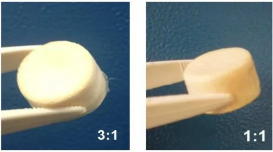
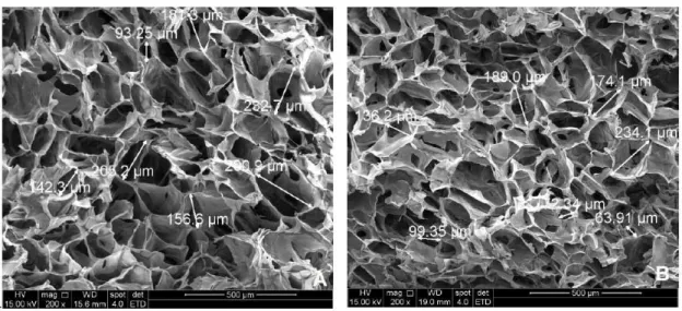
The Chitosan/PVA/BG scaffolds showed final dimensions of 18x10mm, spongy consistency and homogene-ous physical structure (Figure 1). The scaffolds pore morphology was analyzed by SEM and is shown in Figure 2, where it can be noted a network of well-defined, open and interconnected pores with thin walls. Overall, the pores located on the periphery of the scaffold showed a slight flattening on its surface, possibly caused by sample handling during the fracture to obtain the specimen, although fracture was conducted im-mersed in liquid nitrogen.
968 It was observed homogeneous and organized porosity similar for the 1:1 and 3:1 scaffolds, although the first presented more uniform pores (Figure 3). The pore size in the regions analyzed ranged from 64 to 234 µm in 1:1 scaffold, and 93 to 360 μm in 3:1 scaffold. Both scaffolds presented satisfactory pore size range, and opened, interconnected pores (Figure 4), essential for tissue ingrowth according to the literature [27]. The presence of pores of different sizes is very important since bone tissue grows through interconnect-ed pores in the range from 100 to 200 µm, while cell adhesion and vascular formation occurs in less than 100 µm pores.
Figure 2: Chitosan/PVA/BG scaffolds SEM images (A) 3:1 and (B) 1:1 ratios.
969 Figure 4: SEM images showing the pores architecture and interconnectivity of the 3:1 (A) and 1:1 (B) scaffolds.
3.2 Fourier Transform Infrared Spectroscopy
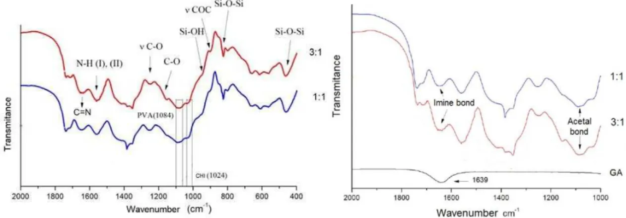
Figure 5 presents the FTIR spectra of the scaffolds normalized at the 1450cm-1 band. The typical bands cor-responding to both polymers in the blend are identified. It was observed by FTIR spectra characteris-tic peaks of amino group binding of Chitosan and broad and superimposed peaks associated with stretching vibration of Si-O-Si group on PVA (1084 cm-1) and Chitosan (1029 cm -1) chemical groups. The band at 966 cm-1 corresponds to Si-OH stretching. The shape of 1648 cm-1 band, associated with the imine bond (C = N), and 1558 cm-1 band, associated with the amino group (NH2) are similar to that found by Costa-Jr. et al [21], and Monteiro Jr. [28]. The band at 1720 cm-1, related to free aldehyde groups (-COH) appears in the spectra of 3:1 scaffold. However, the spectrum on the 1:1 scaffold, this band does not appear, indicating a possible crosslinking with the amino group, forming the C=O connection [22,29]. Dias et al. [30], identified in the FTIR spectrum of his work, the characteristic bands of PVA (1084 cm-1) and Chitosan (1024 cm-1) intensified by the presence of BG and the band 1644 cm-1, associated with the links of the C=N imine (Schiff base) formed by the amino group of Chitosan with aldehyde group of glutaraldehyde. The presence of glutaralde-hyde causes an increase in the intensity of band 1562 cm-1 associated with ethylenic bonds; and 2922 cm-1 frequency related to the stretching of CH groups. This fact can be attributed to increased contributions of glutaraldehyde molecule in quitosana-glutaraldehyde reaction that promotes the increase in crosslinking of the chain. The increasing intensity of the bands relating to the imine bond also suggests that GA crosslinking preferably occurs via Schiff base at carbon 2 glicossidic ring over the link with the hydroxyl groups on car-bons 3 and 6 [22]. Ma et al. [31] reported that the potent cytotoxicity of glutaraldehyde can be reduced with the presence of Chitosan due to the large number of amino groups in its molecular chain, serving as a bridge, increasing the efficiency of crosslinking by glutaraldehyde. The band 1100 cm-1 is related to the glutaralde-hyde crosslinking of the PVA, forming bridges acetals. The 1650 cm -1 and 1638 cm -1 bands are associated with the formation of the imine from the amine group of Chitosan during crosslinking by glutaraldehyde [32].
970
3.3 Porosity evaluation
The results of porosity and density are presented in Table 2. The matrices obtained showed high porosity as expected, both samples showing 97% total porosity. During the execution of the method, the matrix began to swell slightly as shown in Figure 6. To decrease the swelling effect on the measurements, the time of immer-sion in water was fixed as 20 minutes.
Table 2: Results obtained for porosity and density of scaffolds.
Scaffold Papparent (%) Ptotal (%) ρvol (g/cm3) ρtr (g/cm3)
3:1 50 ±5 97.5±0.3 0.04 ±0.02 1.6
1:1 56 ±3 97.2 ±0.5 0.04 ±0.01 1.5
Figure 6: Representation of the swelling presented by 3:1 and 1:1 scaffolds before immersion in deionized water (A) and after 20 minutes immersion in deionized water (B) for the test of apparent porosity.
3.4 Degradation test
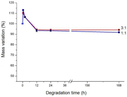
The hybrid matrices designed for tissue regeneration are expected to degrade at the same proportion that the regeneration of damaged tissue occurs. The most common signs that degradation is occurring are the mass loss and deterioration of the mechanical properties of the material [33]. Figure 7 shows the mass loss that occurred with time during the degradation test. Both scaffolds gained mass in the first 0.5 hour (3:1 gained 10.4% and 1:1 gained 14.0%), possibly due to the formation of a carbonated hydroxyapatite layer on the sur-face of the scaffold.
971 The ratio of weight loss was very similar for the matrices 3:1 and 1:1. The mass loss presented by the matrices during the degradation test in vitro was around 10% after a week of testing, with macroscopic preservation of their physical structure. The mass loss was more intense for both scaffolds up to 12 hours. After this period, degradation of the matrices was quite slow. The degradation occurs probably by the solva-tion of ionic groups and by depolymerizasolva-tion of the polymer chains [23]. Degradation of the 3:1 scaffold was slightly lower than that of the 1:1; which can be explained by the preference of glutaraldehyde cross-linking of the amino group of Chitosan with the formation of the group C=N, rather than forming acetal bridge with PVA [23].
3.5 Mechanical test
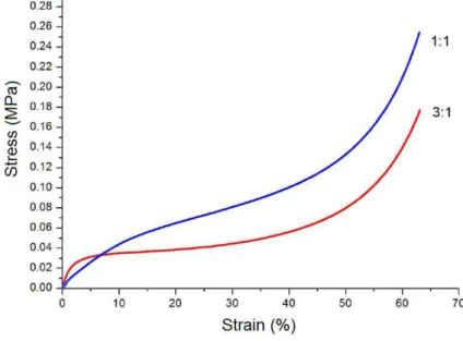
The stress strain compression curves obtained from the compression test of 3:1 and 1:1 scaffolds are present-ed in Figure 8. A strain limit (around 63%) was establishpresent-ed to avoid damage of the equipment. The average elastic modulus resulted in values of 2.2 and 0.9 MPa for 3:1 and 1:1 scaffolds respectively. Maximum stress supported at the strain limit established was 0.18 and 0.26 MPa, for 3:1 and 1:1 scaffolds respectively. The 3:1 scaffold showed higher modulus of elasticity and lower maximum stress in relation to 1:1, that is, the higher PVA content in the sample 1:1 compared to sample 3:1 affected differently these two properties. It should be pointed out that the toughness, as measured by the area under the stress-strain curve, is markedly higher for sample 1:1.
Figure 9: Stress strain compression curves for dried samples.
3.6 Cytotoxicity
The cytotoxicity assay aims to detect the potential of a material or device to produce lethal or sub lethal ef-fects in biological systems at the cell level. This assay should be applied to all categories of biomaterials. The release of toxic sub products of the biomaterial can damage the cells or reduce the rate of cell culture growth. Compared to the positive control (human fibroblasts in culture medium) the cells cultured directly in pres-ence of 1:1 and 3:1 scaffolds showed about 20% and 15% of viability respectively. This low viability is probably due to residual products inside the scaffolds pores. A biomaterial can be considered toxic to be used in biological systems under 50% of cell viability. However, when both scaffolds were soaked in PBS for 24 hours, the cells presented more than 90% of viability. The assay showed that both samples were toxic as pro-duced and not toxic after treatment with PBS, showing this approach was suitable for neutralization of these samples.
4. CONCLUSIONS
im-972 mersion in PBS cell viability increased and was similar to control for both samples 1:1 and 3:1.Therefore, the 1:1 and 3:1 hybrids Chi/PVA with bioactive glass 20% (w/w) cross-linked with glutaraldehyde and dried by lyophilization showed characteristics suitable for application in tissue engineering.
5. ACKNOWLEDGEMENTS
The authors gratefully acknowledge the financial support of CNPq, Capes and Fapemig.
6. BIBLIOGRAPHY
[1] AMINI, A.R., LAURENCIN, C.T., NUKAVARAPU, S.P., “Bone Tissue Engineering: Recent Advances
and Challenges”, Critical Reviews in Biomedical Engineering, v.40, pp. 363-408, 2012.
[2] ZOHORA, F.T., AZIM, A.Y.M.A., “Biomaterials as porous scaffolds for tissue engineering applications:
a review”, European Scientific Journal, v.10, pp. 1857– 1881, 2014.
[3] CHEUNG, H.Y., LAU, K.T., LU, P.T., et al., “A critical review on polymer-based bio-engineered
mate-rials for scaffold development”, Composites Part B – Engineering, v. 38, pp. 291-300, 2007.
[4] PETROVIC, V., ZIVKOVIC, P., et al., “Craniofacial bone tissue engineering”, Oral Surgery Oral Medi-cine, Oral Pathology Oral Radiology, v.114, pp. e1-e9, 2012.
[5] MOTA, J., YU, N., CARIDADE, S.G., et al., “Chitosan/bioactive glass nanoparticle composite me
m-branes for periodontal regeneration,” Acta Biomaterialia, v. 8, n. 11, pp. 4173–4180, 2012.
[6] HOKUGO, A., TAKAMOTO, T., TABATA,Y., “Preparation of hybrid scaffold from fibrin and biode-gradable polymer fiber”, Biomaterials, v.27, pp. 61-67, 2006.
[7] JONES,J.R.,“Reprint of: Review of bioactive glass: From Hench to hybrids”, Acta Biomater., v.23, pp.53–82, 2015.
[8] RAHAMAN, M.N, DAY, D.E., SONNY BAL, B., et al, “Bioactive glass in tissue engineering”, Acta Biomater., v.7, pp.2355–2373, 2011.
[9] DUTRA, C.E.A., PEREIRA, M.M., SERAKIDES, R., et al., “In vivo evaluation of bioactive glass foams associated with platelet-rich plasma in bone defects”, Journal of Tissue Engineering and Regenerative Medi-cine , v.2, pp.221-227, 2008.
[10] PEREIRA, M. M., JONES, J. R, OREFICE, R. L, et al., “Preparation of Bioactive Glass-Polyvinyl Al-cohol Hybrid Foams by the Sol-Gel Method”, Journal of Materials Science. Materials in Medicine, v.16, pp.1045 - 1050, 2005.
[11] REZWAN, K., CHEN, Q.Z., BLAKER, J.J., et al., “Biodegradable and bioactive porous poly-mer/inorganic composite scaffolds for bone tissue engineering”, Biomaterials, v. 27, pp. 3413-3431, 2006. [12] WANG, Y., YANG, C., X. CHEN, X., et al., “Development and characterization of novel biomimetic composite scaffolds based on bioglass-collagen-hyaluronic acid-phosphatidylserine for tissue engineering
applications”, Macromolecular Materials and Engineering, v.291, pp. 254–262, 2006.
[13] RODRIGUES, C.V.M., SERRICELLA, P., LINHARES, A.B.R., et al., “Characterization of a bovine collagen-hydroxyapatite composite scaffold for bone tissue engineering”, Biomaterials, v. 24, pp. 4987–4997, 2003.
[14] LEVENGOOD, S.K.L. and ZHANG, M.Q., “Chitosan-based scaffolds for bone tissue engineering”, Journal of Materials Chemistry B, v. 2, pp. 3161–3184, 2014.
[15] CROISIER, F. and JÉRÔME, C., “Chitosan-based biomaterials for tissue engineering”, European Poly-mer Journal, v. 49, pp. 780–792, 2013.
[16] NANDI, S.K., KUNDU, B. and BASU, D., “Protein growth factors loaded highly porous chitosan sca
f-fold: a comparison of bone healing properties”, Materials Science and Engineering C, v. 33, pp. 1267–1275, 2013.
[17] MAJI, K., DASGUPTA, S., PRAMANIK, K., et al., “Preparation and Evaluation of Gelatin-Chitosan-Nanobioglass 3D Porous Scaffold for Bone Tissue Engineering”, International Journal of Biomaterials, v.2016, Article ID 9825659, pp.1-14, 2016.
[18] MARTINO, A.D., SITTINGER, M., RISBUD, M.V., “Chitosan: a versatile biopolymer for orthopaedic tissue-engineering”, Biomaterials, v. 26, pp. 5983–5990, 2005.
[19] ZHANG, Y., NI, M., ZHANG, M., RATNER, B., “Calcium phosphate-chitosan composite scaffolds for
973 [20] LEMOS, E.M.F. , PATRICIO, P.S., DONICCI, C.L., et al, “Comparison of the Effect of Sol-Gel and Coprecipitation Routes on the Properties and Behavior of Nanocomposite Chitosan-Bioactive Glass
Mem-branes for Bone Tissue Engineering”, Journal of Nanomaterials , v. 2015, p. 1-8, 2015.
[21] COSTA-JUNIOR, E. S., MANSUR, H.S. “Preparação e caracterização de blendas de quitosana/ p o-li(álcool vinílico) reticuladas quimicamente com glutaraldeído para aplicação em engenharia de tecido”, Química Nova, v. 31, n. 6, p. 1460- 1466, 2008.
[22] COSTA-JUNIOR, E. S., PEREIRA, M. M., MANSUR, H. S., “Properties and biocompatibility of Ch
i-tosan films modified by blending with PVA and chemically crosslinked”, Journal of Materials Science: Ma-terials in Medicine, v. 20, pp. 553- 561, 2009.
[23] COSTA-JÚNIOR, E.S., PEREIRA, M.M., MANSUR, H.S., “Properties and biocompatibility of ch
i-tosan films modified by blending with PVA and chemically crosslinked”, Journal of Materials Science. Ma-terials in Medicine, v. 20, pp. 553-561, 2009.
[24] O’BRIEN, F.J., HARLEY, B.A., YANNAS, I.V., et al., “Influence of freezing rate on pore structure in freeze-dried collagen-GAG scaffolds”, Biomaterials, v. 25, pp. 1077-1086, 2004.
[25] JENNINGS, T.A., Lyophilization Introduction and Basic Principles., CRC Press, Florida, 1999. [26] MOSMANN, T. Rapid Colorimetric Assay for Cellular Growth and Survival: Application to Prolifera-tion and Cytotoxicity Assays, Journal of lmmunological Methods, v. 65, pp. 55-63, 1983.
[27] MIZUNO, K., KIDO, H., NARITA, T., et al., “Control of degradation rate of porous biodegradable pol-ymers., In: Proceedings of the 8th polymer for advanced technologies international symposium, pp. 13-16, Budapest, Hungary, 2005.
[28] MONTEIRO JR., O. A. C., AIROLDI, C., “Some studies of crosslinking Chitosan–glutaraldehyde inter-action in a homogeneous system”, International Journal of Biological Macromolecules, v 26, n. 2–3, pp. 119– 128, 1999.
[29] MANSUR, H. S., COSTA, H. S., “Nanostructured poly(vinyl alcohol)/bioactive glass and poly (vinyl alcohol)/Chitosan/bioactive glass hybrid scaffolds for biomedical applications”, Chemical Engineering Jour-nal, v. 137, p. 72– 83, 2008.
[30] DIAS, S.L., MANSUR, H.S., DONNICI, C.L., et al., “Synthesis and characterization of chitosan-polyvinyl alcohol-bioactive glass hybrid membranes”, Biomatter, v. 1, pp. 114-119, 2011.
[31] MA, L., GAO, C., MAO, Z., et al., “Collagen/Chitosan porous scaffolds with improved biostability for
skin tissue engineering”, Biomaterials, v. 24, pp. 4833-4841, 2003.
[32] RAO, P. S., SMITHA, B., SRIDHAR, S., et al., “Preparation and performance of poly(vinyl alco-hol)/polyethyleneimine blend membranes for the dehydration of 1,4-dioxane by pervaporation: Comparison with glutaraldehyde cross-linked membranes”, Separation and purification Technology, v. 48, n. 3, p. 244- 254, 2006.
[33] COSTA, H.S., MANSUR, A.A.P., PEREIRA, M.M., et al., “Engineered Hybrid Scaffolds of Poly(vinyl alcohol)/Bioactive Glass for Potential Bone Engineering Applications: Synthesis, Characterization,