Contents lists available atScienceDirect
Acta Tropica
journal homepage:www.elsevier.com/locate/actatropica
Evaluating a point-of-care circulating cathodic antigen test (POC-CCA) to
detect
Schistosoma mansoni
infections in a low endemic area in north-eastern
Brazil
Fernando Schemelzer Moraes Bezerra
a,⁎, Joames Kau
ffi
mann Freitas Leal
a, Mariana Silva Sousa
a,
Marta Cristhiany Cunha Pinheiro
a, Alberto Novaes Ramos Jr.
a, Vanessa Silva-Moraes
b,
Naftale Katz
ba
Parasitology and Mollusks Biology Research Laboratory, Universidade Federal do Ceará, Rua Capitão Francisco Pedro 1210, Fortaleza, Ceará, 60.430-272, Brazil bLaboratory of Schistosomiasis, Instituto René Rachou, Fundação Oswaldo Cruz (Fiocruz), Avenida Augusto de Lima 1715, Belo Horizonte, Minas Gerais, 30190-002,
Brazil
A R T I C L E I N F O
Keywords: Schistosomiasis Diagnosis POC-CCA Kato-Katz
Low endemicity setting Brazil
A B S T R A C T
Schistosomiasis is still a public health problem in Brazil. The Kato-Katz test is the most frequently used diag-nostic method forSchistosoma mansoniinfection. However, it lacks sensitivity in areas of low prevalence. We have assessed the positivity rate ofS. mansoniinfection in Bananeiras, a village on Capistrano, Ceara, Brazil by performing a point-of-care test in urine to determine the circulating cathodic antigens (POC-CCA), and we compared thefindings with those of the Kato-Katz technique for egg detection in stool and an enzyme-linked immunosorbent assay for specific antibodies against adult worms (SWAP-ELISA) in serum before treatment (baseline). Additionally, the POC-CCA and Kato-Katz test results were compared at one and two years post-treatment, and only POC-CCA strips were utilised for follow-up testing on urine samples at 3–6 weeks. Only one
sample of stool and urine was collected per event. Overall, 258 individuals were investigated at the baseline. The POC-CCA test detected 10 (3.9%) positive cases; however, this amount increased to 30 (11.6%) when con-sidering trace readings as positive (t + ), whereas the Kato-Katz method found only 4 (1.6%) positive cases and the SWAP-ELISA detected 105 (40.7%) positive cases. The consistency observed between a single POC-CCA (t + ) or (t-) and the Kato-Katz (three slides) was poor (Kappa indexes < 0.20). The highest positivity rate as determined by CCA and Kato-Katz was found in adults. At the baseline, a praziquantel treatment was ad-ministered to all individuals regardless of their infection status. According to the POC-CCA test, 93% of the previous positive cases became negative by the third week after the treatment; this rate reached 100% at the sixth week assessment. The follow-up showed that of the 175 individuals evaluated at one year post-treatment, only one (0.6%) showed‘trace’results, and all the individuals were negative for eggs in the stool. At two years, all 185 examined individuals were negative by the Kato-Katz method, and 11 (5.9%) presented traces by POC-CCA. Our results indicate that a single POC-CCA test reveals a significantly higher number of positive cases than the Kato-Katz technique for diagnosingS. mansoniin a low endemic setting, when trace results are considered as positive cases. Nevertheless, the true significance of the trace is not clear. Thesefindings reinforce the need to associate different tools for improved schistosomiasis diagnosis in individuals with low parasite burdens.
1. Introduction
According to data obtained by the national prevalence survey (2010–2015), an estimated 1% positive rate ofSchistosoma mansoniwas found in Brazil, where schistosomiasis remains a significant public health problem (Katz et al., 2017). The parasitological detection ofS.
mansoniby Kato-Katz faecal microscopy is used extensively for diag-nosis due to its practicability, efficacy, and low cost. However, this method has some disadvantages. It has low sensitivity in areas of low endemicity, and consequently, stool analyses must be repeated several times (Cavalcanti et al., 2013;Enk et al., 2008;Pinheiro et al., 2012; Siqueira et al., 2011).
https://doi.org/10.1016/j.actatropica.2018.03.002
Received 22 July 2017; Received in revised form 6 March 2018; Accepted 7 March 2018 ⁎Corresponding author.
E-mail addresses:[email protected](F.S.M. Bezerra),[email protected](J.K.F. Leal),[email protected](M.S. Sousa), [email protected](M.C.C. Pinheiro),[email protected](A.N. Ramos),vanessasmoraes@cpqrr.fiocruz.br(V. Silva-Moraes),nkatz@cpqrr.fiocruz.br(N. Katz).
Available online 08 March 2018
0001-706X/ © 2018 Published by Elsevier B.V.
Alternatively, antibody detection has been suggested to complement the parasitological method and to increase the diagnostic sensitivity. In fact, in low endemic areas and/or after large-scale chemotherapy, the combination of stool microscopy with serological methods has been proposed for the detection of Schistosomiasis mansoni cases in low transmission areas (Alarcón de Noya et al., 2007;Carneiro et al., 2012; Espirito-Santo et al., 2014;Gomes et al., 2014).
Assays for the detection of circulatingSchistosomaantigens (adult worm gut-associated antigens) were described. The circulating cathodic antigen (CCA) and the circulating anodic antigen (CAA) have both been used to diagnose active infections (Corstjens et al., 2014). To improve the techniques used to diagnose schistosomiasis in thefield,van Dam et al. (2004)developed a lateralflow strip immunoassay to detect CCA in urine, and they declared that this test has high sensitivity and spe-cificity for the study of schistosomiasis. The laboratory format of the urine CCA strip test has been developed into a commercially available rapid Point-of-Care test (POC-CCA), and various studies have been conducted in endemic areas of the African and Asian continents to determine the efficacy andfield applicability of this test for schistoso-miasis diagnosis. A summary of 5 countries’evaluations of the POC-CCA test, which was funded by the Schistosomiasis Consortium for Operational Research and Evaluation (SCORE), demonstrated that this method is valuable for detectingS. mansoniin endemic areas (Adriko et al., 2014;Colley et al., 2013;van Dam et al., 2015).
A systematic review indicated that when the Kato-Katz prevalence is below 50%, the POC-CCA test is much more sensitive than the Kato-Katz test, and thus the prevalence determined by the POC-CCA test is consistently much higher (Kittur et al., 2016).
Until the present, only a few studies have been published to show the results of POC- CCA trials in Brazil. A study performed in a low prevalence area (Estreito de Miralta, state of Minas Gerais, Brazil) compared the results of two parasitological techniques, the Kato-Katz and Saline Gradient and the POC-CCA. An analysis of two and 24 slides using the Kato-Katz technique showed positive rates of 10.6% (15/141 individuals) and 19.1% (27/141), respectively. The Saline Gradient technique yielded a positive rate of 17.0%. Considering the results of both parasitological techniques, the positive rate was 24.1%. The POC-CCA test showed a positive rate of 22.7% (32/141- trace as positive), and, when trace results were considered negative, the positivity rate was only 2.1% (Siqueira et al., 2016).
Other studies evaluating the POC-CCA test in low-endemicity set-tings are essential, because in these setset-tings, the infection intensity is less than 100 EPG and the diagnosis fails when parasitological stool sample tests are used.
The present study aimed to evaluate the performance of the POC-CCA underfield-based conditions in a population from a low endemic area located in the north-eastern region of Brazil for the diagnosis ofS. mansonibefore and after treatment.
2. Material and methods
2.1. Ethics statement, recruitment, and treatment
The study protocol was approved by the Universidade Federal do Ceará ethical committee (application No. 302.204). It was conducted according to Resolution No. 466/12 of the Brazilian Health Council. District health authorities and the entire community were informed about the purpose, procedures, and potential risks and benefits of the study. Before enrolment, informed consent was obtained from the vo-lunteers. Parents/legal guardians provided informed consent for their children to participate.
At the baseline, praziquantel (PZQ) treatment (Farmanguinhos, Ministry of Health, Brazil) was administered to all individuals regardless of their infection status. This treatment consisted of a single dose of 60 mg/kg for children (≤15 years old) and 50 mg/kg for adults, as recommended by the Brazilian Health Ministry (Ministério da Saúde, 2014).
2.2. Study area and population
The study was conducted between 2013 and 2015 in the community of Bananeiras, a rural locality in the Capistrano municipality, in Ceará state, Brazil (geographical co-ordinates 4° 28′ 20″S latitude, 38° 54′
14″W longitude). Capistrano extends for 222.6 km2and is located at
155 m altitude, approximately 93 km south of Fortaleza, the capital of Ceará. The area has been known to be endemic forhave endemicS. mansonisince 1976. In a region with a semi-arid climate, the river that crosses the village (Aracoiaba River) remains dry for most of the year. Theflood season runs between the months of December and March. Our door-to-door census was performed in March of 2013, and it identified 297 people aged 2 or older in the Bananeiras community. The pre-valence rate of schistosomiasis reported in 2010 was 1.6% and there has been no specific treatment for this disease in the past two years.
2.3. Inclusion criteria
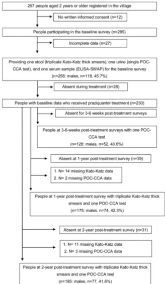
To be included in this study, all the inhabitants of Bananeiras had to meet the following criteria at the baseline: 1) aged≥2 years at re-cruitment; 2) informed consent; 3) provided stool, urine, and serum samples at sufficient/suitable amounts for triplicate Kato-Katz thick smears, for a single point-of-care circulating cathodic antigen (POC-CCA) cassette test and for a serology test; and 4) no recent treatment for schistosomiasis (at least within the past two years). All the people who had been recruited for the study at the baseline (n = 258) were con-sidered for follow-up if the treatment was performed. Theflowchart of the study population is shown inFig. 1.
2.4. Sample collection
Five cross-sectional surveys were implemented. Urine, stool, and blood samples were collected at the baseline from each individual en-rolled in the study; at 3 and 6 weeks after the PZQ administration, only a urine sample was collected; and at one and two years post-treatment, both urine and stool samples were collected. Only one sample of stool and urine was collected at each time point.
At the baseline, one day before the collection day, two plastic containers labelled with unique identification numbers (IDs) (stool and urine containers) were delivered to all the study participants. For child participants, the containers were delivered to the children's mothers or guardians; venous blood samples were also taken at that moment. Individual serum samples were obtained after blood sample cen-trifugation at 3000g for 5 min, and these samples were stored at − 20 °C. On the following day, the individuals were invited to return the containersfilled with a fresh morning urine sample and a lime-sized stool sample to thefieldworkers stationed at Bananeiras Health Center, where the samples were processed on the same day.
2.5. Microscopy
Microscopy was performed using the Helm Test®manufactured by
Biomanguinhos-Fiocruz (Rio de Janeiro, RJ, Brazil). Three Kato-Katz thick smear slides were prepared per stool sample using 41.7 mg tem-plates, and they were examined forS. mansoni(Katz et al., 1972). The infection intensity was expressed as eggs per gram of stool (EPG) for everyone, using the arithmetic mean ofS. mansonifaecal egg counts (FECs) and multiplied by a factor of 24. For quality control, 10% of the Kato-Katz thick smears were re-examined by a senior technician from Rene Rachou Research Center, Oswaldo Cruz Foundation, Minas Gerais, Brazil.
2.6. Circulating cathodic antigen (CCA) test
treatment/33,955: 1 year post-treatment/50,173: 2 years post-treat-ment, Rapid Medical Diagnostics, Pretoria, South Africa) performed at room temperature according to the manufacturer’s instructions on the
day of the sample collection. In brief, one drop of urine was added to the well of the testing cassette and allowed to absorb. Once fully ab-sorbed, one drop of buffer (provided with the CCA test kits) was added. The test results were read visually 20 min later. In cases when the control bands did not develop, the test was considered invalid. Invalid tests were repeated using the same urine sample. Valid tests were scored as negative, trace (weak band) or positive. All the tests were read independently by two investigators, and in case of discordant results, a third independent investigator examined it until an agreement was reached. Urine samples collected at all the time points in the study were treated as described above.
2.7. Serology
The ELISA assay was performed to detect an IgG antibody against soluble worm antigen preparation (ELISA-SWAP) according to the methodology described by Grenfell et al. (2013). In summary, Max-iSorpTM surface microtitre plates (NUNC, Denmark) were sensitised with 100μL/well of 1μg/mL of SWAP diluted in 0.05 M
carbonate-bicarbonate buffer, pH 9.6, for 16 h at 4 °C. The plates were washed three times with 0.15 M PBS, pH 7.2, with 0.05% Tween 20 (LGC Biotecnologia, BR) (washing buffer), and the non-specific sites were blocked with 10% foetal bovine serum (FBS) in washing buffer at 37 °C for 1 h. After another round of washing, 100μL of sera diluted 1:100 in PBS was added to each well in triplicates and the plates were incubated at room temperature (RT) for 1 h. The plates were then subjected to more washing and then incubated at RT for 1 h with IgG-HRP (Southern Biotech, USA) diluted 1:60,000 in washing buffer. The plates were washed again and 100μL of TMB/H2O2substrate was added to each
well. The reaction was stopped after 20 min of incubation in the dark by adding 50μL/well of 2 N sulphuric acid. The results were obtained as absorbance values at 450 nm in a microplate reader (Bio-Rad Labora-tories 3550, JA). The sensitivity, specificity, cut-offvalues, receiver operating characteristic (ROC) curves, likelihood ratios and positive predictive values were determined by Prism 5 software based on true individual egg positives and negatives.
2.8. Statistical analysis
Data were entered into Excel (Microsoft Corp., Redmond, WA, USA) spreadsheets and analysed with SPSS software version 22 (IBM Corp.; Armonk, USA). The proportions were tested using chi-square and dif-ference-of-proportions tests. Consistency between binary variables was established by determining the kappa statistics (k) as follows: k = 0 indicating no agreement; k = 0–0.2 indicating poor agreement; k = 0.21–0.4 indicating fair agreement; k = 0.41–0.6 indicating mod-erate agreement; k = 0.61–0.8 indicating substantial agreement; and
k = 0.81–1.0 indicating almost perfect agreement (Landis and Koch,
1977). A 5% level of significance was adopted for all the inferential procedures.
3. Results
3.1. Study group characteristics and adherence
Fig. 1shows the individuals’adherence at providing stool, urine,
and blood samples for a set of diagnostic tests to detectS. mansoniat the time points of the study. The village census revealed 297 people aged 2 years or older, all of whom were invited to participate. Of the 285 in-dividuals who participated in the baseline cross-sectional survey, twenty-seven were excluded due to incomplete data A complete spe-cimen set (stool, urine, and blood) was available for 258 (90.5%) of the 285 individuals enrolled in the study at baseline, so all three tests could be performed. Among them, 45.7% (n = 118) were males and the median age of the cohort was 30 years (age range: 2–87 years). At three and six weeks post-treatment, 128 individuals were re-examined using one POC-CCA test. Among them, 40.6% (n = 52) were males and the median age of the cohort was 19 years (age range: 2–87 years).
3.2. Positivity rates of S. mansoni infection before treatment
Table 1shows the results for the Kato-Katz method, POC-CCA and SWAP-ELISA forS. mansoni.
According to the three Kato-Katz thick smears examined per in-dividual, the observed positivity rate forS. mansoniwas 1.6% (n = 4). The positivity rate ofS. mansoniincreased with the increasing number of examined smears (p = 0.681), but no significant difference was found. All the positive individuals were classified as having a very low-intensity infection with an arithmetic mean of 8 EPG.
When the trace results were scored as positive, the positivity rate of S. mansonibased on the CCA test increased from 3.9% to 11.6%. The positivity rate forS. mansoniby CCA was significantly higher than that obtained using 3 Kato-Katz smears when the trace results were con-sidered positive (p < 0.001), but this was not the case when the trace results were considered negative (p = 0.176) (Table 1). Of the 258
individuals, just one (0.4%) wasS. mansoni-positive according to all the tests, and 133 individuals (51.5%) were negative forS. mansoniin all the assays. A total of 10 (3.9%) were egg-negative but positive ac-cording to the CCA test and the SWAP-ELISA. Ninety-two individuals (35.7%) were positive only by SWAP-ELISA, whereas 19 (7.4%) were positive only by CCA test. Only one individual (0.4%) was positive by Kato-Katz method, but not by any other test. The remaining 2 in-dividuals (0.8%) were positive according to the Kato-Katz method and by the ELISA-SWAP. There were 29 individuals (11.2%) who were cassette CCA-positive, but they were egg-negative. Of those individuals, 10 (34.5%) were positive by SWAP-ELISA.
3.3. Age-dependent positivity rates
The positive cases by microscopy, POC-CCA test and ELISA-SWAP per age group are shown inTable 2. Overall, the highest positivity rate occurred in adults; however, it is worth noting that there were many people under 20 years old who scored as CCA-positive. The ages of the egg-positive individuals ranged from 33 to 47 years. Twenty (66.6%) and 80 (76.2%) positive people were older than 15 years old according to the POC-CCA test and SWAP-ELISA, respectively. The proportion of POC-CCA (t + )-positive tests was higher in the age groups below 40 years old, and all individuals in the 2–9 year-old age group had trace
readings. There was no association between infection and gender (19 females vs. 14 males; p = 0.683).
3.4. Consistency among the different diagnostic methods before treatment
Table 3 shows the consistency between the different diagnostic approaches for diagnosingS. mansoni. Poor consistency was found be-tween the three Kato-Katz thick smears and a single CCA test regardless of whether the trace results were considered negative (k = 0.12) or positive (k = 0.03).
3.5. Post-treatment readings
Of the 128 individuals who had CCA data records 3 and 6 weeks
after treatment,S. mansonieggs were detected by Kato-Katz in 2 (1.6%) individuals during the baseline cross-sectional survey. A single POC-CCA had trace results considered as negative, in 8 individuals (6.2%). When considering trace results as positive, a considerably higher number of individuals were classified as positive (n = 24, 18.7%).
Three weeks after treatment, of those 128 individuals, a single urine CCA cassette test revealed 2 individuals (1.6%) with S. mansoni, whereas CCA (t + ) found 5 (3.9%). At 6 weeks after treatment eva-luation, regardless of whether the POC-CCA trace results were con-sidered positive or negative, all the individuals were CCA-negative.
Of the 33 individuals who were either Kato-Katz or CCA (t + )-po-sitive at the baseline, 31 swallowed PZQ in the presence of the research team, and they were considered for efficacy evaluation. Among those, 29 (93.5%) were POC-CCA-negative 3 weeks post-treatment. There were twenty-eight individuals who tested negative by Kato-Katz, but they were positive by POC-CCA. Of these, 26 (93%) were POC-CCA-negative upon repeat testing when performed 3 weeks post-treatment. The two remaining POC-CCA-positive cases became negative based on follow-up POC-CCA testing at 6 weeks post-treatment.
At one year post-treatment, of the 175 individuals evaluated by tri-plicate Kato-Katz thick smears and POC-CCA tests, only one (0.6%) was found to be‘trace’and all the individuals were negative upon microscopic
examination. At two years post-treatment, 185 were negative in terms of stool examination and 11 (5.9%) were‘trace’by POC-CCA.
4. Discussion
In Brazil, the profile of schistosomiasis has undergone changes over time due to its control programmes. There is a consensus that there are different prevalence and morbidity profiles in endemic African and Brazilian areas (Coelho et al., 2016). However, in Brazil, despite the reduction in mortality and morbidity due to the success of the Ministry of Health control programmes, schistosomiasis was mentioned in 8756 of the 12,491,280 deaths that were recorded between 2000 and 2011; 6319 as the underlying cause and in 2437 cases as an associated cause of death, thus it remains an important public health problem ( Martins-Melo et al., 2014). In view of recent discussions and the resolution on schistosomiasis elimination by the 65th World Health Assembly (Rollinson et al., 2013;WHO, 2012), the necessity for highly specific and sensitive diagnostic methods for diagnosing S. mansoniafter ex-tensive preventive chemotherapy campaigns is imperative.
The present study attempts to evaluate the performance of the commercially available Point-of-Care Circulating Cathodic Antigen urine cassette test (POC-CCA) for the diagnosis ofSchistosomiasis man-soniin a low endemic area in Ceará State, Brazil. In fact, the positivity rate increased significantly according to the diagnostic approach; the rate ranged from 1.6% by egg detection to 11.6% by the POC-CCA (t + ), meaning there was a seven-fold increase. In fact, studies per-formed in different African settings (Colley et al., 2013;Coulibaly et al., 2011) whereS. mansoniis endemic have repeatedly shown that the POC-CCA is capable of detecting many additional positive cases when compared to Kato-Katz thick smears.
Table 1
Baseline positivity rates ofS. mansoniaccording to each diagnostic method.
Diagnostic approach NO. of infected Individuals
% positive (95% CI)
Single Kato-Katz thick smear 2 0.8 (0.0–1.8) Duplicate Kato-Katz thick smears 3 1.2 (0.0–2.5) Triplicate Kato-Katz thick smears 4 1.6 (0.0–3.1) Single POC-CCA cassette test (t-) 10 3.9 (1.5–6.2) Single POC-CCA cassette test (t + ) 30 11.6 (7.7–15.5)
SWAP-ELISA 105 40.7 (34.7–46.7)
Table 2
Number of positive cases for the respective age group, according to different diagnostic approaches.
Age group Total tested
No. pos/% Positive by microscopy
No. pos/% Positive by POC-CCA (t-)*
No. pos/% Positive by POC-CCA (t + )*
No. pos/% Positive by ELISA-SWAP
2–9 32 0 0 6/18.7 17/53.1
10–19 58 0 2/3.4 7/12.1 18/31.0
20–29 38 0 3/7.9 6/15.8 10/26.3
30–39 43 3/7 2/4.6 7/16.3 15/34.9
40–49 35 1/2.8 0 1/2.8 17/48.6
50–59 23 0 0 0 15/65.2
60 and older 29 0 3/10.3 3/10.3 13/44.8 Total tested 258 4/1.6 10/3.9 30/11.6 105/40.7
*t-, trace negative; t+, trace positive.
Table 3
Consistency between different techniques for the diagnosis ofS. mansoniinfections.
Triplicate Kato-Katz thick smears
K* P-value
Diagnostic test Test result Positive Negative
Single POC-CCA test (t-)
Positive 1 9
Negative 3 245 0.12 0.027 Single POC-CCA test
(t + )
Positive 1 29
Negative 3 225 0.03 0.400
In this study, the consistency observed between the POC-CCA test and Kato-Katz technique was poor, regardless of whether the trace re-sults were considered as negative or positive, which could be partly explained by the very low prevalence observed by microscopy. These results support the findings of studies performed in low S. mansoni endemic areas in Africa (Adriko et al., 2014) and in Brazil (Coelho et al., 2016), which also identified significant disagreement between these methods. Most of the discordant results were CCA-positive but egg-negative, especially when trace results were considered as positive. As a test that is subject to visual reading, POC-CCA is a qualitative method based on individual interpretations. Considering the ambiguity of interpreting the ‘trace’ infection readings (light lines), a two-way
analysis of CCA data with‘trace’results considered infection-negative
and‘trace’results considered infection-positive were performed. The subjectivity and the need for reading standardisation have been dis-cussed (Colley et al., 2013, 2017; Siqueira et al., 2016). Recently, a study in a Brazilian population with a low parasite burden in an en-demic area showed that urine samples with trace results were found in individuals with or without S. mansoni eggs in their stool, after in-tensive examinations. After a 10-fold concentration of the urine samples by lyophilisation, the trace became positive in parasitologically positive cases, but it remained a trace result in parasitologically negative cases (Coelho et al., 2016). Thus, trace readings could not be promptly de-fined as positive or negative. Additionally, this same study showed that hookworm infection influences POC-CCA test results (Coelho et al., 2016), which is inconsistent with previous observations performed in Africa (Shane et al., 2011;Tchuem Tchuenté et al., 2012).
POC-CCA was recently deemed a new tool that could be used in schistosomiasis control strategies (Colley et al., 2017). The authors considered that the results that were Kato-Katz egg-negative/POC-CCA-positive could be explained by the menopause or infertility of Schisto-somafemales, the presence of immature worms, infection caused solely by males and immune fecundity. An important fact, which has not been discussed exhaustively by the authors, is the possibility of a false-po-sitive reaction in the POC-CCA.
Corroborating our data, high prevalence rates ofS. mansoni infec-tion were found in adults in a study performed in Zambia (Lodh et al., 2013). If we followed the control approach for areas with prevalence levels below 10% using parasitological methods, which are aimed at treating only schoolchildren (WHO, 2006), more than half of the po-sitive individuals in this study would remain untreated and would continue to contribute to the transmission of the infection. Never-theless, despite the higher positivity rate found in adults, there were too many positive individuals below 20 years of age. Furthermore, our results show that 13% (POC-CCA) and 11% (SWAP-ELISA) of theS. mansoni-positive individuals were children younger than 6 years old. Thisfinding reinforces the idea that preschool-aged children are at risk of schistosomiasis and that their treatment is also recommended as described elsewhere (Coulibaly et al., 2013, 2012).
Antibody detection is considered highly sensitive, and it has been recommended as a supplementary tool for schistosomiasis diagnosis in individuals with low infection burdens, which are usually hard to detect by parasitological methods (Alarcón de Noya et al., 2007; Grenfell et al., 2013). Our results show that one out of the four egg-positive cases and 19 (63.3%) of the POC-CCA positive (+) cases were antibody-negative. Antibodies can remain in the body for several years after treatment, and individuals with helminth infections can also show cross-reaction. Nevertheless, the negativity of the SWAP-ELISA in 63.3% of CCA-positive (t + ) cases must be further investigated.
The recommended time forS. mansoniassessment after treatment is 15–20 days (Scherrer et al., 2009), but our data and those of others
(Coulibaly et al., 2012, 2013;Lamberton et al., 2014;Legesse and Erko, 2007) still detected CCA from 3 to 6 weeks post-treatment, including or excluding ‘trace’results as positive. Regarding cure controls through
stool examinations, 20 days is considered a too-short period. Brazilian researchers suggest at least one month, with 4 months being considered
the optimal time after treatment, with several stool examinations (Ministério da Saúde, 2014). It is noteworthy to consider the strength of transmission in the area to define the follow-up time after treatment. Once again, African countries show different epidemiological settings when compared to what is found in the Americas, i.e., areas of high transmission for the former and low transmission for the latter.
A lower percentage of individuals showed positive results according to the POC-CCA test at the post-treatment follow-up. Thisfinding could be due to the presence of juvenile worms that were not affected by treatment (Sabah et al., 1986) and/or worms that survived treatment, because the effectiveness of praziquantel is lower than 100% ( Danso-Appiah et al., 2013;Katz et al., 1979,Stothard et al., 2013;Vinkeles Melchers et al., 2014).
Of the 28 cases that showed POC-CCA positivity but were egg-ne-gative before the treatment, 26 (93%) became POC-CCA-neegg-ne-gative at 3 weeks and the remaining 2 cases became negative 6 weeks after pra-ziquantel administration. Studies are still necessary to determine when exactly CCA disappears from the urine after treatment.
The follow-up of the population after one and two years showed some positive POC-CCA reactions that are difficult to interpret. In view of the absence of water in the streams during the observation period, these few positive cases (t + ) could be due to individuals who were infected in other areas, who relapsed after treatment, or who had false-positive results.
The logistical andfinancial implications of using each method have been discussed in several studies (Adriko et al., 2014; Colley et al., 2013;Coulibaly et al., 2013). The POC-based CCA is a user-friendly test that can be performed and read in thefield. No additional equipment is needed for its use, and only a minimum of training if required. Thus, it can be used in remote rural areas that do not have access to a minimal infrastructure and trained staff. The time needed to perform the POC-CCA test is approximately 25 min, which is less time-consuming than the other methods currently employed forS. mansoniscreening. An-other advantage is that the POC-CCA test can detect prepatent infec-tions (de Water et al., 1986), whilst the Kato-Katz method can only detect infections when the adult worms release eggs and the ELISA tests detect only the host’s immunoactivity against the worm, which cannot
differentiate between current and past infections. The disadvantages of POC-CCA compared to the Kato-Katz techniques is that the former is not quantitative, its reading is subjective, trace results remain a problem for interpretation and it does not disclose the presence of other helminths. The current lack of a definitive standard reference test to detect all Schistosomainfections indicates the necessity of combining diagnostic methods to identify individuals with low parasite burdens, particularly in areas with lowS. mansoni infection prevalence. Moreover, further investigations to determine the accuracy of the urine CCA cassette test using highly specific and sensitive diagnostic methods (i.e., polymerase chain reaction (PCR)) (Carneiro et al., 2013;Meurs et al., 2015;Pontes et al., 2002), the detection of Circulating Anodic Antigen by an up-converting phosphor technology-based lateral flow (UPT-LF) assay (Corstjens et al., 2014) or Helmintex (Pinheiro et al., 2012; Favero et al., 2017) is highly recommended in areas with a low prevalence and intensity of infection.
5. Conclusions
Acknowledgements
The authors would like to thank the Central Laboratory of Public Health in Ceará State, the Health Secretariat of the State Government of Ceará and the Health Secretariat of Capistrano municipality for the technical support. We would like to acknowledge Neels van Rooyen from Rapid Medical Diagnostics and Santiago Nicholls from PAHO/ WHO for donating POC-CCA®kits. We also want to give our special
thanks to the population of Bananeiras for their collaboration during thefield work.
References
Adriko, M., Standley, C.J., Tinkitina, B., Tukahebwa, E.M., Fenwick, A., Fleming, F.M., Sousa-Figueiredo, J.C., Stothard, J.R., Kabatereine, N.B., 2014. Evaluation of circu-lating cathodic antigen (CCA) urine-cassette assay as a survey tool for Schistosoma mansoni in different transmission settings within Bugiri District, Uganda. Acta Trop. 136, 50–57.http://dx.doi.org/10.1016/j.actatropica.2014.04.001.
Alarcón de Noya, B., Ruiz, R., Losada, S., Colmenares, C., Contreras, R., Cesari, I.M., Noya, O., 2007. Detection of schistosomiasis cases in low-transmission areas based on coprologic and serologic criteria The Venezuelan experience. Acta Trop. 103 (1), 41–49.
Carneiro, T.R., Pinheiro, M.C.C., de Oliveira, S.M., Hanemann, A.L., Queiroz, J.A., Bezerra, F.S., 2012. Increased detection of schistosomiasis with Kato-Katz and SWAP-IgG-ELISA in a Northeastern Brazil low-intensity transmission area. Rev. Soc. Bras. Med. Trop. 45 (4), 510–513.
Carneiro, T.R., Peralta, R.H., Pinheiro, M.C.C., Oliveira, S.M., Peralta, J.M., Bezerra, F.S., 2013. A conventional polymerase chain reaction-based method for the diagnosis of human schistosomiasis in stool samples from individuals in a low-endemicity area. Mem. Inst. Oswaldo Cruz 108 (8), 1037–1044. http://dx.doi.org/10.1590/0074-0276130202.
Cavalcanti, M.G., Silva, L.F., Peralta, R.H., Barreto, M.G., Peralta, J.M., 2013. Schistosomiasis in areas of low endemicity: a new era in diagnosis. Trends Parasitol. 29 (2), 75–82.http://dx.doi.org/10.1016/j.pt.2012.11.003.
Coelho, P.M., Siqueira, L.M., Grenfell, R.F., Almeida, N.B., Katz, N., Almeida, Á, Carneiro, N.F., Oliveira, E., 2016. Improvement of POC-CCA interpretation by using lyophili-zation of urine from patients with schistosoma mansoni low worm burden: towards an elimination of doubts about the concept of trace. PLoS Negl. Trop Dis. 10 (6), e0004778.http://dx.doi.org/10.1371/journal.pntd.0004778.
Colley, D.G., Binder, S., Campbell, C., King, C.H., Tchuem Tchuenté, L.A., N'Goran, E.K., Erko, B., Karanja, D.M., Kabatereine, N.B., van Lieshout, L., Rathbun, S., 2013. Afi ve-country evaluation of a point-of-care circulating cathodic antigen urine assay for the prevalence of Schistosoma mansoni. Am. J. Trop. Med. Hyg. 88 (3), 426–432.http:// dx.doi.org/10.4269/ajtmh.12-0639.
Colley, D.G., Andros, T.S., Campbell, C.H., 2017. Schistosomiasis is more prevalent than previously thought: what does it mean for public health goals, policies, strategies, guidelines and intervention programs? Infect. Dis. Poverty 6, 63.http://dx.doi.org/ 10.1186/s40249-017-0275-5.
Corstjens, P.L., De Dood, C.J., Kornelis, D., Fat, E.M., Wilson, R.A., Kariuki, T.M., Nyakundi, R.K., Loverde, P.T., Abrams, W.R., Tanke, H.J., Van Lieshout, L., Deelder, A.M., Van Dam, G.J., 2014. Tools for diagnosis, monitoring and screening of Schistosoma infections utilizing lateral-flow based assays and upconverting phosphor labels. Parasitology 141 (14), 1841–1855.http://dx.doi.org/10.1017/
S0031182014000626.
Coulibaly, J.T., Knopp, S., N'Guessan, N.A., Silué, K.D., Fürst, T., Lohourignon, L.K., Brou, J.K., N'Gbesso, Y.K., Vounatsou, P., N'Goran, E.K., Utzinger, J., 2011. Accuracy of urine circulating cathodic antigen (CCA) test for Schistosoma mansoni diagnosis in different settings of Côte d'Ivoire. PLoS Negl. Trop. Dis. 5 (11), e1384.http://dx.doi. org/10.1371/journal.pntd.0001384.
Coulibaly, J.T., N'gbesso, Y.K., Knopp, S., Keiser, J., N'Goran, E.K., Utzinger, J., 2012. Efficacy and safety of praziquantel in preschool-aged children in an area co-endemic for Schistosoma mansoni and S. haematobium. PLoS Negl. Trop. Dis. 6 (12), e1917. http://dx.doi.org/10.1371/journal.pntd.0001917.
Coulibaly, J.T., N'Gbesso, Y.K., Knopp, S., N'Guessan, N.A., Silué, K.D., van Dam, G.J., N'Goran, E.K., Utzinger, J., 2013. Accuracy of urine circulating cathodic antigen test for the diagnosis of Schistosoma mansoni in preschool-aged children before and after treatment. PLoS Negl. Trop. Dis. 7 (3), e2109.http://dx.doi.org/10.1371/journal. pntd.0002109.
Danso-Appiah, A., Olliaro, P.L., Donegan, S., Sinclair, D., Utzinger, J., 2013. Drugs for treating Schistosoma mansoni infection. Cochrane Database Syst. Rev. CD000528. http://dx.doi.org/10.1002/14651858.CD000528.pub2.
Enk, M.J., Lima, A.C., Drummond, S.C., Schall, V.T., Coelho, P.M., 2008. The effect of the number of stool samples on the observed prevalence and the infection intensity with Schistosoma mansoni among a population in an area of low transmission. Acta Trop. 108 (2–3), 222–228.http://dx.doi.org/10.1016/j.actatropica.2008.09.016. Espirito-Santo, M.C., Sanchez, M.C., Sanchez, A.R., Alvarado-Mora, M.V., Castilho, V.L.,
Gonçalves, E.M., Luna, E.J., Gryschek, R.C., 2014. Evaluation of the sensitivity of IgG and IgM ELISA in detecting Schistosoma mansoni infections in a low endemicity setting. Eur. J. Clin. Microbiol. Infect. Dis. 33 (12), 2275–2284.http://dx.doi.org/10. 1007/s10096-014-2196-6.
Favero, V., Frasca Candido, R.R., De Marco Verissimo, C., Jones, M.K., St Pierre, T.G., Lindholz, C.G., Da Silva, V.D., Morassutti, A.L., Graeff-Teixeira, C., 2017.
Optimization of the Helmintex method for schistosomiasis diagnosis. Exp. Parasitol. 177, 28–34.http://dx.doi.org/10.1016/j.exppara.2017.04.001.
Gomes, L.I., Enk, M.J., Rabello, A., 2014. Diagnosing schistosomiasis: where are we? Rev. Soc. Bras. Med. Trop. 47 (1), 3–11. http://dx.doi.org/10.1590/0037-8682-0231-2013.
Grenfell, R.F., Martins, W., Enk, M., Almeida, A., Siqueira, L., Silva-Moraes, V., Oliveira, E., Carneiro, N.F., Coelho, P.M., 2013. Schistosoma mansoni in a low-prevalence area in Brazil: the importance of additional methods for the diagnosis of hard-to-detect individual carriers by low-cost immunological assays. Mem. Inst. Oswaldo Cruz 108 (3).http://dx.doi.org/10.1590/S0074-02762013000300011.pii: S0074-02762013000300328.
Katz, N., Chaves, A., Pellegrino, J., 1972. A simple device for quantitative stool thick-smear technique in Schistosomiasis mansoni. Rev. Inst. Med. Trop. Sao Paulo 14 (6), 397–400.
Katz, N., Rocha, R.S., Chaves, A., 1979. Preliminary trials with praziquantel in human infections due to Schistosoma mansoni. Bull. World Health Organ. 57 (5), 781–785. Katz, N., et al., 2017. Current situation of Shistosomiasis mansoni and soil-transmitted
helminth. XXIV FLAP 2017, Santiago de Chile, Accepted November 2017. Kittur, N., Castleman, J.D., Campbell Jr., C.H., King, C.H., Colley, D.G., 2016. Comparison
of schistosoma mansoni prevalence and intensity of infection, as determined by the circulating cathodic antigen urine assay or by the kato-Katz fecal assay: a systematic review. Am. J. Trop. Med. Hyg. 94 (3), 605–610.http://dx.doi.org/10.4269/ajtmh. 15-0725.
Lamberton, P.H., Kabatereine, N.B., Oguttu, D.W., Fenwick, A., Webster, J.P., 2014. Sensitivity and specificity of multiple Kato-Katz thick smears and a circulating cathodic antigen test for Schistosoma mansoni diagnosis pre- and post-repeated-praziquantel treatment. PLoS Negl. Trop. Dis. 8 (9), e3139.http://dx.doi.org/10. 1371/journal.pntd.0003139.
Landis, J.R., Koch, G.G., 1977. The measurement of observer agreement for categorical data. Biometrics 33 (1), 159–174.
Legesse, M., Erko, B., 2007. Field-based evaluation of a reagent strip test for diagnosis of Schistosoma mansoni by detecting circulating cathodic antigen in urine before and after chemotherapy. Trans. R. Soc. Trop. Med. Hyg. 101 (7), 668–673.
Lodh, N., Mwansa, J.C., Mutengo, M.M., Shiff, C.J., 2013. Diagnosis of Schistosoma mansoni without the stool: comparison of three diagnostic tests to detect Schistosoma mansoni infection fromfiltered urine in Zambia. Am. J. Trop. Med. Hyg. 89 (1), 46–50.http://dx.doi.org/10.4269/ajtmh.13-0104.
Martins-Melo, F.R., Pinheiro, M.C.C., Ramos Jr., A.N., Alencar, C.H., Bezerra, F.S., Heukelbach, J., 2014. 2014, trends in Schistosomiasis-related mortality in Brazil, 2000–2011. Int. J. Parasitol. 44 (14), 1055–1062.http://dx.doi.org/10.1016/j. ijpara.2014.07.009.
Meurs, L., Brienen, E., Mbow, M., Ochola, E.A., Mboup, S., Karanja, D.M., Secor, W.E., Polman, K., van Lieshout, L., 2015. Is PCR the next reference standard for the diag-nosis of schistosoma in stool? A comparison with microscopy in Senegal and Kenya. PLoS Negl. Trop. Dis. 9 (7), e0003959.http://dx.doi.org/10.1371/journal.pntd. 0003959.
Ministério da Saúde, Brasil, 2014. Vigilância Da Esquistossomose Mansoni: Diretrizes técnicas, 4. ed. Brasília.
Pinheiro, M.C.C., Carneiro, T.R., Hanemann, A.L., Oliveira, S.M., Bezerra, F.S., 2012. The combination of three faecal parasitological methods to improve the diagnosis of schistosomiasis mansoni in a low endemic setting in the state of Ceará, Brazil. Mem. Inst. Oswaldo Cruz 107 (7), 873–876.
Pontes, L.A., Dias-Neto, E., Rabello, A., 2002. Detection by polymerase chain reaction of Schistosoma mansoni DNA in human sérum and feces. Am. J. Trop. Med. Hyg. 66 (2), 157–162.
Rollinson, D., Knopp, S., Levitz, S., Stothard, J.R., Tchuem Tchuenté, L.A., Garba, A., Mohammed, K.A., Schur, N., Person, B., Colley, D.G., Utzinger, J., 2013. Time to set the agenda for schistosomiasis elimination. Acta Trop. 128 (2), 423–440.http://dx. doi.org/10.1016/j.actatropica.2012.04.013.
Sabah, A.A., Fletcher, C., Webbe, G., Doenhoff, M.J., 1986. Schistosoma mansoni: che-motherapy of infections of different ages. Exp. Parasitol. 61 (3), 294–303. Scherrer, A.U., Sjöberg, M.K., Allangba, A., Traoré, M., Lohourignon, L.K., Tschannen,
A.B., N'Goran, E.K., Utzinger, J., 2009. Sequential analysis of helminth egg output in human stool samples following albendazole and praziquantel administration. Acta Trop. 109 (3), 226–231.http://dx.doi.org/10.1016/j.actatropica.2008.11.015. Shane, H.L., Verani, J.R., Abudho, B., Montgomery, S.P., Blackstock, A.J., Mwinzi, P.N.,
Butler, S.E., Karanja, D.M., Secor, W.E., 2011. Evaluation of urine CCA assays for detection of Schistosoma mansoni infection in Western Kenya. PLoS Negl. Trop. Dis. 5 (1), e951.http://dx.doi.org/10.1371/journal.pntd.0000951.
Siqueira, L.M., Coelho, P.M., ÁA, Oliveira, Massara, C.L., Carneiro, N.F., Lima, A.C., Enk, M.J., 2011. Evaluation of two coproscopic techniques for the diagnosis of schisto-somiasis in a low-transmission area in the state of Minas Gerais, Brazil. Mem. Inst. Oswaldo Cruz 106 (7), 844–850.
Siqueira, L.M., Couto, F.F., Taboada, D., ÁA, Oliveira, Carneiro, N.F., Oliveira, E., Coelho, P.M., Katz, N., 2016. Performance of POC-CCA®in diagnosis of schistosomiasis
mansoni in individuals with low parasite burden. Rev. Soc. Bras. Med. Trop. 49 (3), 341–347.http://dx.doi.org/10.1590/0037-8682-0070-2016.
Stothard, J.R., Sousa-Figueiredo, J.C., Navaratnam, A.M., 2013. Advocacy, policies and practicalities of preventive chemotherapy campaigns for African children with schistosomiasis. Expert Rev. Anti Infect. Ther. 11 (7), 733–752.http://dx.doi.org/10. 1586/14787210.2013.811931.
Vinkeles Melchers, N.V., van Dam, G.J., Shaproski, D., Kahama, A.I., Brienen, E.A., Vennervald, B.J., van Lieshout, L., 2014. Diagnostic performance of Schistosoma real-time PCR in urine samples from Kenyan children infected with Schistosoma hae-matobium: day-to-day variation and follow-up after praziquantel treatment. PLoS Negl. Trop. Dis. 8 (4), e2807.http://dx.doi.org/10.1371/journal.pntd.0002807. WHO, 2006. Preventive Chemotherapy in Human Helminthiasis. Coordinated Use of
Anthelminthic Drugs in Control Interventions: a Manual for Health Professionals and Programme Managers. World Health Organization, Geneva.
WHO, 2012. Limination of Schistosomiasis. Report by the Secretariat. World Health Organization, Geneva.
de Water, R., Fransen, J.A., Deelder, A.M., 1986. Ultrastructural localization of the
circulating cathodic antigen in the digestive tract of various life-cycle stages of Schistosoma mansoni. Z. Parasitenkd. 72 (5), 635–646.
van Dam, G.J., Wichers, J.H., Ferreira, T.M., Ghati, D., van Amerongen, A., Deelder, A.M., 2004. Diagnosis of schistosomiasis by reagent strip test for detection of circulating cathodic antigen. J. Clin. Microbiol. 42 (12), 5458–5461.