Radiological profile of the ideal candidate for lung volume
reduction surgery to treat emphysema: a systematic
review*
PAULA ANTONIA UGALDE FIGUEROA**, MARTHA SILVIA MARTINEZ-SILVEIRA, EDUARDO PONTE***, AQUILES CAMELIER***, JORGE L. PEREIRA-SILVA***
*Study carried out at the Universidade Federal da Bahia, Salvador. BA.
Correspondence to: Paula Antonia Ugalde Figueroa. Rua Emilio Odebrecht 254/102. CEP: 41830-300 Salvador, BA. Phone. 55 71 3203-3488. E-mail: [email protected]
Submitted: 27 October 2004. Accepted, after review: 18 February 2005. J Bras Pneumol 2005; 31(3): 197-204.
Key words: Pulmonary emphysema. Lung surgery. Tomography X-ray computed. Review.
Background: Lung volume reduction surgery is an alternative treatment for advanced pulmonary emphysema. Radiological evaluation of the type and distribution of emphysema, together with the results of pulmonary function testing, seem to be the main criteria used in deciding whether or not the procedure is indicated.
Objective: To determine the extent of scientific evidence available regarding the radiological profile of the ideal candidate for lung volume reduction surgery.
Method: A systematic review of the literature from January 1994 to January 2004 using the following databases: MEDLINE, EMbase, LILACS, The Cochrane Library and EBM Reviews.
Results: Of 208 articles identified, 16 met the study criteria. Two were randomized (one multicentric, named the 'National Emphysema Treatment Trial' and including 1218 patients, and the other including only 30 patients). The other 14 articles were observational studies. The National Emphysema Treatment Trial identified a subgroup of patients with favorable prognoses when submitted to lung volume reduction surgery. This group consisted of patients with advanced heterogeneous pulmonary emphysema with upper lobe predominance, diffuse pulmonary distention and low exercise capacity. The pattern of the results obtained in the remainder of the studies was consistent with the individual analyses, despite their heterogeneity. In the observational studies, surgical benefit, mortality rates and quality of life were assessed.
198
Figueiroa, PAU, et al.
Radiological profile of the ideal candidate for lung volume reduction surgery to treat emphysema: a systematic review
INTRODUCTION
Chronic obstructive pulmonary disease is a p r e v e n t a b l e a n d t r e a t a b l e d i s e a s e s t a t e characterized by airflow limitation that is not fully reversible. The airflow limitation is usually progressive and is accompanied by an abnormal pulmonary inflammatory response to noxious particles and gases, primarily caused by cigarette smoking(1).
Pulmonary emphysema, which is one of the main components of chronic obstructive pulmonary disease, is defined as an abnormal permanent enlargement of the airspaces distal to the terminal bronchioles, followed by the destruction of their walls, with no evident fibrosis(2). The major functional disturbance caused by emphysema is the loss of lung elastic recoil of the lungs, resulting in reduced expiratory airflow and air trapping, which are responsible for lung hyperinflation(3). Emphysema is usually caused by cigarette smoking, although other types of environmental exposure may be involved in the pathogenesis of the disease(4). In some cases, emphysema is accompanied by α-1 antitrypsin deficiency, which can aggravate the condition in smokers(4). Emphysema is a chronic, progressive, incapacitating disease and can cause intense prolonged suffering for patients and their families, as well as great expenditure of health resources. Emphysema is one of the major causes of premature mortality in the modern world(5). In 1997, according to data collected in the USA, there were 16,365,000 outpatient consultations and 448,000 hospitalizations directly related to chronic obstructive pulmonary disease(6).
In Brazil, according to data from the Sistema de Informações Hospitalares do Sistema Único de Saúde (SIH/SUS, Hospital Information Service of the Unified Health System) of the Ministry of Health, 66,711,853 Brazilian reals (US$22,846,526) were spent due to 182,035 hospitalizations of patients older than 49 years of age and diagnosed with chronic obstructive pulmonary disease(7).
Although clinical treatment may result in symptom relief and reduce the duration of each exacerbation, there is no definite proof that it can alter the natural course of the disease or reduce mortality(1). Lung volume reduction surgery (LVRS) is a therapeutic alternative that can provide symptom relief, increase exercise capacity and
improve quality of life if surgical candidates are selected carefully(8).
In order to determine the potential surgical benefit, is necessary to use imaging techniques to perform qualitative and quantitative evaluations(9). Through a systematic review, we attempted to determine whether the radiological profile, characterized by the type, heterogeneity, anatomical distribution and distension of the emphysema, as well as by the degree of emphysema severity, would be predictive of a positive surgical outcome.
METHODS
Studies regarding LVRS were selected from the following databases: MEDLINE, EMbase, LILACS, The Cochrane Library and EBM Reviews. Studies published from January 1994 to January 2004 were reviewed. Keywords used in the systematic review were, in accordance with the database terminology: "lung volume reduction surgery"; "LVRS"; "Lung/ Surgery"; "Pulmonary surgical procedures"; "pneumoplasty"; pneumonectomy"; "computed tomography"; "Tomography"; "X-ray, computed" "Tomography"; "X-Ray"; "pulmonary emphysema". The best keywords for use in the search strategy were selected on the basis of a careful study of the National Library of Medicine Medical Subject Headings, EMbase database keywords and Biblioteca Virtual em Saúde - Descritores em Ciências da Saúde (Virtual Library of Health - Health Science Keywords) sites. Studies of interest were selected after the titles and the abstracts had been read. At that stage, the full versions of all potentially relevant studies were obtained.
In order to be included in this systematic review, studies must have been published between January 1994 and January 2004 and had to meet at least one of the following criteria: having either a randomized or observational design; involving patients diagnosed with advanced pulmonary emphysema and submitted to LVRS, regardless of the approach taken; assessing the prognostic value of pre or postoperative radiological evaluation (chest X-rays or computed tomography) of LVRS candidates; correlating functional and radiological findings with surgical outcomes; evaluating postoperative follow-up treatment, including functional and radiological parameters; determining morbidity, mortality and quality of life of patients submitted to LVRS. Studies in English, Portuguese and Spanish were reviewed.
Review articles, studies of bullous emphysema and studies that involved laser ablation for the surgical treatment of emphysema were excluded.
Two independent reviewers made the final selection and the independent analysis of each of the studies. Information such as demographics, sample size, study characteristics, methodology, interventions, results and follow-up evaluations, were collected from every selected study. If there was disagreement, the studies were reviewed, aiming at a consensual position and, in case a consensus was not reached, a third reviewer was included in the process.
Considering the objective of this systematic review, we followed the established criteria(12-14), formulating specific questions for each of the reviewed studies and, if those questions were answered by the methodology of a given study, it was immediately selected. The Cochrane Collaboration helped define variables used to evaluate not only the scientific quality of a study
but its level of evidence as well(15). These variables were allocation (appropriate, inappropriate or indefinite); blind study for the intervention and results; loss analysis and follow-up evaluation.
The risk of bias in a study is directly related to the previously defined criteria. Table 1 shows the classification regarding the risk of bias in a randomized study, in accordance with the Cochrane Reviewer Handbook(15).
In general, the same sources of bias seen in randomized studies can be applied to cohort studies(15).
The authors assume that the patients involved in the studies included in the present study were not blinded as to the study design since written informed consent is required prior to the performance of the surgical procedure.
The statistical analysis was descriptive, based on simple frequency and score distribution
RESULTS
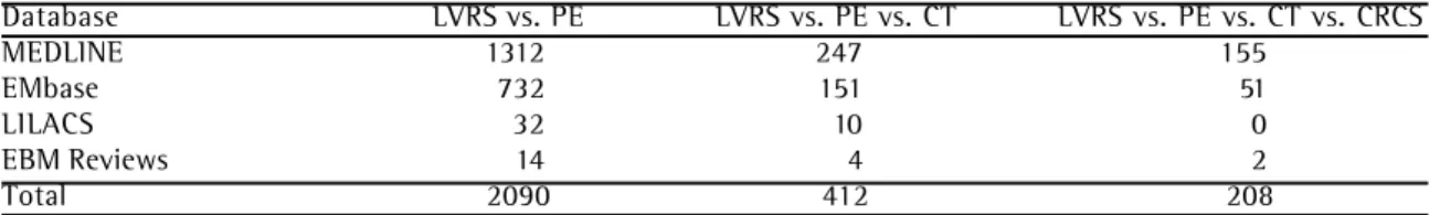
Using the above-mentioned search strategy, we identified a total of 208 articles in the following databases: MEDLINE, EMbase, LILACS, The Cochrane Library and EBM Reviews (Table 2). After their titles and abstracts had been read, 38 were selected. These 38 were submitted to the previously defined inclusion and exclusion criteria(8,16-30), resulting in the exclusion of 22 articles (Table 3).. Therefore, the final selection comprised 16 articles, all of which were fully analyzed and accepted by both reviewers. The 16 remaining articles clearly referred to the theme "radiological profile of the ideal candidate for lung volume reduction surgery", evaluated the prognostic value of the type and distribution of the pulmonary emphysema, and correlated those data with the surgical outcome. Of the 14 observational studies selected, 13 were TABLE 1
The Cochrane Collaboration classification regarding the risk of bias in randomized studies
Individual bias risk Interpretation Relationship with individual criteria
A. Low Plausible bias that is unlikely All criteria met
to severely affect the results
B. Moderate Plausible bias, results can become doubtful One or more criteria partially met
200
Figueiroa, PAU, et al.
Radiological profile of the ideal candidate for lung volume reduction surgery to treat emphysema: a systematic review
cohort studies, 5 of which prospective(8, 18-21), 8 of which were retrospective(22-29), and 1 was a case series study(30).
The 13 cohort studies(8,18-29) were paired for group analysis since they presented the same design, evaluated the same disease and intervention, measured the prognosis, described the follow-up evaluation of all participants and calculated the losses. Table 4 shows the individual characteristics of each of these studies.
Of the 16 selected articles, only 2 were randomized studies; the National Emphysema Treatment Trial (16) presented low risk of bias, and that conducted by Cassina et al.(17) presented a moderate risk of bias (Table 5).
The Cassina et al. study(17) was a randomized study, carried out from March 1995 to November 1996, comprising 30 patients subdivided into two
distinct groups. Group 1 comprised 12 consecutive patients diagnosed with pulmonary emphysema due to α-1 antitrypsin deficiency, whereas group 2 comprised 18 patients with heterogeneous emphysema related to smoking. Patients from both groups were submitted to LVRS. There was a follow-up period of 2 years, and all losses were registered. The objective was to compare functional results during the follow-up period between patients diagnosed with α-1 antitrypsin deficiency and those with heterogeneous emphysema related to smoking. This was because, up to that point, α-1 antitrypsin deficiency was used as an exclusion criterion in many clinical trials or because this condition had not been studied separately. All participants were submitted to the same clinical evaluation protocol and preoperative pulmonary rehabilitation program. In-hospital mortality was nil in both groups. However,
TABLE 3
Article exclusion criteria
Exclusion criteria Reference
Review article Kazerooni EA et al., 1997, 1998, 1999; Ramsey SD et al, 2003; Gierada DS et al., 2002; Bloch KE et al., 2002; Russi EW et al., 1999
Computed tomography not Szekely LA et al., 1997; Ingenito EP et al., 2001; Sugi K et al., used as a predictor for 2001; Baldwin JC et al, 2000; Maki DD et al, 1999; Kotloff RM et
surgical treatment al, 2001
Radiological profile of ideal Bae KT et al., 1997; Cederlund K et al, 2002; Brenner M et al, candidate for surgery not 1997; Cleverley JR et al., 2000; Wisser W et al., 1998; Hunsaker AR
evaluated et al., 1998;
Editorials Gevenois PA et al., 2001; Salzman SH et al., 2000
Artcle in Italian Bonfioli C et al., 1997
TABLE 2
Search results by database and by strategy
Database LVRS vs. PE LVRS vs. PE vs. CT LVRS vs. PE vs. CT vs. CRCS
MEDLINE 1312 247 155
EMbase 732 151 51
LILACS 32 10 0
EBM Reviews 14 4 2
Total 2090 412 208
201
Jor
n
al
B
ra
sil
ei
ro
de
Pn
eu
m
ol
ogi
a 3
1
(3
)
M
ai
/J
u
n
de
2
0
0
5
from August 1994 to FEV1 of emphysema
April 1998 from 40% to 20% of predicted values, smoking, á-1 antitrypsindeficiency detected by CT functional evaluation, normal cardiologic evaluation walk test, CT
Coxson, 2003 21 patients from June Patients who completed radiological, Undefined LVRS FEV1, exercise testing, 3 months 1994 to June 1997 cardiopulmonary and physiological radiological pattern
evaluation
Yusen, 2003 200 consecutive patients Severe heterogeneous pulmonary Another airway disease, severe comorbidities, Preoperative FEV1, dyspnea, morbidity, 5 years from 1993 to 1998 emphysema, adequate cardiologic pulmonary hypertension > 45mmHg, pulmonary mortality, quality of life
evaluation unilateral LVRS unilateral or candidates rehabilitation for bullectomy and LVRS
Hamacher, 1999 37 consecutive patients Adequate radiologic, physiologic á-1 antitrypsindeficiency LVRS and type Respiratory function testing 2 years from August 1994 to and cardiopulmonary evaluation of emphysema and survival rate
December 1998 detected by CT
Weder, 1997 37 consecutive patients Severe emphysema, dyspnea upon Smokers, bullous disease LVRS and type Respiratory function testing 3 months from August 1994 to minimal exertion or at rest of emphysema and survival
December 1996 detected by CT
Gierada, 1997 46 consecutive patients, Severe emphysema, without Bullous disease LVRS and type Respiratory function testing 6 months from January 1993 to severe comorbidities of emphysema and quality of life
February 1996 detected by CT
Slone, 1997 50 consecutive patients Pulmonary rehabilitation, Smoking LVRS Respiratory function testing 6 months from January 1993 to severe emphysema and radiological improvement
October 1994
Thurnheer, 1999 70 consecutive patients Severe emphysema, dyspnea upon Hypercapnia (PaCO2 > 55 mmHg), LVRS Respiratory function testing 3 months from August 1994 to minimal exertion or at rest advanced coronary disease, and radiological evaluation
November 1997 DLCO < 20% and bullous disease
Gierada, 2000 70 consecutive patients Adequate radiologic, physiologic Inadequate distribution of the emphysema LVRS Respiratory function testing 6 months from December 1993 to and cardiopulmonary evaluation for the study or poor physical condition and radiological evaluation
May 1995
Pompeo, 2000 52 consecutive patients Severe pulmonary emphysema, Suppurative lung disease, LVRS and type Respiratory function testing No from October 1995 to preoperative pulmonary bullous disease, asthma, metastatic of emphysema and radiological evaluation
March 1998 rehabilitation cancer, smoking detected by CT
Rogers, 2000 35 consecutive patients Severe pulmonary emphysema, Oxygen arterial saturation < 84% LVRS and type Respiratory function testing 3 months from October 1994 to Adequate radiologic, physiologic for 3 min in cycle ergometry without load of emphysema and radiological evaluation
202
Figueiroa, PAU, et al.
Radiological profile of the ideal candidate for lung volume reduction surgery to treat emphysema: a systematic review
the number of complications was significantly higher in group 1. Functional improvement for the patients in group 1 peaked at 6 months. However, after a one-year follow-up period, there was a significant decline in pulmonary function, which returned to basal levels. On the other hand, functional improvement for the patients in group 2 was consistent and continued for at least two years.
The National Emphysema Treatment Trial(16) was a multicentric, randomized, controlled study, in which patients were subdivided into two treatment groups, and was carried out from January 1998 to July 2002. The study comprised a total of 1218 patients. Patients diagnosed with advanced bilateral pulmonary emphysema were selected. After randomization, 610 patients were submitted to clinical treatment and 608 to both
clinical and surgical treatment. All patients were submitted to the same clinical evaluation protocol and the same pulmonary rehabilitation program (16 to 20 sessions, over a 6- to 10-week period). The study identified a subgroup of patients with favorable prognoses when submitted to LVRS. This group consisted of patients with advanced heterogeneous pulmonary emphysema presenting upper lobe predominance, diffuse pulmonary distension and low exercise capacity.
Functional improvement and the improvement in patient quality of life after 6, 12 and 24 months favored the surgical group. Within 24 months, exercise capacity significantly improved in the surgical group when compared to those patients submitted to clinical treatment alone (16% vs. 3%; p < 0.001). Improvement in exercise capacity TABLE 5
Characteristics of the randomized studies
Reference
Cassina, 1998 NETT, 2003 Study design
Randomized, uncontrolled; 30 participants, 12 with de á-1 antitrypsin deficiency and 18
with acquired emphysema. One study group submitted to LVRS Randomized, controlled; multicentric; 1218 participants, 608 in the surgical group and 610 in the clinical group Comparison
Functional results over a two-year follow-up period
LVRS versus best clinical treatment, both groups were submitted to pulmonary rehabilitation prior to treatment
Inclusion and exclusion criteria
Severe emphysema FEV1< 1 L, dyspnea score > 2, poor quality of life, heterogeneous emphysema, excluding bullous
Emphysema or bronchiectasis, active smoker, body mass index < 18 kg/m2 and hypercapnia Criteria defined by the NETT in 1999(21)
Intervention
LVRS X-ray CT LVRS X-ray CT Outcome
Mortality, morbidity, complications and respiratory function testing
Mortality, morbidity, maximal exercise capacity, evaluation of pulmonary function and quality of life
Follow-up period
6, 12 and 24 months 5 years
was observed in 28%, 22% and 15% of the patients at postoperative months 6, 12 and 24, respectively, compared to 4%, 5% and 3% of the patients in the clinical treatment group (p < 0.001). In conclusion, in surgical group patients diagnosed with heterogeneous pulmonary emphysema presenting upper lobe predominance, diffuse pulmonary distention and low exercise capacity, functional improvement was greater and mortality rates were lower. The greatest surgical benefit was observed in those patients whose symptoms improved to the point of increasing their exercise capacity. For those patients, even a minimal functional improvement can have a significant impact on their quality of life.
DISCUSSION
Lung volume reduction surgery is a procedure that should be indicated only when strict selection criteria are met since treatment success fundamentally depends on precise identification of good candidates for surgery(13).
In the present study, we tried to use a nonquantitative systematic review in order to determine whether there was an ideal radiological pattern that correlated with the postintervention prognosis. The National Emphysema Treatment Trial(16) is the only study using a methodology appropriate for evaluating this question. The article by Cassina et al.(17) was also a randomized study. However, the allocation of patients was inadequate and the patient sample was small (n = 30), insufficient to reject the type 2 error hypothesis, potentially affecting the interpretation of results. In addition, the authors evaluated a heterogeneous population of patients, if we consider that the evolution and the biological behavior of emphysema caused by α-1 antitrypsin deficiency differ from the characteristics of emphysema associated with smoking. Nevertheless, the study helped us conclude that patients diagnosed with heterogeneous emphysema related to smoking presented higher survival rates and functional improvement when submitted to LVRS than did patients diagnosed with emphysema caused by α-1 antitrypsin deficiency who were also submitted to that type of surgery.
The lack of randomized studies in the literature prevented us from performing a quantitative analysis of the systematic review, or meta-analysis. The other 14 studies were observational studies
and were considered in this review. All came to the same conclusion: patients with severe, apical and heterogeneous emphysema have lower mortality rates and higher improvement in their pulmonary function and quality of life. The main bias that resulted from this type of study was related to the lack of a control group, which reduced the acceptance of these conclusions in clinical practice. Since they were observational studies, they did not allow the comparative analysis of outcome measures after an intervention, such as mortality, functional evaluation and quality of life. Although the conclusions drawn by the authors of these studies do not have the same force or scientific validity as those resulting from randomized studies(15), they should be considered applicable to the population.
In view of the need to evaluate the true efficacy of LVRS, we carried out this systematic review and discovered that there was a lack of appropriately designed studies in the literature. This surgical procedure has typically been recommended based on data collected in observational studies. We found only one A-level randomized study(16) evaluating this theme.
This systematic review allowed us to conclude that the radiological profile, characterized by the type, heterogeneity, distribution and diffuse distention of emphysema, together with the degree of emphysema severity, characterized by pulmonary function testing and the evaluation of exercise capacity, represents the main predictor of a positive surgical outcome. Due to the paucity of studies in the literature, this is a grade-B recommendation. Further studies should be carried out in order to provide this recommendation with a higher degree of scientific consistency.
REFERENCES
1. Celli BR, MacNee W, ATS/ERS Task Force. Standards for the diagnosis and treatment of patients with COPD: a summary of the ATS/ERS position paper. Eur Respir J 2004;23:932-46.
2. Snider GL, Kleinerman J, Thurlbeck WM, Bengali ZK. The definition of emphysema: report of a National Heart, Lung and Blood Institute, Division of Lung Diseases, Workshop. Am Rev Respir Dis 1985;132:182-5. 3. O’Donnell DE, Webb KA. Exertional breathlessness in
patients with chronic airflow limitation: the role of lung
hyperinflation. Am Rev Respir Dis 1993;148:1351-7. 4. S i l v e r m a n E K , S p e i z e r F E . R i s k f a c t o r s f o r t h e
204
Figueiroa, PAU, et al.
Radiological profile of the ideal candidate for lung volume reduction surgery to treat emphysema: a systematic review
5. Murray CJL, Lopez AD. Evidence-based health policy: lessons from the global burden of disease study. Science 1996;274:740-3.
6. National Center for Health Statistics. Series 10. Data from the National Health Interview Survey. Vital and health statistics 10 (issues from 1974 to 1995). [cited 2004 ago 20]. Available from: http://www.cdc.gov/ nchs/products/pubs/pubd/se-ries/sr10/ser10.htm. 7. Brasil. Ministério da Saúde. DATASUS. [cited 2004 ago
20]. Available from: http;//www.datasus. gov.br. 8. McKenna RJ Jr, Brenner M, Fischel RJ, Singh N, Yoong
B, Gelb AF, et al. Patient selection criteria for lung volume reduction surgery. J Thorac Cardiovasc Surg 1997;114:957-64
9. National Emphysema Treatment Trial Research Group. Patients at high risk of death after lung-volume– reduction surgery. N Engl J Med 2001;345:1075-83. 10. Dickersin K, Scherer R, Lefebvre C. Identifying relevant
studies for systematic reviews. BMJ 1994;309:1286-91. 11 . Haynes RB, Wilczynski N, McKibbon KA, WalkerCJ, Sinclair JC. Developing optimal search strategies for detecting clínically sound studies in MEDLINE. J Am Med Inforn Assoc 1994;1:447-58.
12. Jaeschke R, Guyatt G, Sackett DL, Evidence-Based Medicine Working Group. Users’ guides to the medical literature. III. How to use an article about a diagnostic test. A. Are the results of the study valid? JAMA 1994;271:389-91. 13. Guyatt GH, Sackett DL, Cook DJ, Evidence-Based Medicine
Working Group. Users’ guides to the medical literature. II. How to use an article about therapy or prevention. A. Are the results of the study valid? JAMA 1993;270:2598-601. 1 4 . Laupacis A, Wells G, Richardson WS, Tugwell P, Evidence-Based Medicine Working Group. Users’ guides to the medical literature. V. How to use an article about prognosis. JAMA 1994;272:234-7.
1 5 . Clarke M, Oxman AD, (Eds.). Cochrane Reviewers’ Handbook 4.1.6 [updated January 2003]. In: The Cochrane Library, Issue 1, 2003.
1 6 . National Emphysema Treatment Trial Research Group, Fishman A, Martinez F, Naunheim K, Piantadosi S, Wise R. A randomized trial comparing lung-volume-reduction surgery with medical therapy for severe emphysema. N Engl J Med 2003;348:2059-73. 17. Cassina PC, Teschler H, Konietzko N, Theegarten D,
Stamatis G. Two-year results after lung volume reduction surgery in alpha1-antitrypsin deficiency versus smoker’s emphysema. Eur Respir J 1998;12:1028-32.
18. Coxson HO, Whittall KP, Nakano Y, Rogers RM, Sciurba FC, Keenan RJ et al. Selection of patients for lung volume reduction surgery using a power law analysis of the computed tomographic scan. Thorax 2003;58:510-4.
1 9 . Yusen RD, Lefrak SS, Gierada DS, Davis GE, Meyers BF, Patterson GA et al. A prospective evaluation of lung volume reduction surgery in 200 consecutive patients. Chest 2003;123:1026-37.
20. Flaherty KR, Kazerooni EA, Curtis JL, Iannettoni M, Lange L, Schork MA et al. Short-term and long-term outcomes after bilateral lung volume reduction surgery : prediction by quantitative CT. Chest 2001;119:1337-46.
2 1 . Hamacher J, Bloch KE, Stammberger U, Schmid RA, Laube I, Russi EW et al. Two years’ outcome of lung volume reduction surgery in different morphologic emphysema types. Ann Thorac Surg 1999;68:1792-8. 2 2 . Hunsaker AR, Ingenito EP, Reilly JJ, Costello P. Lung volume reduction surgery for emphysema: correlation of CT and V/Q imaging with physiologic mechanisms o f i m p r o v e m e n t i n l u n g f u n c t i o n . R a d i o l o g y 2002;222:491-8.
23. Gierada DS, Yusen RD, Villanueva IA, Pilgram TK, Slone RM, Lefrak SS, et al. Patient selection for lung volume reduction surgery: An objective model based on prior clínical decisions and quantitative CT analysis. Chest 2000;117:991-8. 24. Pompeo E, Sergiacomi G, Nofroni I, Roscetti W, Simonetti
G, Mineo TC. Morphologic grading of emphysema is useful in the selection of candidates for unilateral or bilateral reduction pneumoplasty. Eur J Cardiothorac Surg 2000;17:680-6.
25. Rogers RM, Coxson HO, Sciurba FC, Keenan RJ, Whittall KP, Hogg JC. Preoperative severity of emphysema predictive of improvement after lung volume reduction surgery: use of CT morphometry. Chest 2000;118:1240-7.
26. Thurnheer R, Engel H, Weder W, Stammberger U, Laube I, Russi EW, et al. Role of lung perfusion scintigraphy in relation to chest computed tomography and pulmonary function in the evaluation of candidates for lung volume reduction surgery. Am J Respir Crit Care Med 1999;159:301-10. 2 7 . Gierada DS, Slone RM, Bae KT, Yusen RD, Lefrak SS,
Cooper JD. Pulmonary emphysema: comparison of preoperative quantitative CT and physiologic index v a l u e s w i t h c l í n i c a l o u t c o m e a f t e r l u n g - v o l u m e reduction surgery. Radiology 1997;205:235-42. 2 8 . Slone RM, Pilgram TK, Gierada DS, Sagel SS, Glazer HS,
Yusen RD, et al. Lung volume reduction surgery: comparison of preoperative radiologic features and clínical outcome. Radiology 1997;204:685-93. 2 9 . Weder W, Thurnheer R, Stammberger U, Burge M, Russi
EW, Bloch KE. Radiologic emphysema morphology is associated with outcome after surgical lung volume reduction. Ann Thorac Surg 1997;64:313-20. 30. Nakano Y, Coxson HO, Bosan S, Rogers RM, Sciurba FC,