DOI: 10.5935/2359-4802.20170012
Introduction
Epipericardial fat necrosis (EPFN) is a rare, benign, and self-limiting condition, first described in 1957
by Jackson et al.1 Currently, there are only 33 cases
described in the literature.1-9
The pathophysiology of the disease remains uncertain. Likely predisposing factors include torsion of the vascular pedicle of adipose tissue, structural alterations that make the tissue more vulnerable and obesity, which, like the Valsalva maneuver, can lead to increased capillary pressure, causing hemorrhage into the adipose tissue. There are also reports of association
with chest trauma or infections.9
It presents as an acute pleuritic pain, being an important differential diagnosis of chest pain in healthy patients who maintain stable vital data and unaltered laboratory tests.
EPFN is generally diagnosed by Computed Tomography (CT) and, in some cases, paracardiac
opacification is seen on chest radiography.3
Case Report
Case 1
Male individual, neurosurgeon, 62 years old, healthy, was admitted to the emergency room with ventilator-dependent chest pain for 2 days, associated with dyspnea on medium exertion. Physical and laboratory examination showed no alterations.
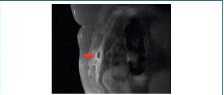
At the chest angiography, there was no vascular filling failure. The remaining assessment showed preserved tomographic aspects. Magnetic resonance imaging (MRI) of the chest identified an elongated image with fat signal intensity in the epipericardial fat, showing enhancement by contrast of the surrounding tissues, suggesting fat infarction with adjacent inflammatory alterations (Figures 1 and 2). The diagnosis of epipericardial fat necrosis was suggested, and the patient was discharged with Non-Steroidal Anti-Inflammatory Drugs (NSAIDs). Symptom remission occurred within 2 days.
Case 2
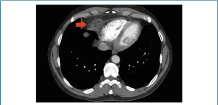
A 60-year-old man, healthy, sought emergency services for ventilator-dependent chest pain associated with recent-onset dyspnea. Laboratory tests and electrocardiogram showed no alterations. At the CT angiography, there was no vascular filling failure. In the mediastinal evaluation, densification of the epipericardial fat was identified with contrast enhancement, suggesting infarction associated with inflammatory alterations (Figures 3 and 4). The patient was discharged with NSAIDs.
Discussion
Necrosis of epipericardial fat is an uncommon or poorly reported cause of chest pain. It generally manifests as pleuritic chest pain and dyspnea, being a differential diagnosis of severe conditions, such as pulmonary thromboembolism and acute myocardial infarction. The pain usually lasts a few days, but may eventually last for months. In cases reported to date, it equally affects both genders and the mean age group is 50 years.
91
International Journal of Cardiovascular Sciences. 2017;30(1):91-94
CASE REPORT
Mailing Address: Núbia Bernardes Carvalho •
Avenida do Contorno, 9.530, Barro Preto. Postal Code: 30140-062. Belo Horizonte, MG, Brazil. E-mail: [email protected]
Epipericardial Fat Necrosis. An Important Differential Diagnosis of Chest Pain
Núbia Bernardes Carvalho,1 Natália de Paula Lopes e Silva,1 Pedro Paulo Nunes Pereira,2 André Volani Morganti,2 Sinval Lins Silva,2 André Silva Rodrigues2
Pósgraduação Ciências Médicas de Minas Gerais (PGCM - MG)1; Hospital Felício Rocho,2 Belo Horizonte, MG − Brazil
Manuscript received July 31, 2016; revised manuscript October 21, 2016; accepted November 28, 2016.
Fat Necrosis / physiopathology; Pericardium; Obesity; Chest Pain; Dyspnea.
92
The EPFN is similar to appendagitis, the necrosis of the epiploic appendix, which presents as localized abdominal pain and is also self-limited.
The difficulty attaining the diagnosis lies in the unspecificity of the signs and symptoms, making imaging tests essential, since the electrocardiogram and the laboratory tests are, in most part, normal. The CT shows an encapsulated, rounded, fat-containing, epipedicardial lesion, associated or not with adjacent pericardial thickening and pleural effusion. The MRI shows an epipericardial lesion with a high T2
sign, typical of adipose tissue edema, with moderate enhancement after administration of gadolinium, suggestive of an active inflammatory process. At the x-ray, which is less specific, paracardiac opacity may be seen, with or without obliteration of the costophrenic sinus. The radiological differential diagnosis includes primary or secondary pericardial neoplasia, pericardial cyst, lung cancer, Morgagni hernia or bronchogenic cyst.
T h e d e f i n i t i v e d i a g n o s i s i s a t t a i n e d b y anatomopathological analysis of the surgically
Figure 1 – T1-weighted image of MRI sagittal section with post-contrast fat saturation.
Figure 2 – T1-weighted image of MRI axial section with post-contrast fat saturation. Patient is in the prone position.
Carvalho et al.
Epipericardial fat necrosis. Case Report Int J Cardiovasc Sci. 2017;30(1):91-94
93
resected fragment, which has fallen into disuse due to the accuracy of the imaging tests and to the benign and self-limiting characteristic of the disease. When performed, the described pathological findings consist
of necrotic adipose cells surrounded by macrophages, neutrophils, or fibrous tissue.
Treatment is carried out in symptomatic patients with non-steroidal anti-inflammatory drugs.
Figure 3 – Axial section of chest computed angiotomography.
Figure 4 – Coronal section of chest computed angiotomography. Carvalho et al.
Epipericardial fat necrosis. Case Report
Int J Cardiovasc Sci. 2017;30(1):91-94
94
Since 2005, when the first case of successful conservative
treatment was described,3 radiology has played a key role
in the diagnosis and follow-up of EPFN.
Conclusion
Necrosis of epipericardial fat is a poorly reported condition due to possible underdiagnosis, and it is important that clinicians, cardiologists and radiologists use it as a differential diagnosis of acute chest pain, after ruling out more severe causes, such as pulmonary thromboembolism or acute myocardial infarction.
The definitive diagnosis is attained by the anatomopathological analysis, but EPFN has been increasingly diagnosed based on imaging tests associated with a suggestive clinical context. The current facilitation of diagnosis, together with a favorable clinical outcome, reinforces the safety of the conservative conduct.
Author contributions
Acquisition of data: Silva SL, Rodrigues AS. Writing of the manuscript: Carvalho NB, Lopes e Silva NP. Critical revision of the manuscript for intellectual content: Pereira PPN, Morganti AV, Rodrigues AS.
Potential Conflict of Interest
No potential conflict of interest relevant to this article was reported.
Sources of Funding
There were no external funding sources for this study.
Study Association
This article is part of the thesis of postgraduate submitted by Núbia Bernardes Carvalho e Natália de Paula Lopes e Silva , from Pós-graduação de Ciências Médicas de Minas Gerais - PGCM - MG.
1. Jackson RC, Clagett OT, McDonald JR. Pericardial fat necrosis: report of three cases. J Thorac Surg. 1957;33(6):723-9.
2. Runge T, Greganti MA. Epipericardial fat necrosis – a rare cause of pleuritic chest pain: case report and review of the literature. Arch Med Sci. 2011;7(2):337-41.
3. Pineda V, Caceres J, Andreu J, Vilar J, Domingo ML. Epipericardial fat necrosis: radiologic diagnosis and follow-up. AJR Am J Roentgenol. 2005;185(5):1234-6.
4. van den Heuvel DA, van Es HW, Cirkel GA, Bos WJ. Acute chest pain caused by pericardial fat necrosis. Thorax. 2010;65(2):188.
5. Giassi KS, Costa AN, Apanavicius A, Bachion GH, Musolino RS, Kairalla
RA. Epipericardial fat necrosis: an unusual cause of chest pain. J Bras Pneumol. 2013;39(5):627-9.
6. Lacasse MC, Prenovault J, Lavoie A, Chartrand-Lefebvre C. Pericardial
fat necrosis presenting as acute pleuritic chest pain. J Emerg Med. 2012;44(2):e269-71.
7. Mazzamuto G, Ghaye B. Epipericardial fat necrosis. JBR–BTR 2012;95(3):154-5.
8. Peres Claro I, Magalhães V, Correia I, Campos P, Sotto-Mayor R, Bugalho
de Ameida A. [Epipericardial fat necrosis - Case report]. Rev Port
Pneumol. 2010;16(3):507-12.
9. Fred HL. Pericardial fat necrosis: a review and update. Tex Heart Inst J. 2010; 37(1): 82–84.
References
Carvalho et al.
Epipericardial fat necrosis. Case Report Int J Cardiovasc Sci. 2017;30(1):91-94