DOI: 10.5935/2359-4802.20170017
Introduction
A l t h o u g h p r i m a r y p e r c u t a n e o u s c o r o n a r y intervention (P-PCI) has been contributing to a decrease in mortality in recent years, ST-segment elevation myocardial infarction(STEMI) is still one of the leading cause of mortality and morbidity all over the world.1 In-hospital mortality rates were reported as 7-10% in some registries.2 Currently, several validated risk factors and scoring systems are used to predict mortality in STEMI patients. In the fibrinolytic era,
thrombolysis in myocardial infarction (TIMI) risk score was developed as a clinical risk score to predict 30-day mortality in patients with STEMI.3 TIMI score was derived and validated among fibrinolytic-eligible patients enrolled in clinical trials, so it can not be easily applied in STEMI patients undergoing P-PCI. Recently, the Global Registry of Acute Coronary Events (GRACE) score has been defined for predicting mortality at 6 months in patients with acute coronary syndromes (ACS), but critically ill patients such as those who died early may be underrepresented.4 In addition
ORIGINAL ARTICLE
Mailing Address: Erkan Yildirim
Gata Kardiyoloji Ad. Postal Code: 06010, Etlik, Ankara – Turkey E-mail: dr_erkanyildirim@yahoo.com.tr; erkan_2157@yahoo.com
The Relationship Between Gensini Score and In-Hospital Mortality in Patients with
ST-Segment Elevation Myocardial Infarction
Erkan Yildirim,1 Atila Iyisoy,1 Murat Celik,1 Uygar Cagdas Yuksel,1 Cengizhan Acikel,1 Baris Bugan,2 Yalcin Gokoglan1
Academia Médica Militar Gulhane1, Departamento de Cardiologia, Ankara; Hospital Militar Girne2 – Turquia
Manuscript received May 17, 2016; revised manuscript August 08, 2016; accepted January 25, 2017.
Abstract
Background: To date, several validated patient-based risk scores have been established to predict mortality and morbidity in patients presenting with ST-segment elevation myocardial infarction (STEMI). The Gensini score was originally developed to quantify the severity of coronary artery disease (CAD).
Objectives: We intend to assess the association between severity of CAD assessed by Gensini score and in-hospital mortality in patients with STEMI undergoing primary percutaneous coronary intervention (P-PCI).
Methods:A total of 539 patients presenting with acute STEMI, who underwent P-PCI within the first 12 hours from the onset of symptoms, were included. The severity of CAD was expressed as the sum of the Gensini scores for each lesion. Patients’ demographic variables, medical histories and clinical features, as well as in hospital major adverse events were obtained from the medical reports.
Results:Of these 539 patients, 416 (77.2%) were male and mean age was 59.14 ± 12.68 years. In-hospital mortality rate was 5.4% (29 patients; 16 men). Mortality rate was 10.5% in female patients and 3.8% in males (P = 0.004). Mean Gensini scores were significantly different in the comparison between patients who survived (54.54 ± 26.34) and those who died (80.17 ± 26.51) (P = 0.001). The multivariable Cox proportional hazards regression analysis model revealed that the Gensini score (P = 0.037), female gender (P = 0.039), serum urea levels (P = 0.041), uric acid levels (P = 0.008) and LVEF (P = 0.001) were independently associated with in-hospital mortality in patients with STEMI undergoing P-PCI.
Conclusion: The Gensini score is independently associated with in-hospital mortality in STEMI patients treated with P-PCI. Therefore, it might play an important role in risk stratification of STEMI patients. (Int J Cardiovasc Sci. 2017;30(1):32-41)
to these clinical scores, some coronary angiography based scoring systems such as Gensini, SYNTAX
(synergy between percutaneous coronary intervention
with Taxus and cardiac surgery) and ACC/AHA have
been established to assess the severity of lesions and provide some prognostic information for patients with coronary artery disease (CAD). Although these scoring systems provide quantitative evaluation, the valuable detailed information derived from angiography is not sufficiently used. In clinical practice, there is a need for an initial stratification of STEMI patients, which aims to identify those at higher risk and decrease the incidence of major adverse cardiovascular events through more appropriate targeting of preventive measures.
The Gensini scoring system is an objective method to determine the severity of CAD according to angiographic findings.5 It was originally developed to quantify the severity of CAD; however, subsequent studies have demonstrated its ability to identify patients who are at high risk of adverse events who are treated with PCI.6 However, little is known about the association between the severity of CAD assesed by the Gensini score and in-hospital mortality in patients with STEMI undergoing P-PCI.
Objective
The aim of the present study was to evaluate the predictive role of Gensini score to detect in-hospital mortality in STEMI patients treated with P-PCI.
Methods
Our single-center study was retrospective and non-randomized (prospective researching the information, retrospectively). A total of 539 consecutive patients presenting with first acute STEMI who underwent P-PCI within the 12 hours from the onset of symptoms, between 2004 and 2013, were included in our study. Patients` demographic variables, medical histories and clinical features, as well as in hospital major adverse events were obtained from medical reports. Since our institute is a tertiary center, P-PCI was conducted as the preferred reperfusion strategy for most STEMI cases. Briefly, the diagnosis of STEMI was made through the criteria of classical symptoms of coronary ischemia (chest pain lasting > 30 minutes), detection of > 1 mm ST-segment elevation in at least two contiguous leads and elevation in cardiac biomarkers, and defined by
the guidelines of the American College of Cardiology and the European Society of Cardiology.7 All STEMI patients were admitted and followed up in intensive coronary care unit and hospital stay was defined as this first admission period of STEMI patients. Previous CABG (coronary artery bypass graft) or PCI, previous STEMI history, reperfusion with thrombolytic drug, known malignancy and severe liver diseases, insufficient data from the clinical recordings and angiographic recordings were all defined as exclusion criteria.
The severity of CAD was calculated using the Gensini score, in which the calculation is based on the evaluation of the number of stenotic segments along with their respective degrees of luminal narrowing and localization within the coronary tree.5 The severity of the disease was expressed as the sum of the scores for each lesion. Two investigators blinded to the patient’s clinical data analyzed the results of the coronary angiography. The study protocol was approved by the local ethics committee (2013-157).
Statistical analysis
Data was processed through SPSS 16.0 statistical package (SPSS Inc, Chicago, IL, USA). All data are expressed as numbers and percentages and continuous variables were tested for normal distribution using the Kolmogorov-Smirnov test. Continuous data with normal distribution are presented as mean and standard deviation and compared by the unpaired T-test, and non-normally distributed data are presented as median and interquartile range and compared through the Mann-Whitney U test. Categorical data are presented as counts and percentages and compared using the chi-square test. Logistic regression analysis was performed to identify independent predictors of in-hospital mortality. For the multivariate analysis, a forward stepwise logistic regression was performed using the variables which were significant at the univariate analysis, together with risk factors. P values < 0.05 were considered statistically significant.
Results
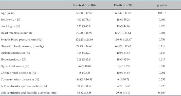
Table 1 – Baseline demographical and clinical features of the groups
Survival (n = 510) Death (n = 29) p value
Age (years) 58.58 ± 12.52 68.96 ± 11.52 0.001*
Sex (men), n (%) 400 (%78.4) 16 (%55.2) 0.004
Smoking, n (%) 233 (%45.7) 13 (%44.8) 0.928
Heart rate (beats/minute) 79.90 ± 16.99 86.51 ± 24.64 0.004
Systolic blood pressure, (mmHg) 132.23 ± 24.96 116.96 ± 24.67 0.756
Diastolic blood pressure, (mmHg) 77.73 ± 14.60 69.29 ± 17.43 0.119
Diabetes mellitus n (%) 116 (%22.7) 10 (%34.5) 0.146
Hypertension, n (%) 218 (%42.8) 19 (%65.5) 0.017
Hyperlipidemia, n (%) 96 (%18.8) 5 (%17.29) 0.832
Chronic renal disease, n (%) 18 (%3.5) 10 (%34.5) 0.001
Coronary artery disease, n (%) 84 (%%16.5) 6 (%20.7) 0.553
Left ventricular ejection fraction, (%) 50.98 ± 8.50 34.72 ± 5.66 0.028
Left ventricular end diastolic diameter, (mm) 48.52 ± 5.58 29.38 ± 4.17 0.436*
*Data without normal distribution compared through Mann-Whitney U test.
Table 2. In-hospital mortality rate was 5.4% (29 patients; 16 men). Twenty-nine patients (5.3%) died during the hospital stay. Causes of death for these 29 patients were: malignant cardiac arrhythmias such as VT/VF in 8 patients (27.5%), cardiogenic shock in 8 patients (27.5%), acute pulmonary edema in 4 patients (13.7%), right heart failure in 3 patients (10.3%), bleeding complications in 2 patients (6.8%), septic shock in 1 patient (3%), acute mitral regurgitation in 1 patient (3%), ventricular septal rupture in 1 patient (3%), and ventricular free wall rupture in 1 patient (3%). Mortality rate was 10.5% for female patients and 3.8% for males (P = 0.004). Of these 539 patients who underwent P-PCI, 454 (84%) were treated successfully with BMS. From the remaining 85 patients, 18 (4%) were treated with DES, 26 (5%) with plain PTCA, in 18 (3%) patients the procedure failed, and 23 (5%) patients underwent CABG. In-hospital mortality did not differ between the groups on different therapies during P-PCI (P > 0.05).
In the initial statistical analysis, we found that some variables significantly differed between patients who survived and those who died. Female gender, history of hypertension, chronic renal disease, age, admission blood glucose, urea, creatinine and uric
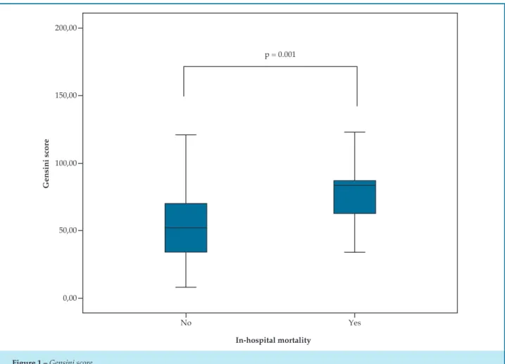
acid levels, heart rate, leukocytes, hemoglobin, platelet and neutrophil count, and LVEF were found to be statistically significantly different between the two groups. (P < 0.05 for all). Angiographic data is shown in Table 3. Mean Gensini scores were significantly different in the comparison between patients who survived (54.54 ± 26.34) and those who died (80.17 ± 26.51) (P = 0.001) (Figure 1). The infarction-related vessel was the left anterior descending coronary artery (LAD) in 224 patients (41.5%), the circumflex coronary artery (CFX) in 104 patients (19.2%), and the right coronary artery (RCA) in 196 patients (36.3%). In-hospital mortality did not differ between the groups of different infarction-related vessels (P > 0.05).
Table 2 – Laboratory findings of the groups
Survival (n = 510) Death (n = 29) p value
Glucose (mg/dL) 149.37 ± 68.17 199.42 ± 103.91 0.048*
Urea (mg/dL) 38.09 ± 17.82 59.03 ± 35.34 0.001*
Creatinine (mg/dL) 1.05 ± 0.49 1.41 ± 0.66 0.001*
Uric acid (mg/dL) 5.52 ± 2.01 7.59 ± 2.97 0.002*
High density lipoprotein, (mg/dL) 42.13 ± 23.01 40.00 ± 7.82 0.670
Low density lipoprotein, (mg/dL) 113.74 ± 36.70 112.95 ± 46.49 0.881*
Triglycerides (mg/dL) 140.06 ± 100.43 128.20 ± 76.43 0.569*
Albumin (g/dL) 3.40 ± 0.54 3.15 ± 0.57 0.085*
Leukocytes (n/μL) 11.695.39 ± 4.031.71 14.646.42 ± 7.062.39 0.017*
Hemoglobin (g/dL) 13.77 ± 1.97 12.55 ± 2.07 0.006*
Hematocrit (%) 41.19 ± 5.18 37.68 ± 5.60 0.202
Platelet (x103/μL) 248.37 ± 70.35 253.25 ± 100.67 0.010
Neutrophil (n/μL) 7.953.01 ± 3.384.94 10.045.93 ± 6.286.28 0.032*
Lymphocyte (n/μL) 2531.72 ± 2362.84 2407.87 ± 2173.31 0.279*
*Data without normal distribution is compared by Mann-Whitney U test.
gender (P = 0.039), urea levels (P = 0.041), uric acid levels (P = 0.008) and LVEF (P = 0.001) were independently associated to in-hospital mortality in patients with STEMI undergoing P-PCI (Table 5).
We independently examined 25 randomly chosen angiograms, visually estimated lesion scores, and calculated Gensini scores. The intra-observer and inter-observer correlation coefficient was 0.96 and 0.95 for the Gensini score, indicating good reproducibility and reliability.
Discussion
The main finding of this study was that the Gensini score is independently associated to in-hospital mortality in STEMI patients treated with P-PCI. Nonetheless, the female gender, higher serum urea and uric acid levels, and lower LVEF were also associated to in-hospital mortality in this group of patients.
STEMI is the most important part of ACS. Consistently with our data, in-hospital mortality rates ranged between 6% and 14%.8 Many factors such as previous MI, door to balloon time, presence
p = 0.001
No Yes
In-hospital mortality
200,00
150,00
100,00
50,00
0,00
Gensini score
Figure 1 – Gensini score
Table 3 – Angiographic data
Survival (n = 510) Death (n = 29) p value
Coronary artery involvement
LAD, n (%) 210 (41.1) 14 (48.2)
0.148
CFX, n (%) 104 (20.3) 5 (17.2)
RCA, n (%) 196 (38.4) 10 (34.4)
Gensini score 54.54 ± 26.34 80.17 ± 26.51 0.001
LAD: left anterior descending coronary artery; CFX: circumflex coronary artery; RCA: right coronary artery
(Stent-Primary Angioplasty in Myocardial Infarction),10 and MCRS (The Mayo Clinic Risk Score)14 included angiographic variables in the scoring algorithm. The CADILLAC risk score was able to accurately predict 30-day and 1-year mortalities after P-PCI in STEMI patients. Baptista et al.15 applied the PAMI
Table 4 – Results of univariable logistic regression analysis
Variable OR p value
Female gender 2.955 0.005
Gensini score 1.029 0.001
Urea 1.026 0.001
Uric acid 1.363 0.001
LVEF 0.797 0.001
History of hypertension 1.007 0.020
History of chronic renal disease 14.36 0.001
Age 1.076 0.001
Admission blood glucose 1.007 0.001
Creatinine 1.811 0.001
Uric acid 1.363 0.001
Heart rate 1.022 0.048
Systolic blood pressure 0.974 0.002
Diastolic blood pressure 0.962 0.004
Leucocytes count 1.000 0.004
Neutrophil count 1.156 0.001
Hemoglobin 0.733 0.002
Hematocrit 0.879 0.001
Albumin 0.424 0.044
Table 5 – Effects of several variables on in-hospital mortality by the multivariable regression analysis
Variable Adjusted OR 95% CI p value
Female gender 7.780 1.10-54.66 0.039
Gensini score 1.033 1.00-1.06 0.037
Urea 1.030 1.00-1.06 0.041
Uric acid 1.544 1.11-2.13 0.008
LVEF 0.761 0.65-0.88 0.001
*By entering gender, age, history of hypertension, chronic renal disease, admission blood glucose, urea, creatinine, uric acid, heart rate, SBP, DBP, leukocytes count, neutrophil count, hemoglobin, hematocrit, albumin, LVEF and the Gensini score
In addition to those scores, which were established from combination of both clinical and angiographic variables, some scoring systems based solely on
The purpose of coronary scoring systems is to quantify the severity of coronary stenosis. Different coronary arteries carry different volumes of blood to the heart, and coronary scores take this into account. The degree of stenosis was also considered in these scoring systems. Overall, the individual ability of these angiography based scores to predict mortality is uncertain, and an important limitation is that these scores have been largely limited to elective patients.19 Although the SYNTAX score is frequently used for predicting mortality in patients with STEMI undergoing P-PCI, the percent diameter stenosis is not considered in scoring and a distinction is made only between occlusive (100%) and non-occlusive (50-99% stenosis) disease. Furthermore, stenosis is
considered severe when it causes ≥ 50% reduction in
the luminal diameter by visual assessment in vessels
≥ 1.5 mm. However, in the Gensini score, lesions causing
< 50% reduction in the luminal diameter in vessels < 1.5 mm diameter are considered in the scoring algorithm. The Gensini score was originally developed to quantify the severity of CAD. It has been widely used in clinical trials to assess the extent and severity of CAD.However, subsequent studies have demonstrated its ability to identify patients treated by PCI who are at high risk of adverse events. Nevertheless, little is known about the correlation between the Gensini score and short-term mortality in STEMI patients.20-23 There are few recently published studies that have evaluated the Gensini score in the context of P-PCI. In a recent report, the Gensini score was found to be associated with lower MACE during hospital stay and at 6 months after PCI in acute STEMI patients.24 Thus, the authors concluded that it could be used to predict short-term MACE in STEMI patients during the post PCI period. Acet et al.25 found that the TIMI risk index is significantly related to the Gensini score in predicting the extent and severity of CAD in patients with STEMI.
This study represents the analysis of the relationship between the Gensini score and in-hospital mortality in patients with STEMI undergoing P-PCI. The current study has demonstrated that STEMI patients with higher Gensini scores, regardless of other clinical variables, are at increased risk of in-hospital mortality. According to our study, a 1 point increase in the Gensini score is related to a 3% increase of in-hospital mortality risk in patients with STEMI undergoing P-PCI. Several factors could account for this finding: the Gensini score concentrates on the LMCA, proximal LAD, and mid-LAD arteries, which are assigned relatively higher weighting factors.
Occlusion of these coronary arteries causes large areas of infarction which may be related to higher mortality rates. One of the limitations of using the Gensini score for risk stratification is the absence of any clinical variables. This deficiency can be successfully addressed through its combination with clinical-based risk models. In our study, we also found that female gender, higher urea and uric acid levels and lower LVEF were associated to in-hospital mortality in STEMI patients undergoing P-PCI. These findings were consistent with previous studies in literature.20-23 In the univariate regression analysis, differently from other variables, hemoglobin, hematocrit, albumin and LVEF were found to be negatively correlated to in-hospital mortality, meaning that lower values in these variables were associated to a higher mortality. However, this association was no longer significant after the multivariate analysis, except for LVEF. Mortality rate was 10.5% (13/123) in women and 3.8% (16/416) in men. Higher in-hospital mortality in women is often attributed to a delayed hospital admission, a higher clustering of comorbidities, older age and more bleeding complications after interventions.26-28 Our study highlights the importance of adjusting mortality to these variables. We did not find a difference in the presence of diabetes, even though others have reported that this tends to be more common.
The result of our study was an improvement of existing conventional risk factors and scoring systems for STEMI patients. A particular strength of our study is that all patients admitted to hospital with a diagnosis of STEMI were included, unlike reports from some registries, in which only data from selected patients were analysed. Improving the prediction of mortality is a major challenge and selecting the proper therapy is difficult, but we think that the combination of established risk factors such as TIMI and GRACE with angiographic score assessment provides the best information to predict in-hospital mortality in STEMI patients undergoing P-PCI. Readily available risk factors such as hypertension, smoking, diabetes, LDL, family history of MI and clinical variables such as age, gender, door to balloon time and biomarkers should also be considered to improve the prediction of mortality.
Limitations
to all patients such as those who present with other form of ACS or those who were not treated with PCI. The relatively small sample size of the current study indicates the need to validate the findings with a larger patient cohort. Door to balloon time and onset of the infarction are important parameters which may also be associated to in hospital mortality. However, due to our retrospective design and insufficient medical reports, a considerable amount of patient data about the onset of the infarction or door to balloon time were lacking or unreliable. Therefore, these parameters could not be studied. In clinical practice, angiographic scores are calculated by visual lesion assessment (rather than laboratory determination), which would likely lead to greater interobserver variability. Also, we did not compare the Gensini score to other angiographic score systems such as the SYNTAX, which is frequently used to predict mortality in STEMI patients undergoing P-PCI. Further prospective randomized studies with a large sample and attendance of multicentre are required to confirm this hypothesis more effectively.
Conclusions
It is clear that there is a clinical necessity for an accurate and useful scoring system to identify patients at a higher risk for in-hospital mortality, who require more intensive care. The Gensini score may have a valuable role in the
risk stratification of STEMI patients undergoing P-PCI. However, the Gensini score should be improved through a combination that includes clinical, procedural and laboratory variables.
Author contributions
Conception and design of the research: Yildirim E, Iyisoy A, Yuksel UC. Acquisition of data: Yildirim E, Celik M, Yuksel UC, Bugan B, Gokoglan Y. Analysis and interpretation of the data: Yildirim E, Iyisoy A, Celik M, Yuksel UC, Bugan B, Gokoglan Y. Statistical analysis: Yildirim E, Celik M, Acikel C. Writing of the manuscript: Celik M, Bugan B. Critical revision of the manuscript for intellectual content: Yildirim E, Iyisoy A, Yuksel UC, Acikel C, Gokoglan Y. Supervision: Iyisoy A.
Potential Conflict of Interest
No potential conflict of interest relevant to this article was reported.
Sources of Funding
There were no external funding sources for this study.
Study Association
This article is part of the thesis of Doctoral submitted by Erkan Yildirim, from Gülhane Military Medical Academy.
1. Keeley EC, Boura JA, Grines CL. Primary angioplasty versus intravenous
thrombolytic therapy for acute myocardial infarction: a quantitative review of 23 randomised trials. Lancet. 2003;361(9351):13-20.
2. Brodie BR, Stuckey TD, Hansen C, Muncy D. Benefit of coronary reperfusion before intervention on outcomes after primary angioplasty for acute myocardial infarction. Am J Cardiol. 2000;85(1):13-8.
3. Morrow DA, Antman EM, Charlesworth A, Cairns R, Murphy SA, de Lemos JA, et al. TIMI risk score for ST-elevation myocardial infarction: a convenient, bedside, clinical score for risk assessment at presentation: an intravenous nPA for treatment of infarcting myocardium early II trial substudy. Circulation. 2000;102(17):2031-7.
4. Fox KA, Dabbous OH, Goldberg RJ, Pieper KS, Eagle KA, Van de Werf F, et al. Prediction of risk of death and myocardial infarction in the six months after presentation with acute coronary syndrome: prospective
multinational observational study (GRACE). BMJ. 2006;333(7578):1091.
5. Gensini GG. A more meaningful scoring system for determining the severity of coronary heart disease. Am J Cardiol. 1983;51(3):606.
6. Huang G, Zhao JL, Du H, Lan XB, Yin YH. Coronary score adds prognostic information for patients with acute coronary syndrome. Circ J. 2010;74(3):490-5.
7. O'Gara PT, Kushner FG, Ascheim DD, Casey DE Jr, Chung MK, de Lemos JA, et al; American College of Cardiology Foundation/American
Heart Association Task Force on Practice Guidelines. ACCF/AHA guideline for the management of ST-elevation myocardial infarction: a report of the American College of Cardiology Foundation/American
Heart Association Task Force on Practice Guidelines. Circulation.
2013;127(4):e362-425. Erratum in: Circulation. 2013;128(25):e481.
8. O'Gara PT, Kushner FG, Ascheim DD, Casey DE Jr, Chung MK, de Lemos JA, et al; American College of Emergency Physicians; Society for Cardiovascular Angiography and Interventions. ACCF/AHA guideline for the management of ST-elevation myocardial infarction: a report of the American College of Cardiology Foundation/American Heart Association Task Force on Practice Guidelines. J Am Coll Cardiol. 2013;61(4):e78-140.
9. Oduncu V, Erkol A, Turan B, Akgün T, Karabay CY, Tanboğa IH, et al.
Predictors and long-term prognostic significance of angiographically visible distal embolization during primary percutaneous coronary intervention. Turk Kardiyol Dern Ars. 2013;41(6):486-94.
10. Addala S, Grines CL, Dixon SR, Stone GW, Boura JA, Ochoa AB, et al. Predicting mortality in patients with ST-elevation myocardial infarction treated with primary percutaneous coronary intervention (PAMI risk score). Am J Cardiol. 2004;93(5):629-32.
11. De Luca G, Suryapranata H, van 't Hof AW, de Boer MJ, Hoorntje JC, Dambrink JH, et al. Prognostic assessment of patients with acute myocardial infarction treated with primary angioplasty: implications for early discharge. Circulation. 2004;109(22):2737-43.
12. Halkin A, Singh M, Nikolsky E, Grines CL, Tcheng JE, Garcia E, et al. Prediction of mortality after primary percutaneous coronary intervention
for acute myocardial infarction: the CADILLAC risk score. J Am Coll
Cardiol. 2005;45(9):1397-405.
13. Eagle KA, Lim MJ, Dabbous OH, Pieper KS, Goldberg RJ, Van de Werf F, et al; GRACE Investigators. A validated prediction model for all forms of acute coronary syndrome: estimating the risk of
6-month postdischarge death in an international registry. JAMA.
2004;291(22):2727-33.
14. Singh M, Rihal CS, Lennon RJ, Spertus J, Rumsfeld JS, Holmes DR Jr. Bedside estimation of risk from percutaneous coronary intervention: the new Mayo Clinic risk scores. Mayo Clin Proc. 2007;82(6):701-8. Erratum in: Mayo Clin Proc. 2013;88(5):532.
15. Baptista SB, Farto e Abreu P, Loureiro JR, Thomas B, Nédio M, Gago S, et al. PAMI risk score for mortality prediction in acute myocardial indarction treated with primary angioplasty. Rev Port Cardiol. 2004;23(5):683-93.
16. Sianos G, Morel MA, Kappetein AP, Morice MC, Colombo A, Dawkins K, et al. The SYNTAX Score: an angiographic tool grading the complexity of coronary artery disease. EuroIntervention. 2005;1(2):219-27.
17. Wykrzykowska JJ, Garg S, Girasis C, de Vries T, Morel MA, van Es GA, et al. Value of the SYNTAX score for risk assessment in the all-comers population of the randomized multicenter LEADERS (Limus Eluted from A Durable versus ERodable Stent coating) trial. J Am Coll Cardiol. 2010;56(4):272-7.
18. Capodanno D, Capranzano P, Di Salvo ME, Caggegi A, Tomasello D, Cincotta G, et al. Usefulness of SYNTAX score to select patients with left main coronary artery disease to be treated with coronary artery bypass graft. JACC Cardiovasc Interv. 2009;2(8):731-8.
19. Lev EI, Kornowski R, Vaknin-Assa H, Porter A, Teplitsky I, Ben-Dor I, et al. Comparison of the predictive value of four different risk scores for outcomes of patients with ST-elevation acute myocardial infarction undergoing primary percutaneous coronary intervention. Am J Cardiol. 2008;102(1):6-11.
20. Fang J, Alderman MH. Gender differences of revascularization in patients with acute myocardial infarction. Am J Cardiol. 2006;97(12):1722-6.
21. Sadeghi HM, Stone GW, Grines CL, Mehran R, Dixon SR, Lansky AJ, et al. Impact of renal insufficiency in patients undergoing primary angioplasty for acute myocardial infarction. Circulation. 2003;108(22):2769-75.
22. Lazzeri C, Valente S, Chiostri M, Picariello C, Gensini GF. Uric acid in the early risk stratification of ST-elevation myocardial infarction. Intern Emerg Med. 2012;7(1):33-9.
23. Kruk M, Przyluski J, Kalinczuk L, Pręgowski J, Kaczmarska E, Petryka
J, et al. Risk is not flat. Comprehensive approach to multidimensional risk management in ST-elevation myocardial infarction treated
with primary angioplasty (ANIN STEMI Registry). Postepy Kardiol
Interwencyjnej. 2013;9(3):212-20.
24. Qin W, Yang Y, Li X, Men L, Guo J, Liu F, et al. [Value of mean platelet volume and Gensini score on predicting short-term outcome in acute ST segment elevation myocardial infarction patient post emergency
percutaneous coronary intervention]. Zhonghua Xin Xue Guan Bing Za
Zhi. 2015;43(1):22-5.
25. Acet H, Ertas F, Bilik MZ, Aydın M, Yüksel M, Polat N, et al. The relationship of TIMI risk index with SYNTAX and Gensini risk scores in predicting the extent and severity of coronary artery disease in patients
with STEMI undergoing primary percutaneous coronary intervention.
Ther Adv Cardiovasc Dis. 2015;9(5):257-66.
26. Jneid H, Fonarow GC, Cannon CP, Hernandez AF, Palacios IF, Maree AO, et al; Get With the Guidelines Steering Committee and Investigators. Sex differences in medical care and early death after acute myocardial infarction. Circulation. 2008;118(25):2803-10.
27. Milcent C, Dormont B, Durand-Zaleski I, Steg PG. Gender differences in hospital mortality and use of percutaneous coronary intervention in acute myocardial infarction: microsimulation analysis of the 1999 nationwide French hospitals database. Circulation. 2007;115(7):833-9.
28. Benamer H, Tafflet M, Bataille S, Escolano S, Livarek B, Fourchard V, et al; CARDIO-ARHIF Registry Investigators. Female gender is an independent predictor of in-hospital mortality after STEMI in the era of primary PCI: insights from the greater Paris area PCI Registry.