D e te ctio n o f m ultiple m yco plasm a
infe ctio n in ce ll culture s by PCR
Departamento de Microbiologia, Instituto de Ciências Biomédicas, Universidade de São Paulo, São Paulo, SP, Brasil
J. Timenetsky, L.M. Santos, M. Buzinhani and E. Mettifogo
Abstract
A total of 301 cell cultures from 15 laboratories were monitored for mycoplasma (Mollicutes) using PCR and culture methodology. The infection was detected in the cell culture collection of 12 laboratories. PCR for Mollicutes detected these bacteria in 93 (30.9%) samples. Although the infection was confirmed by culture for 69 (22.9%) samples, PCR with generic primers did not detect the infection in five (5.4%). Mycoplasma species were identified with specific primers in 91 (30.2%) of the 98 samples (32.6%) considered to be infected.
Mycoplasmahyorhinis was detected in 63.3% of the infected samples,
M. arginini in 59.2%, Acholeplasma laidlawii in20.4%, M. fermentans
in 14.3%, M. orale in 11.2%, and M. salivarium in 8.2%. Sixty (61.2%) samples were co-infected with more than one mycoplasma species. M. hyorhinis and M. arginini were the microorganisms most frequently found in combination, having been detected in 30 (30.6%) samples and other associations including up to four species were detected in 30 other samples. Failure of the treatments used to elimi-nate mycoplasmas from cell cultures might be explained by the occurrence of these multiple infections. The present results indicate that the sharing of non-certified cells among laboratories may dis-seminate mycoplasma in cell cultures.
Co rre spo nde nce
J. Timenetsky
Departamento de Microbiologia ICB, USP
Av. Professor Lineu Prestes, 1374 05508-900 São Paulo, SP Brasil
E-mail:joti@ usp.br
Publication supported by FAPESP.
Received April 8, 2005 Accepted April 10, 2006
Ke y words •Mycoplasma
•Cell culture
•Multiple infection
•Mycoplasma detection
Intro ductio n
Mycoplasmas (Mollicutes) inhabit plants, insects, animals, and humans. Most belong to the normal flora of their hosts; however, some are primary pathogens and many pos-sess an opportunistic character. There is some controversy about other species as the cause of syndromes of unknown etiology or of diseases in immunosuppressed hosts. These bacteria can also seriously interfere with bio-medical research by infecting laboratory
ani-mals or cell cultures (1,2).
inter-ference with viral replication, chromosome modifications, or cell transformation may occur (12). The identification of these phe-nomena in accidental or experimental infec-tions may contribute to the understanding of the relationship between mycoplasmas and the host cell (13).
The first isolation of mycoplasma from a cell culture was reported in 1956 (14). At present, about 20 species isolated from this system have been reported. Acholeplasma laidlawii, Mycoplasma arginini, M. orale,
M. salivarium, M. fermentans, and M. hyor-hinis are detected in about 95% of cell cultures accidentally infected with this mi-croorganism. Species such as Ureaplasma urealyticum, M. pneumoniae and M. pirum
are rarely present in cell cultures and some were isolated only once (15-18).
In most cases, mycoplasma infection originates from contaminated animal serum, but contaminated aerosols in the laboratory contribute to its dissemination. The inci-dence of mycoplasmas in cell cultures de-pends on sampling, institution and duration of the evaluation period (19,20). A. laidlawii
and M. arginini are of bovine origin and M. hyorhinis originates from swine (21).
The presence of mycoplasmas of swine origin in bovine serum is justified by the contamination of this product in mixed slaughterhouses (22). Trypsin, a compound of swine origin, is used in cell cultures and mycoplasmas have never been isolated from cultures containing it because of the myco-plasmacidal activity of this compound (22), but their DNA has been detected in our laboratory (Timenetsky J, personal commu-nication). M. orale and M. salivarium in-habit the human oropharynx and usually initially infect cell cultures through the aero-sol generated by mouth pipetting (3,12).
Mycoplasmas usually infect specific hosts, but in the case of animals some spe-cies may be found in different animal hosts. The human host barrier is rarely crossed by mycoplasmas of animal origin. However,
cell cultures might be infected with species of human or animal origin (23). The litera-ture proposes that the sharing of non-certi-fied cells between laboratories might be the main route of dissemination of mycoplasma (21). The frequency of infection of cell cul-tures with mycoplasma is approximately 30% (21), but the distribution of species causing multiple infections is still unknown. The aim of the present study was to determine the most frequent association of mycoplasmas in cell cultures.
Mate rial and Me thods
Micro o rganism s
The following mycoplasma ATCC strains were used as control: U. urealyticum T960,
A. laidlawii P8, M. pneumoniae-FH, M. genitalium G37, M. hominis PG-21, M. hyorhinis BTS7, M. orale CH19299, M. salivarium PG-20, M. buccale CH202247,
M. fermentans PG-18, M. pulmonis PG-34,
M. arginini G230,and M. arthritidis PG-6.
Sampling
A total of 301 cell culture samples were studied, including 219 lineage cells, 47 hy-bridomas, 31 human blood cultures infected with Plasmodium falciparum, and 4 cultures containing the trypomastigote forms of Try-panosoma cruzi. The samples were received from 15 laboratories in the State of São Paulo, Brazil. The cells presented elonga-tion, intracellular inclusions, vacuolizaelonga-tion, dark granulations, acidification of the cul-ture medium, growth reduction, or rupcul-ture of the monolayer. Some cell cultures showed no morphological alterations and were moni-tored for the first time.
Culture
dilution was inoculated onto solid medium. The cultures were incubated for 15 days at 30ºC under aerobic conditions. A subculture of each culture was performed if the initial culture showed no mycoplasma. The micro-organism was presumptively identified based on alterations in the pH of the broth in the absence of turbidity, production of “fried egg” colonies on agar plates and positive subculture after filtration of the initial cul-ture through 0.22-µm membranes (24).
The cell culture samples were also inocu-lated into thioglycolate broth (Difco, De-troit, MI, USA) and BHI agar (Difco) con-taining 5% sheep blood and incubated for 5 days at 37ºC under aerobic and anaerobic conditions.
Polyme rase chain re action
The technique described by Fan et al. (25) was used. Target DNA was extracted from the cell culture samples and from cul-tures of the reference strains by boiling 1.0 mL of each sample.
Nucle otide se que nce of prime rs and
ampli-fie d products (26)
Primers for the generic detection of Mol-licutes. GPO3 5' GGGAGCAAACACGAT AGATACCCT 3', MGSO 5' TGCACCATC TGTCACTCTGTTAACCTC 3 (270 bp).
Specific primers for the detection of: i)
M. salivarium: MSALF 5' ATGGATTG TAAAGTGCTGTTGCTAG 3', MSALR 5' GCGTCAACAGTTCTCTGCCG 3' (434 bp); ii) M. arginini: MAGF 5' GCATGGAA TCGCATGATTCCT 3', GP4R 5' GGTG TTCTTCCTTATATCTACGC 3' (545 bp); iii) M. hyorhinis: MHRHF 5' GAACGGGA TGTAGCAATACATTC 3', MHRHR 5' AGCGGACTGAAGTTGAGCTTCAG 3' (604 bp); iv) M. fermentans: UNI-5' TAAT CCTGTTTGCTCCCCAC 3', FER+ 5' AAGAAGCGTTTCTTCGCTGG 3' (579 bp); v) M. orale: UNI- 5' TAATCCTG
TTTGCTCCCCAC 3', ORA5 5' GGAGCG TTTCGTCCGCTAAG 3' (583 bp): vi) A. laidlawii: UNI- 5' TAATCCTGTTTGC TCCCCAC 3', ACH3 5' AGCCGGACTGA GAGGTCTAC 3' (505 bp).
For the detection of Mollicutes by poly-merase chain reaction (PCR), 50 pmol of each GPO3/MGSO primer, 2 U Taq DNA polymerase (Biotools, São Paulo, SP, Bra-zil), 2.0 mM MgCl2, 100 µM of each deox-yribonucleotide triphosphate (dNTP), 1 µL DNA, and ultrapure water were added to a final volume of 50 µL. The thermocycler (model PTC-100, MJ Research, Inc., Water-town, MA, USA) was programmed for one cycle at 94ºC for 5 min, 35 cycles at 94ºC for 30 s, 55ºC for 30 s, and 72ºC for 30 s, and a final step at 72ºC for 10 min.
Twenty-five picomoles of each primer, 1 U Taq DNA polymerase (Biotools), 1.4, 1.8, and 1.6 mM MgCl2 for M. salivarium, M.
arginini and M. hyorhinis, respectively, 200 µM of each dNTP, 1 µL DNA extracted from the cell culture sample, and ultrapure water to a final volume of 50 µL were used for the detection of M. salivarium, M. arginini and
M. hyorhinis by PCR. The thermocycler was programmed for 40 cycles at 94ºC for 30 s, 55ºC for 30 s, and 72ºC for 60 s and a final step at 72ºC for 5 min.
For the detection of M. fermentans, M. orale and A. laidlawii, the reaction mixture contained 40 pmol of each primer, 1 U Taq
DNA polymerase (Biotools), 1.2 mM MgCl2, 200 µM of each dNTP, 1 µL DNA extracted from the cell culture sample, and ultrapure water to a final volume of 50 µL. The ther-mocycler was programmed for one cycle at 95ºC for 15 min, 30 cycles at 95ºC for 30 s, 64ºC for 90 s, and 72ºC for 90 s, and a final step at 72ºC for 10 min.
A positive control (DNA of the reference strains) and a negative control (ultrapure water) were added to each amplification.
The DNA concentration of each solution was measured by absorbance at 260 nm (GIM GENE QUANT 2, Pharmacia®, Bjork-gatar, Sweden). The solutions were then di-luted up to the 10-9 in decimal steps and submitted to PCR as described above.
Statistical analysis (27)
The culture results were used as refer-ence for comparison with the PCR results. The sensitivity parameters were calculated as follows: positive/negative + true-positive results. Specificity was calculated as follows: negative/positive + true-negative results. Accuracy was obtained as follows: true-positive + true-negative/total number of samples.
Re sults
The mycoplasma reference strains grew on solid and liquid medium and were con-firmed at the species level by PCR. Table 1 shows the general results obtained for the samples evaluated by culture and PCR. My-coplasmas were detected by culture in 69/ 301 (22.9%) of the cell culture samples. No other bacterial genera were identified. The cultured mycoplasmas presented “fried egg” colonies, shifted the pH of the broth without turbidity and were subcultured after filtra-tion. PCR using primers to detect Mollicutes
revealed the presence of targeted DNA in 93/301 (30.9%) samples. Thus, these bacte-ria were present in 24/301 (8.0%) additional samples. Five (5.4%) samples were positive
by culture and negative by PCR using the generic primers. Mollicutes were detected by PCR in 86/219 (39.7%) cell lineage samples, 5/47 (10.6%) hybridoma samples and 2/31 (6.5%) P. falciparum culture samples.
The 98 samples (32.6%) positive by PCR with generic primers or by culture were again submitted to PCR using species-specific primers and the sequences used were able to detect the target Mollicutes (Figure 1). Most 60/98 (61.2%) of the infected cell cultures presented at least two mycoplasma species. Some samples were infected with three or four species. M. hyorhinis was detected in 62/98 (63.3%) of these samples, M. arginini
in 58/98 (59.2%), A. laidlawii in 20/98 (20.4%), M. fermentans in 14/98 (14.3%),
M. orale in 11/98 (11.2%), and M. salivarium
in 8/98 (8.2%). PCR was able to identify the mycoplasma species analyzed in 91/98 (92.9%) samples, while none of these spe-cies were identified in seven (7.1%) samples.
M. hyorhinis and M. arginini were present as the single infecting agent in 12/98 (12.4%) and 10/98 (10.2%) samples, respectively. These species, however, were associated
500 bp 500 bp
1 2 3 4 5 6 7 8 9
Figure 1. Agarose gel electrophoresis of the PCR as-say performed as positive control to detect 16SrRNA of
the searched mycoplasmas in pure culture. Lanes 1
and 9, size markers DNA-1 kb plus (Invitrogen); lane 2,
Mycoplasma salivarum - 434 bp; lane 3, Acholeplasma
laidlawii - 505 bp; lane 4, M. arginini - 545 bp; lane 5, M. fermentans - 579 bp; lane 6, M. orale - 583 bp; lane 7,
M. hyorhinis - 604 bp; lane 8, negative control (water).
Table 1. Detection of Mollicutes in cell culture
samples by PCR and culture methodologies.
Culture PCR
Positive Negative Total
Positive 64 5 69
Negative 29 203 232
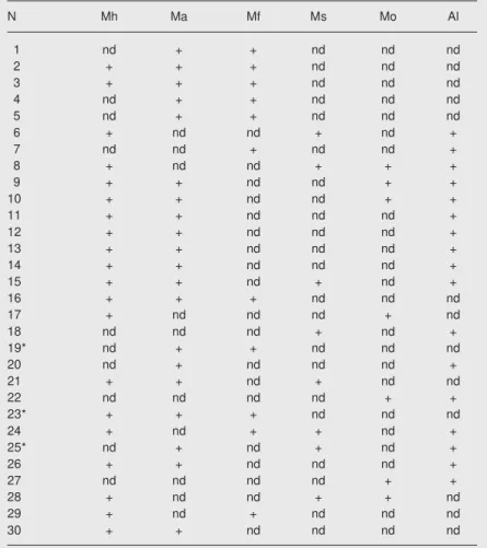
in 30/98 (30.6%) positive samples. M. fer-mentans, A. laidlawii and M. orale were also detected as single agents in 2/98 (2.0%), 3/ 98 (3.1%) and 4/98 (4.1%) samples, respectively, but they were associated with other species in 12/98 (12.3%), 17/98 (17.4%) and 7/98 (7.1%) positive samples, respectively. M. salivarium was only de-tected in association with other species. Mycoplasmas were identified infecting cell cultures in 12 laboratories and were present in association in four. Table 2 also shows mycoplasma associations other than M. hyorhinis and M. arginini detected in cell cultures.
The sensitivity of culture compared to PCR using primers for the detection of
Mollicutes was 68%, specificity was 97%, and accuracy was 89%. The respective primer concentrations were 1 ng/µL for the detec-tion of M. arginini, 1 pg/µL for M. salivarium
and M. orale, and 100 fg/µL for M. hyorhinis,
M. fermentans and A. laidlawii. The primers used for the detection of Mollicutes showed a sensitivity of 100 fg/µL. Figure 2 illus-trates the sensitivity of the primers in detect-ing the DNA target of M. hyorhinis.
D iscussio n
Accidental infection of cell cultures with mycoplasma (Mollicutes), especially when unnoticed, usually invalidates the results of biomedical research. The cell cultures may not die but remain altered and inadequate for experimentation (28).
The frequency of mycoplasma infection
500 bp
1 2 3 4 5 6 7 8 9 10 11 12 13
Figure 2. Agarose gel electrophoretic analysis of the PCR assay performed with MHRHF/MHRHR and dif-ferent concentrations of DNA/µL obtained by dilution
from a culture of Mycoplasma hyorhinis. Lane 1, 1000
ng; lane 2, 100 ng; lane 3, 10 ng; lane 4, 1 ng; lane 5,
100 pg; lane 6, 10 pg; lane 7, 1 pg; lane 8, 100 fg; lane
9, negative control (water); lane 10,10 fg; lane 11, 1 fg;
lane 12, 100 atg; lane 13, size markers DNA-1 kb plus
(Invitrogen).
Table 2. Cell cultures in which more than one species of mycoplasma other than
Mycoplasma hyorhinis and Mycoplasma arginini was detected by PCR.
N Mh Ma Mf Ms Mo Al
1 nd + + nd nd nd
2 + + + nd nd nd
3 + + + nd nd nd
4 nd + + nd nd nd
5 nd + + nd nd nd
6 + nd nd + nd +
7 nd nd + nd nd +
8 + nd nd + + +
9 + + nd nd + +
10 + + nd nd + +
11 + + nd nd nd +
12 + + nd nd nd +
13 + + nd nd nd +
14 + + nd nd nd +
15 + + nd + nd +
16 + + + nd nd nd
17 + nd nd nd + nd
18 nd nd nd + nd +
19* nd + + nd nd nd
20 nd + nd nd nd +
21 + + nd + nd nd
22 nd nd nd nd + +
23* + + + nd nd nd
24 + nd + + nd +
25* nd + nd + nd +
26 + + nd nd nd +
27 nd nd nd nd + +
28 + nd nd + + nd
29 + nd + nd nd nd
30 + + nd nd nd nd
Thirty cultures of 98 tested (30.6%) were positive. N = number of the cell culture sample: 1-5: L929, 6: CTY, 7: TCTY, 8-11: HeLa, 12-15: PLF, 16: AT, 17: CHO, 18: BHK, 19: OX3, 20: MDBK, 21: NHI3T3, 22: HT29, 23: C15.6, 24: T end, 25: N418, 26:
HEK, 27: VERO, 28: PK136, 29 and 30: WEH. Mh = M. hominis; Ma = M. arginini; Mf =
M. fermentans; Ms = M. salivarium; Mo = M. orale; Al = Acholeplasma laidlawii; nd =
non-detected mycoplasma species. +Detected species specific mycoplasma.
in cell cultures can reach 100% (29), but the outcome depends on the sampling variables. The 32.6% rate of cell culture infection ob-served in the present study agrees with the literature regarding the frequency of tested samples but not regarding infected laborato-ries. Twelve of the 15 laboratories studied were found to be infected. This finding is important because all are research laborato-ries, except for one which only commercial-izes cells. This type of information has not been available in Brazil before this report and it is hoped that these data may contribute to the control of this infection.
The laboratory that commercializes cells takes specific care and possesses a rigid quality control system, including the moni-toring for mycoplasmas. Most laboratories surveyed in the present study maintain cells for research or laboratory diagnosis. Some of these cells were labeled as originating from the institution that commercializes cells. However, these cells were probably infected with mycoplasmas over time due to their continuous manipulation. This situation, if not analyzed, may compromise the reputa-tion of the institureputa-tion that provides the cell culture.
Primary infection of cell line cultures with one mycoplasma species is the most widely reported situation in the literature. The presence of various species is related to primary cell cultures; however, this possi-bility was not specifically examined in the present study.
The species detected in the present study derived from animals or humans or both. The diversity of mycoplasma species in the same cell culture indicates the occurrence of different initial infection sources. The sub-culturing of a cell culture among laborato-ries over time due to successive sharing may explain the detection of multiple mycoplasma species. Mycoplasmal diversity accumulates over time mainly due to failure to control the infection.
Several methods for the detection of
mycoplasmas in cell cultures have been re-ported. The use of two methods has been the most recommended strategy to minimize false results. PCR in combination with cul-ture is the most widely recommended proce-dure (30). In the present study, PCR detected more infected samples than culture. Five positive cultures were not confirmed by PCR for Mollicutes but their identification was possible by specific PCR. The low amount of mycoplasmas or their DNA in the cell culture sample, the presence of inhibitors or failure of the diagnostic method may explain the contradictory nature of the results ob-tained by culture and PCR (23).
PCR permits the detection of femtogram amounts of mycoplasma DNA, correspond-ing to one bacterial cell. However, the sensi-tivity of specific PCR varies from species to species.
The isolation of more than one myco-plasma species is time consuming even for a specialized laboratory. In this respect, PCR permits not only the diagnosis of mycoplas-mas in cell cultures but also allows the deter-mination of the distribution of species (21).
M. orale, M. hyorhinis and A. laidlawii
are the species most frequently found in cell cultures (29). They were also detected in the present study but at different frequencies. M. hyorhinis was the most frequent, followed by M. arginini, findings that are in contrast to the literature (23,31).
M. fermentans is considered to be a nor-mal inhabitant of the human urogenital tract. It is a fastidious species, a fact that impaired its isolation in the past. In 1986, M. fermen-tans was associated with the development of AIDS in HIV-positive individuals, a fact that, in turn, attracted the interest of the scientific community. Subsequently, M. fer-mentans has been detected in or associated most frequently with tissues and blood of individuals with poorly studied diseases or diseases of unknown etiology (1,32). The increase in the frequency of M. fermentans
grow-ing use of human blood cells or tissues for primary culture (23).
M. hyorhinis and M. fermentans were detected co-infecting a cell culture inocu-lated with Coxiella burnetii, without evi-dence of morphological alterations caused by this association (data not shown). Myco-plasma infections in cell cultures that do not cause cell death or alterations in cellular organization, on the other hand, may repre-sent models for the study of the biology of these bacteria and of the cell cultures. In fact, in the last decade the number of experi-ments in this field, especially on mycoplas-mas of human or animal interest, has been increasing (13).
A cell culture infected with mycoplas-mas must be promptly eliminated and the non-infected cells should be carefully pre-served. An attempt can be made to treat cell cultures of special value for research if in-fected with mycoplasmas. Antibiotics or anti-bodies are the most widely used methods; however, it should be remembered that these compounds are not necessary mycoplasma-eliminating agents when they infect cells and that the biology of mycoplasmas varies among species (33). In this context, analysis
of the toxicity of the antimicrobial agent as well as the sensitivity of mycoplasma to the drug should be performed (34,35). An at-tempt to eliminate a multiple mycoplasma infection in cell culture is not considered, a fact that might explain the failures of treat-ment of mycoplasma-infected cell cultures. The sensitivity of mycoplasma to antibiotics is variable and the treatment is directed at a cell culture infected with a single myco-plasma species (21). On the other hand, the use of antibiotics to prevent the growth of microorganisms in cell cultures may hide the agent for years and impair its character-ization.
Mycoplasmas are sensitive to the ha-bitual processes of decontamination used in a laboratory. Some persistent laboratory neg-ligence such as the production of aerosols or other inadequate techniques contributes to the dissemination of mycoplasmas among cell cultures (21). Comments by laboratory staff confirm failures in the control of cell cultures, as well as disregard about myco-plasma infection in this system. This context compromises the efforts made to improve the quality of biomedical research.
Re fe re nce s
1. Baseman JB, Tully JG. Mycoplasmas: sophisticated, reemerging,
and burdened by their notoriety. Emerg Infect Dis 1997; 3: 21-32.
2. Nicolson GL, Gan R, Haier J. Multiple co-infections (Mycoplasma,
Chlamydia, human herpes virus-6) in blood of chronic fatigue
syn-drome patients: association with signs and symptoms. APMIS 2003;
111: 557-566.
3. Rottem S, Barile MF. Beware of mycoplasmas. Trends Biotechnol
1993; 11: 143-151.
4. Balish MF, Krause DC. Cytoadherence and the cytoskeleton. In:
Razin S, Herrmann R (Editors), Molecular biology and pathogenicity
of mycoplasmas. New York: Kluwer Academic/Plenum Publishers;
2002. p 491-518.
5. Dimitrov DS, Franzoso G, Salman M, Blumenthal R, Tarshis M,
Barile MF, et al. Mycoplasma fermentans (incognitus strain) cells
are able to fuse with T lymphocytes. Clin Infect Dis 1993; 17 (Suppl
1): S305-S308.
6. Lo SC, Hayes MM, Kotani H, Pierce PF, Wear DJ, Newton PB III, et
al. Adhesion onto and invasion into mammalian cells by
Myco-plasma penetrans: a newly isolated mycoplasma from patients with
AIDS. Mod Pathol 1993; 6: 276-280.
7. Copperman R, Morton HE. Reversible inhibition of mitosis in
lym-phocyte cultures by non-viable Mycoplasma. Proc Soc Exp Biol Med
1966; 123: 790-795.
8. Miyazaki K, Takaku H, Umeda M, Fujita T, Huang WD, Kimura T, et al. Potent growth inhibition of human tumor cells in culture by
argi-nine deiminase purified from a culture medium of a Mycoplasma
-infected cell line. Cancer Res 1990; 50: 4522-4527.
9. Pollack JD, Williams MV, McElhaney RN. The comparative
metabo-lism of the Mollicutes (Mycoplasmas): the utility for taxonomic
clas-sification and the relationship of putative gene annotation and
phy-logeny to enzymatic function in the smallest free-living cells. Crit
Rev Microbiol 1997; 23: 269-354.
10. Chambaud I, Wroblewski H, Blanchard A. Interactions between
mycoplasma lipoproteins and the host immune system. Trends
Microbiol 1999; 7: 493-499.
11. D’Orazio JA, Cole BC, Stein-Streilein J. Mycoplasma arthritidis
mi-togen up-regulates human NK cell activity. Infect Immun 1996; 64:
12. Razin S, Yogev D, Naot Y. Molecular biology and pathogenicity of
mycoplasmas. Microbiol Mol Biol Rev 1998; 62: 1094-1156.
13. Rottem S. Interaction of mycoplasmas with host cells. Physiol Rev
2003; 83: 417-432.
14. Robinson LB, Wichelhausen RH. Contamination of human cell
cul-tures by pleuropneumonialike organisms. Science 1956; 124:
1147-1148.
15. Boolske G. Survey of mycoplasma in cell cultures and a comparison
of detection methods. Zentralbl Bakteriol Mikrobiol Hyg 1990; 269:
331-340.
16. Hay RJ, Macy ML, Chen TR. Mycoplasma infection of cultured cells.
Nature 1989; 339: 487-488.
17. McGarrity GJ, Kotani H, Butter GH. Mycoplasmas and tissue culture
cells. In: Maniloff J (Editor), Mycoplasmas, molecular biology and
pathogenesis. Washington: American Society for Microbiology;
1992. p 445-454.
18. Tang J, Hu M, Lee S, Roblin R. A polymerase chain reaction based
method for detecting Mycoplasma/Acholeplasma contaminants in
cell culture. J Microbiol Methods 2000; 39: 121-126.
19. Barile MF, Rottem S. Mycoplasmas in cell cultures. In: Kahane I,
Adoni A (Editors), Rapid diagnosis of mycoplasmas. New York:
Plenum; 1993. p 155-193.
20. McGarrity GJ, Sarama J, Vanaman V. Cell culture techniques. ASM
News 1985; 51: 170-183.
21. Smith A, Mowles J. Prevention and control of mycoplasma infection
of cell cultures. In: Tully JG, Razin S (Editors), Molecular and
diagnostic procedures in mycoplasmology. San Diego: Academic
Press; 1996. p 445-451.
22. Barile MF, Hopps HE, Grabowski MW. Incidence and sources of mycoplasma contamination: a brief review. In: McGarrity GJ, Murphy
DG, Nicholes WW (Editors), Mycoplasma infection of cell cultures.
New York: Academic Press; 1978. p 35-45.
23. Tully JG. Mollicute-host interrelationships: Current concepts and
diagnostic implications. In: Tully JG, Razin S (Editors), Molecular
and diagnostic procedures in mycoplasmology. San Diego:
Aca-demic Press; 1996. p 1-21.
24. Del Giudice RA, Tully JG. Isolation of mycoplasmas from cell cul-tures by axenic cultivation techniques. In: Razin S, Tully JG
(Edi-tors), Molecular and diagnostic procedures in mycoplasmology.
New York: Academic Press; 1995. p 411-418.
25. Fan HH, Kleven SH, Jackwood MW. Application of polymerase
chain reaction with arbitrary primers to strain identification of
Myco-plasma gallisepticum. Avian Dis 1995; 39: 729-735.
26. van Kuppeveld FJ, van der Logt JT, Angulo AF, van Zoest MJ, Quint WG, Niesters HG, et al. Genus- and species-specific identification
of mycoplasmas by 16S rRNA amplification. Appl Environ Microbiol
1992; 58: 2606-2615.
27. Hopert A, Uphoff CC, Wirth M, Hauser H, Drexler HG. Specificity and sensitivity of polymerase chain reaction (PCR) in comparison with other methods for the detection of mycoplasma contamination
in cell lines. J Immunol Methods 1993; 164: 91-100.
28. Drexler HG, Uphoff CC, Dirks WG, MacLeod RA. Mix-ups and
mycoplasma: the enemies within. Leuk Res 2002; 26: 329-333.
29. McGarrity GJ, Sarama J, Vanaman V. Factors influencing
microbio-logical assay of cell-culture mycoplasma. In Vitro 1979; 15: 73-81.
30. Uphoff CC, Drexler HG. Comparative PCR analysis for detection of
mycoplasma infections in continuous cell lines. In Vitro Cell Dev Biol
Anim 2002; 38: 79-85.
31. Uphoff CC, Drexler HG. Detection of mycoplasma contaminations in
cell cultures by PCR analysis. Hum Cell 1999; 12: 229-236.
32. Blanchard A, Montagnier L. AIDS-associated mycoplasmas. Annu
Rev Microbiol 1994; 48: 687-712.
33. Del Giudice RA, Gardella RS. Antibiotic treatment of
mycoplasma-infected cell cultures. In: Tully JG, Razin S (Editors), Molecular and
dagnostic procedures in mycoplasmology. San Diego: Academic
Press; 1996. p 439-443.
34. Taylor-Robinson D, Bebear C. Antibiotic susceptibilities of
myco-plasmas and treatment of mycoplasmal infections. J Antimicrob
Chemother 1997; 40: 622-630.
35. Uphoff CC, Drexler HG. Comparative antibiotic eradication of
myco-plasma infections from continuous cell lines. In Vitro Cell Dev Biol