ORIGINAL
RES
EAR
CH
Correspondence to: Marco Orsini – Programa de Pós-Graduação em Ciências da Reabilitação – Praça das Nações, 34, Bonsucesso – CEP: 21041-021 – Rio de Janeiro (RJ), Brazil – E-mail: [email protected]
Presentation: Jul. 2013 – Accepted for publication: Feb. 2014– Financing source: none – Conflict of interests: nothing to declare – Approval at the Ethics Committee n. 119.818 (UNISUAM).
ABSTRACT | The Charcot-Marie-Tooth (CMT) disease is a peripheral hereditary neuropathy with progressive dis-tal muscle atrophy and weakness, mainly in lower limbs, that evolves limiting the gait and balance. The objective of the study was to analyse the immediate effects of us-ing Ankle-Foot Orthosis (AFO) in the gait’s kinematics and balance in patients with CMT. Nine individuals were evaluated by Tinetti scales and Dynamic Gait Index (DGI) and gait’s kinematics parameters through the motion capturing system. These evaluations were done before and during the use of AFO. Two-Way repeated analysis of variance was done to try the main or interaction effects related to “orthoses” and “repetition”. A significant interac-tion effect was observed between the gait cycle and use the AFO to the average velocity (Wilks’ Lambda=0.156, p=0.030, η2=0.844) like significant main effects in the
an-kle joint to the gait cycle (Wilks’ Lambda=0.091, p=0.008,
η2=0.909) and the use of AFO (Wilks’ Lambda=0.444,
p=0.013, η2=0.556). It was observed a significant change
in the DGI scale during the use of AFO (p<0.05). The use of AFO promoted immediate effects on gait kinematics and in balance reactions. The results suggest that the use of AFO is an efficient strategy to stabilize the ankle joint, besides avoiding foot drop in patients with CMT.
Keywords | Charcot-Marie-Tooth Disease; Orthotic Devices; Gait; Postural Balance.
Immediate effects of using ankle-foot orthoses
in the kinematics of gait and in the balance
reactions in Charcot-Marie-Tooth disease
Efeitos imediatos do uso de órteses tornozelo-pé na cinemática da
marcha e nas reações de equilíbrio na doença de Charcot-Marie-Tooth
Efectos inmediatos del uso de una ortesis para tobillo-pie en la cinemática de la
marcha y en las reacciones de equilibrio en la enfermedad de Charcot-Marie-Tooth
Rouse Barbosa Pereira1, Lílian Ramiro Felício1, Arthur de Sá Ferreira1, Sara Lúcia de Menezes1,
Marcos Raimundo Gomes de Freitas2, Marco Orsini1,2
Study conducted in the Graduate Program in Rehabilitation Sciences at Centro Universitário Augusto Motta (UNISUAM) – Bonsucesso (RJ), Brazil.
1Graduate Program in Rehabilitation Sciences at UNISUAM – Bonsucesso (RJ), Brazil. 2Universidade Federal Fluminense (UFF) – Niterói (RJ), Brazil.
RESUMO | A doença de Charcot-Marie-Tooth (CMT) é uma neuropatia periférica hereditária caracterizada por atrofia e paresia progressiva da musculatura distal, principalmente em membros inferiores, que evolui com limitações na marcha e no equilíbrio. O objetivo do estudo foi analisar os efeitos imediatos do uso de ÓrteseTornozelo-Pé (OTP) na cinemática da marcha e no equilíbrio de pacientes com CMT. Nove indivíduos foram avaliados pelas escalas de Tinetti e DynamicGait Index (DGI) e os parâmetros cinemáticos da marcha através de cinemetria. As avaliações foram realizadas antes e durante o uso das órte-ses. A análise de variância com medidas repetidas foi realizada para testar o efeito principal e de interação dos fatores “órtese” e “repetição”. Foi observado um efeito de interação significativo entre o ciclo da marcha e o uso da OTP para a média da velo-cidade (Wilks’ Lambda=0,156, p=0,030, η2=0,844), assim como
efeito principal significante na articulação do tornozelo para o ciclo da marcha (Wilks’ Lambda=0,091, p=0,008, η2=0,909) e
para o uso da OTP (Wilks’ Lambda=0,444, p=0,013, η2=0,556).
Foi observada uma modificação significativa na escala DGI du-rante o uso da OTP (p<0,05). O uso de OTP promoveu efeitos imediatos na cinemática da marcha e nas reações de equilí-brio. Os resultados obtidos sugerem que o uso de OTP é uma estratégia eficaz na estabilização do tornozelo, além de evitar o derreamento do pé em pacientes com CMT.
INTRODUCTION
he Charchot-Marie-Tooth disease (CMT) is the most frequent type among hereditary neuromuscular disor-ders. It can be classiied as demyelinating (CMT1) and axonal (CMT2). Other classiications are based on transmission patterns and genetic pool. With a slow and progressive evolution, it is characterized by amyotrophy, paresis, supericial and profound hypoesthesia, and distal hypo- or arelexia1,2. In the lower limbs, it afects
espe-cially the muscles of the distal third, causing functional impairments during gait and balance reactions3,4.
he pattern of gait in individuals with CMT is cha-racterized by foot-dragging during the swing phase, with increased compensation of hip and knee lexion. Deicits in plantar lexion are also observable, in addi-tion to a decrease in the length and width of steps, in-creased base of support, and steppage gait5,6.
Rehabilitation resources have been proposed with the purpose of improving gait6,7, in special the use of
Ankle-Foot Orthoses (AFO)8. AFOs are commonly prescribed
for musculoskeletal disorders of the ankle and/or subtalar joints for the purposes of stabilizing knee joints during the swing phase and avoiding plantar lexion during the initial contact phase9,10. Although the adherence to this
device is still low among patients with CMT, its pres-cription seems relevant, as it promotes improved balance reactions and gait performance in this population11-15.
Kinematic analyses of gait have been considered determinant for the conduction of therapy treatments and the prescription of orthoses, as well as for pre- and post-treatment assessments16. hese analyses enable
the identiication of biomechanical damage caused by muscle weakness, along with proprioceptive deicits and their repercussion on the functional aspects of gait
and balance reactions in individuals with CMT17. he
Tinetti scale and the Dynamic Gait Index (DGI) are also applicable to the scientiic-clinical context, as they are imbued with psychometric characteristics that are easily handled, low costs and good reliability, and are thus useful in assessing balance and mobility, in ad-dition to predicting the risk of falls18-21.
he choice for this rehabilitation modality — applying AFOs to the lower limbs — is based on the evidence of some scientiic studies in which gait was analyzed among individuals with CMT and an associa-tion was found between the use of AFO and improve-ments in gait and repercussions concerning balance and protection reactions10-14. However, up until the present
moment, we have not found any scientiic studies by au-thors who investigated the immediate efects of AFOs on gait and their implications in the risk of falling and postural instability in individuals with CMT, which is the proposal of the present study.
By investigating the immediate efects of AFOs among patients with CMT, we aim at stimulating the use of this device in this population, where adheren-ce is still very low. From a clinical perspective, patients with CMT who use AFOs usually experience the di-sease more severely, as the physical and psychological discomfort caused by this device lead many patients to abandon treatment11,13,15. Furthermore, rehabilitating
patients with CMT by means of using AFOs can lower costs in public health, since CMT is a chronic, pro-gressive and incurable disease8. In the Uniied Health
System, orthoses and other orthopedic devices are good intervention options, due to the low costs incurred in their production and because they contribute in cutting down rehabilitation expenses, in addition to reducing the potential future demand for other health services22.
RESUMEN | La enfermedad de Charcot-Marie-Tooth (CMT) es una neuropatía periférica hereditaria caracterizada por atrofia y paresia progresiva de la musculatura distal, principalmente en los miem-bros inferiores, que evoluciona con limitaciones en la marcha y en el balance. El objetivo de esto estudio fue analizar los efectos inme-diatos del uso de ortesis para tobillo-pie (OTP) en la cinemática de la marcha y en el balance de pacientes con CMT. Nueve sujetos fueron evaluados por las escalas de Tinetti y DynamicGait Index
(DGI) y los parámetros cinemáticos de la marcha por la cinemetria. Las evaluaciones fueron realizadas antes y durante el uso de la ortesis. El análisis de variancia con medidas repetidas fue realizado para testar el principal efecto y de interacción de los factores “orte-sis” y “repetición”. Se observó un efecto de interacción significativo
entre el ciclo de la marcha y el uso de OTP para la media de ve-locidad (Wilks’ Lambda=0,156, p=0,030, η2=0,844), como también
el principal efecto significativo en la articulación del tobillo para el ciclo de la marcha (Wilks’ Lambda=0,091, p=0,008, η2=0,909) y
para el uso de una OTP (Wilks’ Lambda=0,444, p=0,013, η2=0,556).
Se observó una modificación significativa en la escala DGI durante el uso de una OTP (p<0,05). El uso de una OTP promovió efectos inmediatos en la cinemática de la marcha y en las reacciones de balance. Los resultados obtenidos sugieren que el uso de OTP es una eficaz estrategia para estabilizar el tobillo, y evita la curvatura del pie en pacientes con la CMT.
We believe that the use of AFOs reduces excessive hip/knee lexion during the swing phase as well as postural instability. It also minimizes alterations found in variables of gait, such as decreases in the length and speed of steps and increases in the duration of steps in patients with CMT.
METHODOLOGY
his is a quasi-experimental study conducted with a convenience sample that counted 10 patients diagnosed with CMT who sought the neurology department at the Antônio Pedro University Hospital of Universidade Federal Fluminense (HUAP/UFF) between July 2012 and April 2013. he inclusion criteria were: being diag-nosed with CMT; age range between 18 and 60 years; and having a referral for AFO use. he exclusion cri-teria were: presence of pain, joint blockages or impair-ments caused by orthopedic surgery in the lower limbs; AFO use in the last six months prior to the study; clini-cal conditions (diabetes or severe heart problems) that afected independent mobility; and other neurological diseases that interfered with gait and/or balance. One of the patients was excluded from the study for expe-riencing pain in the lower limbs during the assessment with AFO. herefore, 9 individuals (n=9) participated in the study, 4 men and 5 women, with an age average of 41 years (SD11), average body mass of 71 kg (SD14) and average height of 1.64 m (SD0.07).
We complied with Resolution 196/96 of the National Health Council, and the research study was approved by the Ethics Committees of Centro Universitário Augusto Motta (UNISUAM) (report number 119.818) and of HUAP/UFF (report number 118.488).
Instruments
Kinematic gait data: kinematics
We used Qualisys Track Manager (QTM) 1.6.0.x, a mo-vement analysis system with an acquisition frequency of 240 Hz, imbued with three interconnected Prorelex
infrared cameras. he data were captured and processed by the software QTM.
Balance: Tinetti Gait and Balance Assessment Tool
he Tinetti Scale was translated and culturally adapted to Brazil by Gomes, with formal evidence of its validity
and reliability18. his scale assesses balance and gait with
scores that vary from 0 to 28 points, in which a score lower than 19, and between 19 and 24 points represent, respectively, high and moderate risks of falls19.
Qualitative gait data: Dynamic Gait Index
he DGI is a reliable tool used to assess human gait and balance that was translated and culturally adapted to the Portuguese language by Castro et al.20. It is
compo-sed of eight tasks that involve gait in diferent sensory contexts. Its score varies from 0 to 24 points, in which 19 or less points indicate a high risk of falls21.
Muscle function: the Medical Research Council
he individuals’ muscle function was assessed through the instrument devised by the Medical Research Council (MRC), which is composed of scores that vary from 0 to 5 points for each muscle group tested. Its ad-vantage is that it is a test of quick application and a sim-ple way to assess muscle function. Moreover, it is cost-free and does not require any equipment to be applied23.
Procedures
Anamneses and neurological examinations were ducted to characterize the participants’ clinical con-dition. In the neurological examination, their muscle function was assessed through the MRC instrument, followed by evaluations of their supericial and pro-found sensibility, as well as propro-found relexes. Next, we carried out the assessment of gait and balance reactions using the Tinetti and the DGI scales18,20, followed by a
kinematic assessment of gait using the QTM system. Firstly, the participants underwent the assessments wearing their habitual shoes, and after a ten-minute break, they used an AFO along with their regular shoes. All assessments were conducted at the Laboratory for Analysis of Human Movement of UNISUAM.
In the kinematic analysis, only the dominant lower limb was assessed due to the CMT’s symmetric pattern3.
We used seven 15-mm relexive and passive markers at-tached to the participants’ skin with a double-sided tape on the following anatomic spots: posterior superior iliac spine, anterior superior iliac spine, greater trochanter, lateral epicondyle, lateral malleolus, 5th metatarsal, and
posterior facet of the calcaneus16. he participants were
using an AFO at irst. All of them repeated this task ive times. Next, the orthoses were attached and the ive repetitions were performed once again with the AFOs. he variables analyzed were speed, displacement and duration of steps, and angular displacement of the hip, knee and ankle on the sagittal plane. To collect data, the axes and reference coordinates were deined as fol-lows: axis X (lateral-medial), axis Y (anterior-posterior) and axis Z (superior).
STATISTICAL ANALYSIS
he data were analyzed through the software SPSS (ver-sion 17 for Windows). he level of signiicance adopted was p<0.05. he descriptive analysis is presented here in bar graphs and tables, considering the mean±SD for the continuous variables, and median values (minimum; maximum) for the categorical variables. he associa-tion between the variables [age, weight, height, Tinetti index, DGI, speed of step (m/s), Movement Amplitude (MA) of the hip (degrees), knee MA (degrees) and ankle MA (degrees)] was tested through Spearman’s Correlation Coeicient and is displayed on a table. For the comparative analysis, we used the general linear model with repeated measurements for the factors or-thosis (without; with) and repetition (1, 2, 3, 4 and 5) for each dependent variable [stepping speed (m/s), hip MA (degrees), knee MA (degrees) and ankle MA (de-grees)], as well as the Wilcoxon Signed Rank test for the variables Tinetti index and DGI.
RESULTS
In regards to sensitivity, all individuals presented alte-rations in their supericial and profound sensitivity in the distal crural third, varying according to the degree
of compromise (anesthesia or hypoesthesia). Profound relexes in the lower limbs were either null or bilaterally decreased in the entire sample. Crural distal paresis, es-pecially in the dorsal and lexor groupings, was present in all patients assessed. According to the MRC instru-ment, three individuals were at degree three, three at degree two, two at degree one, and only one individual was at degree zero in regards to the dorsilexor muscles.
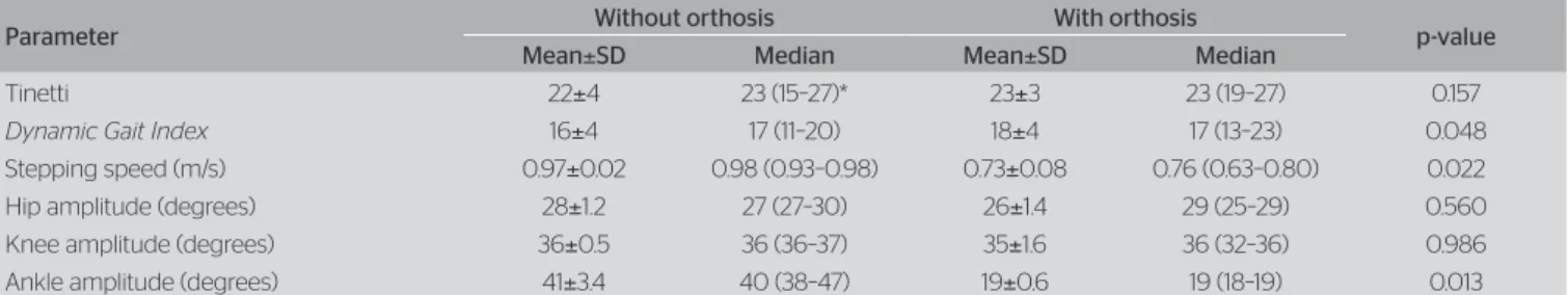
Upon analyzing the sample according to the Tinetti and DGI scales with and without the use of AFO, we observed that the DGI presented signiicant modii-cations between the two conditions (p=0.048). While using the AFO, the participants performed 8.4% better when compared to not using orthoses (Table 1).
he main results concerning the linear and angular data of gait with and without AFO are summarized on Table 1 and Graph 1. A signiicant diference can be observed in the median of stepping speed, which was 25% lower without the use of AFO. We obser-ved a signiicant interaction efect between the cycle of gait and the use of AFO on speed average (Wilks’ Lambda=0.156, p=0.030, η2=0.844), as well as a main
efect pertaining to the cycle (Wilks’ Lambda=0.144, p=0.014, η2=0.886) and to the use of AFO (Wilks’
Lambda=0.496, p=0.022, η2=0.504). As for hip MA, we
did not observe any signiicant main efects regarding gait cycle (Wilks’ Lambda=0.369, p=0.214, η2=0.631)
and AFO use (Wilks’ Lambda=0.956, p=0.560,
η2=0.044), and did not detect any signiicant interaction
efect between these two factors (Wilks’ Lambda=0.544, p=0.466, η2=0.456). In regards to knee MA, we did
not observe any signiicant main efects regarding gait cycle (Wilks’ Lambda=0.325, p=0.162, η2=0.675) and
AFO use (Wilks’ Lambda=1.000, p=0.986, η2=0.000),
with no interaction efect between them (Wilks’ Lambda=0.309, p=0.144, η2=0.691). Concerning ankle
MA, we observed a signiicant main efect on gait cycle (Wilks’ Lambda=0.091, p=0.008, η2=0.909) and
AFO use (Wilks’ Lambda=0.444, p=0.013, η2=0.556).
However, we did not observe a signiicant interaction
Table 1. Scores obtained through the Tinetti scale and the Dynamic Gait Index, and kinematic data regarding gait with and without the use of orthosis (n=9)
Parameter Without orthosis With orthosis p-value
Mean±SD Median Mean±SD Median
Tinetti 22±4 23 (15–27)* 23±3 23 (19–27) 0.157
Dynamic Gait Index 16±4 17 (11–20) 18±4 17 (13–23) 0.048
Stepping speed (m/s) 0.97±0.02 0.98 (0.93–0.98) 0.73±0.08 0.76 (0.63–0.80) 0.022 Hip amplitude (degrees) 28±1.2 27 (27–30) 26±1.4 29 (25–29) 0.560 Knee amplitude (degrees) 36±0.5 36 (36–37) 35±1.6 36 (32–36) 0.986 Ankle amplitude (degrees) 41±3.4 40 (38–47) 19±0.6 19 (18–19) 0.013
efect between the cycle of gait and the use of AFO for this joint (Wilks’ Lambda=0.227, p=0.072, η2=0.773).
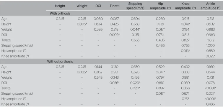
We observed a signiicant correlation concerning AFO use between the following variables: knee MA and ankle MA (r=0.733; p=0.025), hip MA and knee MA (r=0.783; p=0.013), and Tinetti and DGI (r=0.802; p=0.009). When an AFO was not used, we observed signiicant correlations between the following variables: Tinetti and DGI (r=0.698; p=0.036), Tinetti and speed (r=0.751; p=0.020), DGI and speed (r=0.751; p=0.020), speed and ankle MA (r=0.745; p=0.021), speed and hip MA (r=0.762; p=0.017); and hip MA and ankle MA (r=0.933; p<0.001) (Table 2).
DISCUSSION
he alterations observed in the individuals’ muscle func-tion along with alterafunc-tions in sensitivity contributed to the presence of biomechanical alterations perceived du-ring the kinematic analysis of gait4. In this study, we
were able to identify — through kinematic analysis — immediate modiications in the linear and angular data concerning gait with the use of AFO, which conirms
the results found by other researchers17. he authors in
question analyzed the gait of 16 individuals with CMT, in addition to their clinical characteristics, and conclu-ded that kinematics analyses are useful tools to deine the characteristics of gait in individuals with CMT.
In regards to the DGI scale, the results showed that the use of AFOs promoted modiications in the per-formance of gait patterns and balance reactions by all individuals included in the sample, but it was unable to diminish the risk of falls20,21.
he hypothesis that the use of AFOs would redu-ce exredu-cessive hip/knee lexion during the swing phase was not conirmed. We observed a change in the mean of hip/knee MA with the use of AFO, but this mo-diication did not reach the signiicance level adopted (p<0.05). hese results difer from the indings of ano-ther study10 in which the authors investigated the efects
of three AFO types on joint angles during the gait of an individual with CMT, and concluded that the use of AFOs during gait decreased excessive hip and knee lexion during the swing phase. However, the data were collected after one month of daily use of the orthoses, which allowed the patient to adapt to the devices. In the present study, the efect investigated was immediate and did not allow for a phase of adaptation, which can
Graph 1. Averages of the linear and angular data concerning the five repetitions of the gait cycle with and without the use of orthosis. Speed, hip amplitude, knee amplitude and ankle amplitude
1.40
1.20
1.00
0.80
0.60
0.40
0.20
0.00
40.0 35.0 30.0 25.0 20.0 15.0 10.0 5.0 0.0
50.0 45.0 40.0 35.0 30.0 25.0 20.0 15.0 10.0 5.0 0.0
80.0 70.0 60.0 50.0 40.0 30.0 20.0 10.0 0.0
1 2 3 4 5
1 2 3 4 5
1 2 3 4 5
1 2 3 4 5
Repetition
Repetition
Repetition
Repetition
Without orthosis With orthosis
St
epping speed (m/
s)
Hip MA – amplitude (degr
ees)
Ankle MA – amplitude (degr
ees)
Knee MA – amplitude (degr
justify the diferences in the results. Although it did not improve gait signiicantly, the use of an AFO modiied it immediately. he authors of this study hope that the results will serve as a basis that aids professionals in-volved with the physical rehabilitation of patients with CMT to stimulate adhesion to this device (still very low in this population).
he AFOs acted by compensating for the mus-cle weakness of the dorsilexor grouping during the swing phase and by managing plantar lexion during initial support, thus controlling foot-dragging. hese indings are in agreement with those observed by a group of researchers11 that reported the case of a patient
who presented improvements in the performance of gait patterns, with less inadequate movement synergies after using a type of orthosis developed especially for patients with CMT.
Del Bianco and Fatone12 evaluated the
func-tions of a pre-fabricated AFO model made of silico-ne and another osilico-ne composed of posterior springs, as well as their efects on the kinematics of the gait of an individual with CMT. he authors reported that both devices improved gait patterns by correcting deviations during the support and swing phase when compared to solely wearing shoes.
he hypothesis that AFOs could minimize alterations found in the linear variables of gait was not conirmed. he AFOs were not capable of imme-diately minimizing the alterations found in stepping
speed, length and duration. hese results agree with the indings of other researchers13 who characterized
the main diferences in the clinical presentation and function of the gait of two groups of patients with CMT (AFO group = regular AFO use; non-AFO group). hey concluded that the users of AFOs wal-ked more slowly and with more efort. hey justiied these results with the greater severity of the disease in the AFO group, which leads to a poorer perception of capability during gait performance. In this study, however, we observed that, despite a decrease in speed average, there was an increase in the averages of the DGI and Tinetti scales, which represents functional improvements in the gait of the individuals who parti-cipated in our research.
he results of this study demonstrate that the use of AFOs combined with regular shoes modiies the patterns of gait and balance reactions in indivi-duals with CMT, in comparison to wearing only regular shoes. Although the size of the sample is considered respectable, studies with a larger number of individuals and diferent types of orthoses must be stimulated with the purpose of providing more speciic data used to prescribe orthoses to individuals with CMT and stimu-lating its use within this population. Furthermore, using AFOs to treat patients with CMT can reduce rehabi-litation expenses, as low costs are involved in their pro-duction. Moreover, they reduce these patients’ future demands for Physical herapy services.
Table 2. Spearman’s Correlation for age, height, weight, Tinetti scale, Dynamic Gait Index, and kinematic data concerning gait with and without the use of orthoses
Height Weight DGI Tinetti Stepping
speed (m/s)
Hip amplitude (°)
Knee amplitude (°)
Ankle amplitude (°) With orthosis
Age 0.345 0.245 0.080 0.087 0.604 0.260 0.915 0.318 Height – 0.005* 0.914 0.425 0.683 0.139 0.041* 0.932 Weight – – 0.586 0.218 0.044* 0.017* 0.194 0.983 DGI – – – 0.009* 0.135 0.754 0.813 0.983 Tinetti – – – – 0.565 0.405 0.827 0.612 Stepping speed (m/s) – – – – – 0.486 0.765 1.000 Hip amplitude (°) – – – – – – 0.013* 0.559 Knee amplitude (°) – – – – – – – 0.025*
Without orthosis
Age 0.345 0.245 0.144 0.130 0.650 0.529 0.402 0.160 Height – 0.005* 0.852 0.931 0.626 0.041* 0.333 0.544 Weight – – 0.548 0.343 0.456 0.797 0.881 0.731 DGI – – – 0.036* 0.020* 0.810 0.930 0.078 Tinetti – – – – 0.020* 0.897 0.368 <0.001* Stepping speed (m/s) – – – – – 0.017* 0.674 0.021* Hip amplitude (°) – – – – – – 0.152 <0.001*
Knee amplitude (°) – – – – – – – 0.486
CONCLUSION
he use of AFOs promoted immediate changes in gait speed and balance in patients with CMT. However, this device was not capable of immediately minimizing the risk of falls and postural instability in this population. he results obtained suggest that the use of AFOs is an eicient conduct to stabilize ankle joints and manage foot-dragging among patients with CMT. However, it is not possible to extend these conclusions to all indivi-duals with Charcot-Marie-Tooth disease, as the sample studied here was small.
REFERENCES
1. Vieira THF, Aires RD, Mendonça VA, Correa CL. Reabilitação física em um paciente com a doença de Charcot-Marie-Tooth: relato de caso. Rev Neurociênc. 2009;17(3):287-92.
2. Neves ELA, Kok F. Clinical and neurophysiological investigation of a large family with dominant Charcot-Marie-Tooth type 2 disease with pyramidal signs. Arq Neuropsiquiatr. 2011;69(3):424-30.
3. Fávero RA, Polese JC, Oliveira SG, Schuster RC. Análise da marcha e funcionalidade na doença de Charcot-Marie-Tooth: relato de caso. Rev Neurociênc. 2010;18(1):44-9.
4. Vinci P, Perelli S. Footdrop, foot rotation, and plantarflexor failure in Charcot-Marie-Tooth disease. Arch Phys Med Rehabil. 2002;83(4):513-6. 5. Don R, Serrao M, Vinci P, Ranavolo A, Cacchio A, Ioppolo F, et al. Foot
drop and plantar flexion failure determine diferent gait strategies in Charcot-Marie-Tooth patients. Clin Biomech (Bristol, Avon). 2007;22(8):905-16.
6. Ferrarin M, Bovi G, Rabufetti M, Mazzoleni P, Montesano A, Moroni
I, et al. Reliability of instrumented movement analysis as outcome
measure in Charcot-Marie-Tooth disease: results from a multitask locomotor protocol. Gait Posture. 2011;34(1):36-43.
7. Vinci P, Gargiulo P, Panunzi M, Baldini L. Psychological distress in patient with Charcot-Marie-Tooth disease. Eur J Phys Rehabil Med. 2009;45(3):385-9.
8. Pereira RB, Orsini M, Ferreira AS, Silva JG, Corrêa CL, Freitas MRG,
et al. Efeitos do uso de órteses na doença de Charcot-Marie-Tooth:
atualização da literatura. Fisioter Pesqui. 2012;19(4):388-93.
9. Guillebastre B, Calmels P, Rougier PR. Efects of rigid and dynamic ankle-foot orthoses on normal gait. Foot Ankle Int. 2009;30(1):51-6.
10. Burdett RG, Hassell G. Efects of three types of ankle-foot orthoses on the gait and bicycling of a patient with Chartcot-Marie-Tooth disease. J Prosthet Orthot. 2004;16(1):25-30.
11. Vinci P, Paoloni M, Ioppolo F, Gargiulo P, Santilli. Gait analysis in patient with severe Charcot-Marie-Tooth disease: a case study with a new orthotic device for footdrop. Eur J Phys Rehabil Med. 2010;46(3):355-61. 12. Del Bianco J, Fatone S. Comparison of silicone and posterior leaf
spring ankle-foot orthoses in a subject with Charcot-Marie-Tooth disorder. J Prosthet Orthot. 2008;20(4):155-62.
13. Ramdharry GM, Pollard AJ, Marsden JF, Reilly MM. Comparing gait performance of people with Charcot-Marie-Tooth disease who do and do not wear ankle foot orthoses. Physiother Res Int. 2012;17(4):191-9. 14. Guzian MC, Bensoussan L, Viton JM, Bovis VM, Ramon J, Azulay
JP, et al. Orthopaedic shoes improve gait in a Charcot-Marie-Tooth patient: a combined clinical and quantified case study. Prosthet Orthot Int. 2006;30(1):87-96.
15. Vinci P, Gargiulo P. Poor compliance with ankle-foot-orthoses in Charcot-Marie-Tooth disease. Eur J Phys Rehabil Med. 2008;44(1):27-31. 16. Kadaba MP, Ramakrishna HK, Wootten ME. Measurement of
lower extremity kinematics during level walking. J Orthop Res. 1990;8(3):383-92.
17. Newman CJ, Walsh M, O’Sullivan R, Jenkinson A, Bennett D, Lynch B,
et al. The characteristics of gait in Charcot-Marie-Tooth disease types I and II. Gait Posture. 2007;26(1):120-7.
18. Gomes GC. Tradução, adaptação cultural e exame das propriedades de medida da escala “Performance-oriented mobility assessment” (POMA) para uma amostragem de idosos brasileiros institucionalizados. [dissertação]. Campinas: Universidade Estadual de Campinas; 2003.
19. Karuka AH, Silva JAM, Navega MT. Análise da concordância entre instrumentos de avaliação do equilíbrio corporal em idosos. Rev Bras Fisioter. 2011;15(6):460-6.
20. Castro SM, Perracini MR, Ganança FF. Versão brasileira do Dynamic Gait Index. Rev Bras Otorrinolaringol. 2006;72(6):817-25.
21. Chiu YP, Fritz SL, Light KE, Velozo CA. Use of item response analysis to investigate measurement properties and clinical validity of data for the dynamic gait index. Phys Ther. 2006;86(6):778-87.
22. BRASIL. Ministério da Saúde. Secretaria de Assistência à Saúde. Portaria nº 116, de 09 de setembro de 1993. Inclui no Sistema de Informações Ambulatoriais do Sistema Único de Saúde a concessão dos equipamentos de órteses, próteses e bolsas de colostomia. Diário Oficial da União, Brasília, n. 176, 15 set.; 1993.