Contrasting roles of donor and recipient TGFB1 and IFNG gene
polymorphic variants in chronic kidney transplant rejection
Papéis contrastantes das variantes polimórficas dos genes TGFB1 e IFNG do doador e do
receptor na rejeição crônica de transplantados renais
Verônica Porto Carreiro de Vasconcellos Coelho1,2,3, Rafael Ioschpe1, Cristina Caldas1, Monica Spadafora-Ferreira1,4,
João Americo Fonseca5, Maria Regina Alves Cardoso3,6, Selma Aliotti Palacios1, Jorge Kalil1,2,3, Anna Carla Goldberg3,7
ABSTRACT
Objective: To assess the long-term impact (minimum of 3 years follow-up) of polymorphisms in cytokine genes in donor:recipient pairs on the results of the transplant. Methods: We compared genetic cytokine polymorphisms and the primary factors of risk for the development of chronic rejection in paired groups of renal transplant patients with and without chronic allograft nephropathy [CAN].
Results: Multivariate analysis indicated that the presence of the high-production TT genotype (codon 10) of the transforming growth factor beta-1 (TGFB1) was protective in receptors (p=0.017), contrasting with the increased risk when present in donor samples (p=0.049). On the other hand, in the case of the gamma interferon studied, the greater frequency of the high production allele was protective in the analysis of the donor group (p=0.013), increasing the risk of chronic nephropathy of the allograft when present in the recipients (p=0.036). Conclusion: Our results highlight the importance ofTGFB1
genotyping in donors, and indicate that polymorphisms in the gene of this cytokine in donor cells might contribute to the development of chronic allograft nephropathy
Keywords: Chronic renal allograft dysfunction; Genetic polymorphism; Donor genotype; Transforming growth factor beta-1; Interferon-gamma
RESUMO
Objetivo: Avaliar o impacto de longo prazo (com seguimento mínimo de 2 anos) de polimorfismos em genes de citocinas em pares doador:receptor
sobre os resultados do transplante. Métodos: Comparamos os polimorfismos genéticos das citocinas e os principais fatores de risco para o desenvolvimento de rejeição crônica em grupos pareados de pacientes transplantados renais com e sem nefropatia crônica do aloenxerto [CAN].
Resultados: A análise multivariada indicou que a presença do genótipo TT (códon 10) de alta produção do fator de crescimento transformador beta-1 (TGFB1) era protetor nos receptores (p=0,017), em contraste com o risco aumentado quando presente nas amostras de doadores (p=0,049). Por outro lado, no caso do interferon gama estudado, a maior frequência do alelo de alta produção foi protetora na análise do grupo de doadores (p=0,013), mas aumentava o risco de nefropatia crônica do aloenxerto quando presente nos receptores (p=0,036). Conclusão:
Nossos resultados ressaltam a importância da genotipagem de TGFB1
também em doadores, e indicam que polimorfismos no gene desta citocina em células do doador podem contribuir no desenvolvimento da nefropatia crônica do aloenxerto.
Descritores: Disfunção renal crônica do aloenxerto; Polimorfismo genético; Genótipo do doador; Fator transformador de crescimento-beta 1; Interferon gama
INTRODUCTION
In spite of accumulated knowledge, the reasons why some patients, but not others, with similar clinical backgrounds, develop chronic rejection after renal transplantation are still unclear. The inflammatory nature of rejection has led to the query on the
Study carried out at Instituto do Coração, Faculdade de Medicina, Universidade de São Paulo, - USP - São Paulo (SP), Brazil
1 Instituto do Coração, Faculdade de Medicina, Universidade de São Paulo - USP - São Paulo (SP), Brazil
2 Divisão de Imunologia Clínica e Alergia, Faculdade de Medicina, Universidade de São Paulo, - USP - São Paulo (SP), Brazil 3 Instituto de Investigação em Imunologia - Institutos Nacionais de Ciência e Tecnologia, Brazil
4 Laboratório de Imunogenética, Instituto Butantan, São Paulo, Brazil
5 Unidade de Transplante Renal, Faculdade de Medicina, Universidade de São Paulo - USP - São Paulo (SP), Brazil 6 Departamento de Epidemiologia, Faculdade de Saúde Pública, Universidade de São Paulo - USP - São Paulo (SP), Brazil 7 Instituto Israelita de Ensino e Pesquisa Albert Einstein - IIEPAE - São Paulo (SP), Brazil
Autor correspondente: Anna Carla Goldberg - Av. Albert Einstein 627, 2SS, Bloco A - Morumbi - CEP 05652-000 - São Paulo (SP), Brasil. - Tel.: 2151-0941 - e-mail: [email protected]
Received: Aug 16, 2010 - Accepted: Jan 24, 2011
contribution of cytokine gene polymorphisms to the outcome of solid organ grafting, especially in the case of kidneys. Initial studies starting over 10 years ago, in renal transplanted patients, highlighted an association
between the high production -308 TNFA allele and a
low production IL10 genotype with acute rejection (1, 2)
and polymorphic IFNG CA repeat and IL10 genotype
in chronic rejection (2).
Chronic allograft nephropathy (CAN) is
identified by a progressive decline in renal function, and presents with typical histological features. These include the hallmarks of inflammatory processes, such as mononuclear cell infiltration, perivascular and interstitial inflammation, fibrosis, hyperplasia of the intima leading to partial or total decrease of the vascular lumen, tubular atrophy, and even glomerulosclerosis and ischemia. After 10 years,
over 50% of patients will have developed CAN (3)
culminating with a loss of the graft itself. In spite of the ever-increasing improvement of immunosuppressive protocols, CAN still remains a major problem partly as a result of the use of calcineurin inhibitors. In addition, a variety of factors have been reported associated with the development and progression of CAN. Donor/recipient HLA (Human Leukocyte Antigen) disparity, the basis of alloreactivity and acute rejection, is a major risk factor; donor age, graft cold ischemia time, the number of acute rejection episodes, hyperlipoproteinemia, hypertension, and CMV infection episodes have also been established as factors in the progression of chronic allograft
dysfunction (reviewed in detail in (4, 5).
In the initial phases of CAN, increased HLA expression and inflammatory cytokines such as IL-1
(Interleukin 1), IFN-γ (Interferon gamma), and TNF-α
(Tumor necrosis factor alpha), in addition to MCP-1 (Monocyte chemotactic protein 1) are present, as mononuclear cells infiltrate the kidney and adhere to the endothelium. At a later stage, concomitant to the proliferation of myofibroblasts and intimal hyperplasia, cytokines shift to a type 2 profile, which includes IL-4,
IL-10, and TGF-β1 (transforming growth factor beta 1),
as well as PDGF (platelet-derived growth factor) and
EGF (epidermal growth factor)(6). This combination of
factors is responsible for the phenotypic transformation
of fibroblasts into myofibroblasts (7).Endothelium and
smooth muscle cells stain brightly for TNF-α, PDGF,
and TGF-β1 (8). TGF-β1 is also expressed on fibroblasts
and areas of fibrosis (9). Though immunohistochemistry
studies show that TGF-β1 is present in biopsies from
kidneys with either acute or chronic rejection, a clearly enhanced staining of the interstitium is observed in
chronic rejection (10). Finally, cyclosporine A, the major
immunosuppressant drug used in renal transplanted
patients, has been shown to induce TGF-β1 production
in a proximal tubular cell line (11), and a similar effect has
been described for tacrolimus (12). On the other hand,
TGF-β1 has been repeatedly reported as a regulatory
cytokine playing an important role in many models of tolerance, contributing to the immunosuppressive capacity of circulating CD4+CD25+ T lymphocytes in vivo (13) .
OBJECTIVE
In this study, we investigated genetic polymorphisms of some cytokine genes involved in the first steps and in the progression of atherosclerosis, in addition to known effector-phase and regulatory cytokines, aiming to
identify susceptibility genes for CAN (14). Polymorphisms
of candidate cytokine genes were compared between groups of donor/recipient pairs with or without CAN, which were matched as best possible for major risk factors for CAN, such as level of HLA disparities, type and age of donor, number of acute rejection episodes, presence of hypertension, and cytomegalovirus (CMV) infection.
METHODS
Subjects and follow-up
This retrospective case:control study comparing two groups of patients, included patients who underwent renal transplantation and their respective donors at Hospital das Clínicas, University of São Paulo School of Medicine. The Ethics Committee approved this study and subjects gave their informed consent for blood sampling. Renal biopsies were performed according to clinical indications and classified according to Banff
criteria (15). DNA samples from 102 donor/recipient
and some of the more recently transplanted patients received MMF from early on. Demographic features are shown in Table 1.
DNA extraction and genotyping
Blood samples were drawn and DNA extracted by DTAB/CTAB (Dodecyltrimethylammonium bromide/
Cetyltrimethylammonium bromide)(16) or alternatively
by salting-out methods (17) as described elsewhere.
Unless otherwise mentioned, cytokine genotyping of 24 SNPs (single nucleotide polymorphisms) in 18 genes was performed by PCR-SSP (polymerase chain reaction with sequence-specific primers) on ready trays designed for the 13th International Histocompatibility Workshop on Cytokine Polymorphism by the Collaborative Transplant Study center in Heidelberg (http://www.ctstransplant. org/public/reagents.shtml). Briefly, PCR-SSP typing by the Heidelberg kit consisted of 48 PCR primer mixes dispensed in 96-well PCR trays. Master mix (MgCl2 buffer, dNTPs, and glycerol) was combined with 20 U Taq polymerase and 1.2 - 3.0 µg DNA, and dispensed onto the trays. Products were electrophoresed on 2% agarose gel and interpreted as defined by the Workshop protocol. The Heidelberg kit allowed SNP haplotyping
for IL1B (-511 C/T and +3692 C/T), TGFB1 (codon 10
C/T and 25 G/C), TNFA (-238 G/A , and -308 G/A) ,
IL2 (-330 T/G and +160 G/T), IL4 (-1098 T/G, -590 C/T
and -33 C/T), IL6 (-174 G/C, nt565 G/A), IL10 (-1082
G/A,-819 T/C and -590 A/C), and ICAM1 (G6241R
and E465D) genes. The tray also permitted typing for single SNPs in genes coding for IFNG (3´UTR5644
A/T), IL1A (-889 C/T), IL1R (pst1970 C/T), IL1RN
(mspa111100 C/T), IL12B (-1188 A/C), and IL4RA
(+1902 G/A). MCP1 SNP at position -2518 (A/G) was
analyzed by PCR-RFLP, using the following primers: forward CCGAGATGTTCCCAGCACAG and reverse
CTGCTTTGCTTGTGCCTCTT (18).
Statistical analysis
Bivariate analyses were carried out, using CAN as the dependent variable and the different gene polymorphisms investigated as independent variables. Variables significant in the bivariate analyses were the first entered into the multiple logistic regression models, but all other variables were tested. Two criteria were used to keep variables in the final model: statistical significance (p<0.05) or a clear change in the estimates of the effects of some polymorphisms produced by
those not selected in the first step of the analysis (19).
The analyses were performed using STATA software, version 8.0.
RESULTS
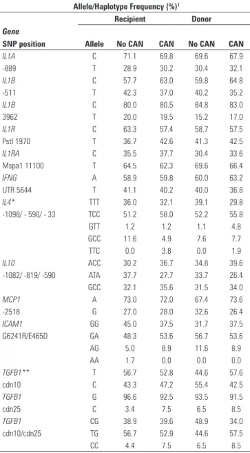
We analyzed 17 different gene polymorphisms, mostly cytokines associated with inflammation and/ or atherosclerosis, in both donor and recipient DNA samples. There was no deviation from expected Hardy-Weinberg proportions in any of the genes analyzed.
Most SNPs analyzed, including those from IL1B (2
SNPs), IL4 (3 SNPs), IL10 (3 SNPs), ICAM1 (2 SNPs),
and MCP1, were equally distributed in groups with
and without CAN, in donor and in recipient samples.
Preliminary analyses led us to discard TNFA, IL2, IL6,
and IL12B as non-informative in our population due to
their very low frequency in the healthy population, and were not tested further. A summary of the allele and haplotype frequencies in all groups is shown in Table 2.
In the case of IL1A and IL1B, which are neighboring
genes only about 60 kb apart, typing of IL1A alleles
in position -889, and IL1B SNPs at -511 and +3962,
disclosed at least 6 different haplotypes. However, in
almost half of the cases, joint IL1A/IL1B haplotypes
could not be unambiguously defined. In other words, a
comparison of IL1A/IL1B haplotype distribution in the
two groups was not possible.
Of all genes analyzed, the sole significant difference disclosed upon Chi-square analysis was the presence of the high producer TT genotype in codon 10 of the
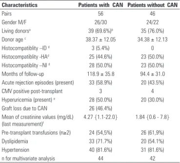
Characteristics Patients with CAN Patients without CAN
Pairs 56 46
Gender M/F 26/30 24/22
Living donorsa 39 (69.6%)b 35 (76.0%)
Donor age c 38.37 ± 12.05 34.38 ± 12.13
Histocompatiblity –ID d 3 (5.4%) 0
Histocompatiblity -HAd 25 (44.6%) 23 (50.0%)
Histocompatiblity –NI d 28 (50.0%) 23 (50.0%)
Months of follow-up 118.9 ± 35.8 94.4 ± 31.0
Acute rejection episodes (present) 33 (58.9%) 20 (43.5%)
CMV positive post-transplant 3 4
Hyperuricemia (present) e 28 (50.0%) 20 (30.0%)
Graft loss due to CAN 26 (46.4%)
Mean of creatinine values (mg/dL) (last measurement)f
4.27 {1.1-22.0} 1.84 {0.6 - 7.8}
Pre-transplant transfusions (n≥2) 24 (54,5%) 26 (61,9%)
Dyslipidemia 33 (71.7%) 20 (54.1%)
Hypertension 40 (81.6%) 31 (81.6%)
n for multivariate analysis 44 42
Table 1. Demographic and clinical features of the patients with and without chronic allograft nephropathy (CAN)
a Obs: 4 (with CAN) and 9 (without CAN) living donors were unrelated (difference not significant)
b Values in parenthesis are percentage of total number of patients in the group
c Values are mean ± standard deviation ( difference not significant by the Student t test for unpaired samples) d ID - HLA identical donor (sibling; 0/6 mismatches)
HA - haploidentical donor 1-3/6 mi’smatches NI - nonidentical donor 4-6/6 mismatches e Information not available in 16 CAN and 6 no CAN patients
TGFB1 gene (χ2 = 6.547, p = 0.0379), present in almost 40% of transplant recipients with CAN, compared to 15% in the group free of CAN.
In the multivariate analysis, the IL1A low
production allele was shown to be marginally protective when present in the graft (p=0.052). Results of the multivariate analysis can be seen in Table 3.
More importantly, the high production TGFB1 TT
genotype (codon 10) was protective in recipients (p=0.017) but conferred increased risk when present in donor samples (p=0.049). Conversely, in the case
of IFNG polymorphism, the high production allele
was protective in the donor analysis (p=0.013), but increased the risk of CAN when present in recipients (p=0.036). Finally, in spite of our careful matching of groups with and without CAN, and in accordance with published literature, acute rejection was confirmed as a risk factor for CAN (p=0.024). Hyperuricemia was analyzed in a smaller sample (67 instead of 86 pairs) and was also shown to be a risk factor for CAN (p=0.013, C.I. 1.628-63.437, data not shown). On the other hand, donor type, donor age, number of HLA
disparities, presence of hypertension, dyslipidemia, number of pre-transplant transfusions, and months of follow-up, also included in the multivariate analysis, were equally distributed, and thus did not impact the result of the analysis.
DISCUSSION
Our case:control analysis of individual gene polymorphisms disclosed a significant increase of the
TGFB1 high production genotype in donors from the
group with CAN. There was a trend toward significance in several other cytokine gene polymorphisms analyzed, however the relatively low number of patients in this study impacts upon this type of analysis. Thus, in order to counterbalance the lower power of the individual analysis we employed a multivariate analysis where all variables were taken into account. This analysis brought forth clear-cut results, confirming known risk factors like acute rejection episodes, as well as discriminating protective and risk-conferring cytokine
gene polymorphisms. This was the case for TGFB1 and
IFNG. Donor high production TGFB1 TT genotype
(codon 10) was confirmed in a multivariate analysis to be associated with CAN, but the same genotype when present in the recipients conferred protection.
In fact, despite TGF-β1´s short-term tubule-repairing
effect in the graft, its dominant intra-graft increased production seems to have an overall negative effect adding to progression of chronic rejection, enhancing
Allele/Haplotype Frequency (%)1 Recipient Donor Gene
SNP position Allele No CAN CAN No CAN CAN
IL1A C 71.1 69.8 69.6 67.9
-889 T 28.9 30.2 30.4 32.1
IL1B C 57.7 63.0 59.8 64.8
-511 T 42.3 37.0 40.2 35.2
IL1B C 80.0 80.5 84.8 83.0
3962 T 20.0 19.5 15.2 17.0
IL1R C 63.3 57.4 58.7 57.5
PstI 1970 T 36.7 42.6 41.3 42.5
IL1RA C 35.5 37.7 30.4 33.6
Mspa1 11100 T 64.5 62.3 69.6 66.4
IFNG A 58.9 59.8 60.0 63.2
UTR 5644 T 41.1 40.2 40.0 36.8
IL4* TTT 36.0 32.1 39.1 29.8
-1098/ - 590/ - 33 TCC 51.2 58.0 52.2 55.8
GTT 1.2 1.2 1.1 4.8
GCC 11.6 4.9 7.6 7.7
TTC 0.0 3.8 0.0 1.9
IL10 ACC 30.2 36.7 34.8 39.6
-1082/ -819/ -590 ATA 37.7 27.7 33.7 26.4
GCC 32.1 35.6 31.5 34.0
MCP1 A 73.0 72.0 67.4 73.6
-2518 G 27.0 28.0 32.6 26.4
ICAM1 GG 45.0 37.5 31.7 37.5
G6241R/E465D GA 48.3 53.6 56.7 53.6
AG 5.0 8.9 11.6 8.9
AA 1.7 0.0 0.0 0.0
TGFB1** T 56.7 52.8 44.6 57.6
cdn10 C 43.3 47.2 55.4 42.5
TGFB1 G 96.6 92.5 93.5 91.5
cdn25 C 3.4 7.5 6.5 8.5
TGFB1 CG 38.9 39.6 48.9 34.0
cdn10/cdn25 TG 56.7 52.9 44.6 57.5
CC 4.4 7.5 6.5 8.5
Table 2. Summary of allele and haplotype frequencies in groups with and without chronic allograft nephropathy (CAN)
1 According to the literature, alleles associated with high production are:
IL1A (-889*T), IL1B (-511*T; +3962*T),IL1RA (C), IFNG (T), TGFB1 (codon 10*T; codon 25*G), IL4 (-590*T; -33*T), IL10(-1082*G, -819*C, -590*C), MCP1 (-2518*G)
UTR = untranslated region ; Pst1 and Mspa1 = restriction enzymes; cdn = codon * IL4, IL10, ICAM1 data are shown in the form of haplotypes
** TGFB1 data are shown as both allele and haplotype frequencies. TT Genotype (cdn 10) χ2 = 6.547, p = 0.0379
Chronic rejection (CAN) OR ORadj 95%CIadj p-value
Acute cellular rejection 1.87 4.12 1.20 - 14.13 0.024
TGFB codon 10 TT recipient genotype 0.72 0.07 0.007 - 0.61 0.017
TGFBcodon 10 TT donor genotype 2.71 7.21 1.01 - 51.29 0.049
IFNG UTR5644 TT recipient genotype 1.16 5.83 1.12 - 30.19 0.036
IFNG UTR5644 TT donor genotype 0.72 0.16 0.04 - 0.68 0.013
Table 3. Multivariate analysis of gene polymorphisms significantly associated with CAN
known variables when looking into multi-factorial susceptibility helps highlight hidden differences. Thus, our study groups were matched for donor type and age, HLA compatibility, presence of hypertension, dyslipidemia, number of pre-transplant transfusions, CMV positivity, and immunosuppressive regimen. Not unexpectedly, however, we were not able to control two well-known risk factors, namely the number of acute
rejection episodes and hyperuricemia (27), which were
significantly increased in the CAN group.
However, the data we have obtained are generally in accordance with the published literature on the subject (28-30).
CONCLUSION
Our results highlight the importance of donor cytokine genotyping and show that cytokine polymorphisms present in the grafted tissue might, indeed, contribute to the development of CAN. The combination of recipient and donor genotyping may help choose additional or alternative therapeutic approaches for renal transplant patients at higher risk, such as the early introduction of MMF or other drugs with a potential curbing effect on the development of CAN.
ACKNOWLEDGMENTS:
This work was supported by FAPESP (São Paulo State Research Foundation, grant # 01/09850-0) and CNPq (National Council for Scientific and Technological Development, grant # 0062/2001-0). ACG, MRAC, JK, and VC are recipients of personal grants from CNPq.
REFERENCES
1. Sankaran D, Asderakis A, Ashraf S, Roberts IS, Short CD, Dyer PA, et al. Cytokine gene polymorphisms predict acute graft rejection following renal transplantation. Kidney Int. 1999;56(1):281-8.
2. Asderakis A, Sankaran D, Dyer P, Johnson RW, Pravica V, Sinnott PJ, et al. Association of polymorphisms in the human interferon-gamma and interleukin-10 gene with acute and chronic kidney transplant outcome: the cytokine effect on transplantation. Transplantation 2001;71(5):674-7.
3. Nankivell BJ, Borrows RJ, Fung CL, O’Connell PJ, Allen RD, Chapman JR. The natural history of chronic allograft nephropathy. N Engl J Med. 2003;349(24):2326-33.
4. Joosten SA, Sijpkens YW, van Kooten C, Paul LC. Chronic renal allograft rejection: pathophysiologic considerations. Kidney Int. 2005;68(1):1-13. 5. Li C, Yang CW. The pathogenesis and treatment of chronic allograft
nephropathy. Nat Rev Nephrol. 2009;5(9):513-9.
6. Shirwan H. Chronic allograft rejection. Do the Th2 cells preferentially induced by indirect alloantigen recognition play a dominant role? Transplantation. 1999;68(6):715-26.
7. Desmouliere A, Geinoz A, Gabbiani F, Gabbiani G. Transforming growth factor-beta 1 induces alpha-smooth muscle actin expression in granulation tissue myofibroblast proliferation and fibrosis. There is
growing recognition of the importance of increased
TGF-β1 not only when CAN is already present, but
also during acute rejection episodes and occurrence of cyclosporine nephrotoxicity, situations clearly associated
with the development of CAN (20-22). An enhancement of
TGFB1 transcription post-transplantation in the donor graft as a result of genetic polymorphism would partly explain these observations. This outcome contrasts with
TGF-β1 production by the recipients´ T lymphocytes,
where it is linked to suppression and down-regulation of inflammatory responses, as is largely reported in
literature (23). Accordingly, Park et al. (24) found the
frequency of TGFB1 lower and of
intermediate-producing genotypes (codon 10 CC and codon 25 GG) to be significantly higher in patients with recurrent acute rejection episodes, whereas high producer genotypes were increased in donors of patients with chronic renal allograft dysfunction. It is interesting to point out that
this same high producer TGFB1 codon 10 T allele, when
present in homozygosis in renal transplant recipients, was reported to be a potential risk for allograft function
decline (25). These heterogeneous data may, at least in
part, reflect the effect of other relevant factors in CAN, including the positive or negative effect of other gene polymorphisms.
The presence of the TT genotype associated with a
high production of IFN-γ in the recipient group sample
was identified as a risk factor in recipients, whereas when present in donor grafts it conferred protection. We do not have, at present, a good explanation for this last
observation. IFN-γ is produced almost exclusively by
NKT, NK, and activated T lymphocytes, and thus the
only source of donor IFN-γ would be donor lymphocytes
still present within the grafts at early time points after
transplantation. Supporting a protective role for IFN-γ, a
possible explanation has been put forth by Halloran et al.
(26) in a study with recipient IFN-γ knockout mice, where
it was shown that IFN-γ was essential to protect allografts
myofibroblasts and in quiescent and growing cultured fibroblasts. J Cell Biol.1993;122(1):103-11.
8. Noronha IL, Daniel V, Rambausek M, Waldherr R, Opelz G. Soluble interleukin-2 receptor (sIL-2R) and tumor necrosis factor plasma levels in renal allograft recipients. Transplant Proc. 1990;22(4):1859-60.
9. Cuhaci B, Kumar MS, Bloom RD, Pratt B, Haussman G, Laskow DA, et al. Transforming growth factor-beta levels in human allograft chronic fibrosis correlate with rate of decline in renal function. Transplantation. 1999;68(6):785-90. 10. Shihab FS, Yamamoto T, Nast CC, Cohen AH, Noble NA, Gold LI, et al.
Transforming growth factor-beta and matrix protein expression in acute and chronic rejection of human renal allografts. J Am Soc Nephrol. 1995;6(2): 286-94.
11. Wolf G, Zahner G, Ziyadeh FN, Stahl RA. Cyclosporin A induces transcription of transforming growth factor beta in a cultured murine proximal tubular cell line. Exp Nephrol. 1996;4(5):304-8.
12. Shihab FS, Bennett WM, Tanner AM, Andoh TF. Mechanism of fibrosis in experimental tacrolimus nephrotoxicity. Transplantation. 1997;64(12): 1829-37.
13. Chen W, Jin W, Hardegen N, Lei KJ, Li L, Marinos N, et al. Conversion of peripheral CD4+CD25- naive T cells to CD4+CD25+ regulatory T cells by TGF-beta induction of transcription factor Foxp3. J Exp Med. 2003;198(12): 1875-86.
14. Smith AJ, Humphries SE. Cytokine and cytokine receptor gene polymorphisms and their functionality. Cytokine Growth Factor Rev. 2009;20(1):43-59. 15. Racusen LC, Solez K, Colvin RB, Bonsib SM, Castro MC, Cavallo T, et al.
The Banff 97 working classification of renal allograft pathology. Kidney Int. 1999;55(2):713-23.
16. Gustincich S, Manfioletti G, Del Sal G, Schneider C, Carninci P. A fast method for high-quality genomic DNA extraction from whole human blood. Biotechniques 1991;11(3):298-300- 2.
17. Bignon JD, Viña MF. HLA class II typing by PCR-SSOP. Charron D, Fauchet R, editors. Paris: EDK Medical and Scientific International Publisher; 1995. 18. Rovin BH, Lu L, Saxena R. A novel polymorphism in the MCP-1 gene regulatory
region that influences MCP-1 expression. Biochem Biophys Res Commun. 1999;259(2):344-8.
19. Clayton D, Hills M. New York: Oxford University Press; 1996.
20. Sharma VK, Ding R, Li B, Bologa RM, Lagman M, Eduafo A, et al. Molecular correlates of human renal allograft rejection. Transplant Proc. 1998;30(5): 2364-6.
21. Pribylova-Hribova P, Kotsch K, Lodererova A, Viklicky O, Vitko S, Volk HD, et al. TGF-beta1 mRNA upregulation influences chronic renal allograft dysfunction. Kidney Int. 2006;69(10):1872-9.
22. Palomar R, Mayorga M, Ruiz JC, Cuevas J, Rodrigo E, Cotorruelo JG, et al. Markers of fibrosis in early biopsies of renal transplants. Transplant Proc. 2005;37(3):1468-70.
23. Huber S, Schramm C, Lehr HA, Mann A, Schmitt S, Becker C, et al. Cutting edge: TGF-beta signaling is required for the in vivo expansion and immunosuppressive capacity of regulatory CD4+CD25+ T cells. J Immunol. 2004;173(11):6526-31.
24. Park JY, Park MH, Park H, Ha J, Kim SJ, Ahn C. TNF-alpha and TGF-beta1 gene polymorphisms and renal allograft rejection in Koreans. Tissue Antigens. 2004 ; 64(6):660-6.
25. Chow KM, Szeto CC, Poon P, Lau WY, Lai FM, Li PK. Transforming growth factor-beta1 gene polymorphism in renal transplant recipients. Ren Fail. 2005;27(6):671-5.
26. Halloran PF, Miller LW, Urmson J, Ramassar V, Zhu LF, Kneteman NM, et al. IFN-gamma alters the pathology of graft rejection: protection from early necrosis. J Immunol. 2001;166(12):7072-81.
27. Perico N, Codreanu I, Caruso M, Remuzzi G. Hyperuricemia in kidney transplantation. Contrib Nephrol. 2005;147:124-31.
28. Hoffmann S, Park J, Jacobson LM, Muehrer RJ, Lorentzen D, Kleiner D, et al. Donor genomics influence graft events: the effect of donor polymorphisms on acute rejection and chronic allograft nephropathy. Kidney Int. 2004;66(4): 1686-93. 29. Hutchinson IV. The role of transforming growth factor-beta in transplant
rejection. Transplant Proc. 1999;31(7A):9S-13S.