Apparent life-threatening event: a review
Evento com aparente risco de morte: uma revisão
Evento con aparente riesgo de muerte (alte): una revisión
Mariana Tresoldi das N. Romaneli1, Emílio Carlos E. Baracat2
Instituição:Universidade Estadual de Campinas (Unicamp), Campinas, SP, Brasil
1Mestre em Ciências pelo Departamento de Pediatria da Faculdade de
Ciências Médicas da Unicamp; Médica Pediatra, Assistente do Hospital de Clínicas da Unicamp, Campinas, SP, Brasil
2Professor Livre-docente na Área de Urgência e Emergência pelo
Departamento de Pediatria da Faculdade de Ciências Médicas da Unicamp; Professor-Associado do Departamento de Pediatria da Faculdade de Ciências Médicas da Unicamp, Campinas, SP, Brasil
ABSTRACT
Objective: To perform a critical review by gathering all the available information about apparent life-threatening events. Data sources: Bibliographic review of the articles published in Portuguese, English and Spanish from the electronic databases Medline, Lilacs and SciELO, using the key-words: apparent threatening events, apparent life-threatening event, infant, apnea, monitoring, and cyanosis. Data synthesis: Apparent life-threatening events define sudden events with, a combination of apnea, color change, and marked change in the muscle tone, that have various underlying causes. The real incidence remains unknown, and it affects infants from 11 to 12 weeks of age. There is no association between apparent life-threatening events and sudden infant death syndrome. There are many possible causes for the events, and they must be investigated even in apparently healthy infants, because the presence of a severe underlying disease associated with the event is possible. If the cause of the apparent life-threatening events is found, it must be treated properly. If there is no explainable cause, the event is considered idiopathic and generally has a benign course.
Conclusions: It is necessary to investigate all the infants taken to the pediatric emergency unit after experiencing an apparent life-threatening event, since there is the risk of mor-bidity caused by an underlying disease or the event itself, as well as subsequent mortality. Consensus guidelines about the investigation in apparently healthy infants who experienced apparent life-threatening events are not available. Most authors recommend that careful observation and hospital monitoring should be performed for at least for 24 hours after the event.
Key-words: infantile apparent life-threatening event; infant; death; apnea.
RESUMO
Objetivo: Realizar uma revisão crítica reunindo informações disponíveis a respeito dos eventos com aparente risco de morte.
Fontes de dados: Revisão bibliográfica dos artigos (em português, inglês e espanhol) obtidos dos bancos de dados eletrônicos Medline, Lilacs e SciELO, utilizando as palavras-chave: eventos com aparente risco de morte, evento com aparente risco de vida infantil, lactente, apneia, monitorização e cianose.
Síntese dos dados: Os eventos com aparente risco de morte são súbitos e caracterizados por uma combinação de apneia, alteração na coloração da pele e tônus muscular, com inúmeras causas subjacentes. Sua incidência verdadeira é desconhecida e a faixa etária mais acometida é de 11 a 12 semanas. Não há correlação entre o evento com aparente risco de morte e a síndrome da morte súbita do lactente, embora já tenham sido consideradas manifestações da mesma doença. Muitas vezes, o lactente tem aparência saudável ao ser avaliado pelo pediatra após apresentar eventos com apa-rente risco de morte, porém, isso não afasta a possibilidade de existir uma doença grave associada ao evento, que deve ser investigada e tratada. Quando não são encontradas as causas, o evento é idiopático, geralmente com boa evolução.
Conclusões: É necessário investigar os lactentes levados ao pronto-socorro após apresentarem eventos com aparente risco de morte, devido ao risco de sequelas e mortalidade. Não há uma padronização das condutas a serem realizadas diante de um lactente com aparência saudável que tenha evento com aparente risco de morte, mas recomenda-se que o paciente seja internado e a causa do evento, investigada. A observação e o monitoramento em ambiente hospitalar devem ocorrer no mínimo 24 horas após o evento.
Endereço para correspondência: Mariana Tresoldi das N. Romaneli Rua Francisco Fadin, 520 – apto. 444 CEP 13140-000 – Paulínia/SP E-mail: [email protected]
Conflito de interesse: nada a declarar
Palavras-chave: evento com aparente risco de vida in-fantil; lactente; óbito; apneia.
RESUMEN
Objetivo: Realizar una revisión crítica, reuniendo las in-formaciones disponibles respecto a los Eventos con Aparente Riesgo de Muerte (ALTE - Apparent life-threatening event).
Fuentes de datos: Revisión bibliográfica de los artículos (en portugués, inglés y español) obtenidos de las bases de da-tos electrónicas MEDLINE, LILACS y SCIELO, utilizándose las palabras clave ALTE, evento con aparente riesgo de vida infantil, lactante, apnea, monitorización y cianosis.
Síntesis de los datos: Los ALTE (apparent life-threatening event) son eventos súbitos y caracterizados por una combinación de apnea, alteración en la coloración de la piel y tono muscular, con innúmeras causas subyacentes. Su incidencia verdadera es desconocida y la franja de edad más acometida es de 11 a 12 semanas. No hay correlación entre ALTE y SIDS (Síndrome de la Muerte Súbita del Lactante), aunque ya hayan sido conside-radas manifestaciones de la misma enfermedad. Muchas veces, el lactante tiene apariencia sana al ser evaluado por el pediatra después de presentar ALTE, pero eso no aleja la posibilidad de que exista una enfermedad grave asociada al evento, la cual se debe investigar y tratar. Cuando no se encuentran causas, el evento es idiopático, generalmente con buena evolución.
Conclusiones: Es necesario investigar los lactantes lleva-dos a la emergencia después de presentar ALTE, por riesgo de secuelas y de mortalidad. No hay una estandarización de las conductas a tomar frente a un lactante con apariencia sana que presentó ALTE, pero se recomienda que se interne el paciente y se investigue la causa del evento. La observación y monitoración en ambiente hospitalaria debe ocurrir por un mínimo de 24 horas después del evento.
Palabras clave: evento con aparente riesgo de vida in-fantil; lactante; óbito; apnea.
Introduction
The term Apparent Life-Threatening Event (ALTE) was irst deined in 1986 by the National Institutes of Health in theUnited States. The deinition was the result of a consensus reached at a conference on infantile apnea and home monitoring, which established the term ALTE and ended the use of terms such as “aborted crib death”
and “near-miss SIDS”. The conference was organized in response to the dilemma caused by the dificulty of dein-ing the differences between ALTE, Sudden Infant Death Syndrome (SIDS) andapnea of infancy, even though the lack of pathophysiologic correlations means distinctions are obligatory(1-4). In Portuguese, terms that back-trans-late as “event possibly threatening life” (episódio de possível ameaça à vida), “event apparently involving risk to life” (evento com aparente risco de vida)” and “event apparently involving risk of death” (evento com aparente risco de morte) have been used as translations of the English term “ap-parent life-threatening event”(5-7).
The following deinitions were established by the 1986 consensus with certain modiications based on the the results of later studies:
• Apnea: Cessation of respiratory air low. The cause of the respiratory pause may be central, muscular, obstruc-tive or mixed. Short (15 seconds), central apnea can be normal at all ages(2).
• Apnea of Prematurity: An unexplained episode of cessa-tion of breathing for 20 seconds or longer, or a shorter respiratory pause associated with bradycardia, cyanosis, pallor, and/or marked hypotonia. The term should be reserved for infants up to 37 weeks’ post-conception gestational age. In some cases apnea of prematurity may persist beyond 37 weeks, particularly in newborn infants with less than 28 weeks’ gestational age(2-3).
• ALTE (Apparent Life-threatening Event): An episode that is frightening to the observer and that is charac-terized by some combination of apnea (central or occa-sionally obstructive), color change (usually cyanotic or pallid but occasionally erythematous or plethoric), ma-rked change in muscle tone (usually mama-rked limpness), choking or vomit relex(2). After an ALTE, the infant’s course is often benign, but there is a risk of subsequent morbidity and mortality caused either by the event itself or by the subjacent disease that provoked the event(3,8-9).
• SIDS (Sudden Infant Death Syndrome): The sudden death of any infant or young child, which is unexplained by history and in which a thorough postmortem exa-mination fails to demonstrate an adequate explanation of cause of death(2).
The objective of this review article is to identify infor-mation on ALTE in the literature and use it to develop an instrument designed to improve the care given to infants who present with ALTE.
Methods
Searches were run on PUBMED, SCIELO and LILACS databases to identify publications in English, Portuguese or Spanish using the following keywords: ALTE, apparent life-threatening event, infant, apnea, monitoring and cyanosis. A total of 59 articles published between 1984 and 2011 and related to ALTE were identiied and 55 of these, covering clinical manifestations, epidemiology, risk factors, admission criteria, clinical course, etiology, treatment, and outpatients follow-up of infants suffering ALTE, were selected for review. These studies included retrospective and prospective designs, case descriptions, review articles, consensus statements and metananlyses (no randomized studies exist) and have a variety of sample sizes and evidence levels.
Results
The results of the major studies are in Table 1 along with the description of the number of patients included, percent-age of death, and ALTE recurrence and main etiologies. We highlight a great variation in the prevalence of the main etiologies (respiratory, gastrointestinal, and idiopathic) in the different studies.
Discussion
Epidemiological aspects of ALTE
Incidence
The true incidence of ALTEin the general population of infants less than 12 months old has not yet been clearly estab-lished(4). Different authors present their data in different ways: some as a proportion of the total number of visits to emergency by children under 1 year; others as a proportion of hospital admissions in the same age group; and still others report the
igures with relation to the number of live births at the same place during a given period. Thus, the incidence of ALTE is variously given as 0.2 to 1.9% of infants less than 1 year old(15-17); as 0.6 to 5.0 in every 1000 live births(4,17-20); and as 2.3 to 4.2% of admissions resulting from visits to an emergency service(6,20). With regard to age at time of ALTE, studies show that the age of peak incidence is from 11 to 12 weeks of life, ranging from events that occur during irst hours of life to events occurring up to the end of the irst year, since the majority of these authors do not consider events affecting children over 1 year to be ALTE(6,8,19,21-23). Notwithstanding this general trend, some samples had peak ALTE incidence at 7–8.5 weeks of age(9,24).
Risk factors
The studies listed the following risk factors for ALTE: age greater than 2 months(25), prior occurrence of ALTE(9), prematurity, late delivery and comorbidities(19). There was not, however, any evidence that apnea of prematurity (which tends to disappear between 34 and 36 weeks’ cor-rected gestational age) is a risk factor for apnea of infancy or, therefore, for ALTE(2,17,24). Notwithstanding, apnea events are more frequent and more symptomatic among infants with post-conceptual ages below 34 weeks and cease after 43 weeks’ corrected gestational age(26). On the other hand, late delivery (at gestational ages greater than 42 weeks) is also considered a risk factor associated with occurrence of ALTE(19).
ALTEand SIDS
Although infants with a history of ALTE used to be considered at increased risk of SIDS and studies show that 7 to 10% of SIDS victims have suffered some type of ALTE previously (15,16,27,28), there does not appear to be any evidence that ALTE and SIDS are facets of the same disease, or even that infants who suffer ALTE are at greater risk of dying from SIDS(1,3,9,16,19,29,30).
Further reinforcing the hypothesis that ALTE and SIDS are different diseases, studies show that ALTE and SIDS have different ages of incidence, with SIDS being more frequent around 3 to 5 months, whereas ALTE’s incidence peaks 1 to 3 months earlier(12-15,18,27,31).
Table 1 - Results dos principal studies analyzed
Author Type of
study n
Mean age (weeks)
Deaths (%)
Recurrent ALTE
(%)
ALTE etiologies (%) Idiopathic ALTE (%)
Anjos(6) prospective 30 15.4 3.3 73.0
Respiratory: 6.6;
Neurological: 13.3; Gastrointestinal: 23.3;
Others: 6.8
50.0
Romaneli(7) retrospective 145 15.0 7.6 44.1
Respiratory: 37.3;
Neurological: 4.2; Gastrointestinal: 4.9; Cardiocirculatory: 4.2;
Others: 14.2
35.2
Davies(9) prospective 65 7.0 0 12.0
Respiratory: 26.5;
Neurological: 9; Gastrointestinal: 26.0; Cardiocirculatory: 3.0;
Others: 12.5
23.0
Etxaniz(17) prospective 50 8.4 2.0 8
Respiratory: 22.0;
Neurological: 4.0; Gastrointestinal: 10.0;
Cardiocirculatory: 2;
Others: 4.0
58
Kiechl-Kohlendorfer(18) prospective 164 8.0 - 37.8
Respiratory: 29.0;
Neurological: 1.0; Gastrointestinal: 22.0; Cardiocirculatory: 2.0;
Others:1.5
44.5
Semmekrot(19) prospective 110 11.4 0 10.0
Respiratory: 24.6;
Neurological: 0; Gastrointestinal: 37.3; Cardiocirculatory: 2.7;
Others: 7.4
28.0
Samuels(21) prospective 157 12.0 1.9 70.7 Neurological: 6.3;
Others: 42.8 50.9
Altman(22) prospective 243 12.0 1.2 46.5
Respiratory: 35.3;
Neurological: 10.6; Gastrointestinal: 29.6; Cardiocirculatory: 0.8;
Others: 23.7
16
Brand(25) retrospective 243 11.7 - 2.0
Respiratory: 33.3;
Neurological: 8.2; Gastrointestinal: 29.2;
Others: 13.3
16
Bonkowsky(32) retrospective 471 9.4 0.4
-Respiratory: 28.2;
Neurological: 9.7; Gastrointestinal: 40.3;
Others: 8.6
13.2
among infants who suffered ALTE(13,29). The “back to sleep”
campaign to persuade carers to put infants to bed in the supine position achieved a 30 to 50% reduction in SIDS mortality, but did not impact on the incidence of ALTE during the same pe-riod, indicating that ALTE and SIDS are distinct entities(3,13,31).
ALTE and acquired morbidity
Infants who suffer ALTE episodes generally enjoy a be-nign course, with no sequelae related either to the event or to its cause and with little chance of the symptoms recur-ring(3,8-10,27). However, around 5% of ALTE cases are severe events involving prolonged periods of apnea and bradycardia and lead to neurological sequelae, such as non-progressive chronic encephalopathy, delayed neuropsychomotor develop-ment, epilepsy and behavioral problems, although it cannot be proven that these sequelae are exclusively caused by the event itself, since they could be associated with the subjacent disease that triggered the ALTE(2,30,32-34).
The rate of recurrence of ALTE can be higher than the incidence of ALTE in the general population, even among infants who did not suffer sequelae from previous episodes(20).
ALTE and mortality
The mortality rate linked with ALTE is unknown, but it is known that it is not safe to assume that an infant is not at risk of a subsequent fatal event once the cause of the original episode has been identiied(2,3,17,34).
Rates of death during follow-up among infants monitored after suffering ALTE vary greatly in the studies analyzed, ranging from zero to 7.6%(7,17-19-21,32).
Pathophysiology of ALTE
The mechanisms that lead to the manifestations of ALTE can be explained by some of the phenomena shown to be causes of consequences of the subjacent disease, such as apnea. Apnea (whether it has a neurological cause or is due to airway obstruction, or both) causes a reduction in oxygenation and diverted blood low, leading to other manifestations of ALTE, such as cyanosis, pallor, lushing and muscle hypotonia(3).
Muscle tone effects like hypotonia, hypertonia and rhythmic movements of the extremities may originate in the central nervous system (CNS) or may be secondary to other processes, such as crying that triggers the vasovagal relex, or even convulsive crises(3). One study has shown that initially there are electroencephalographic abnormalities which are then followed by one or more pauses in respira-tory movements, culminating in a fall in peripheral oxygen
saturation below 60%, leading to clinical cyanosis with sinus tachycardia, lasting 40 seconds on average(35).
Other clinical manifestations of ALTE, such as choking, coughing and the vomit relex are protective relexes triggered by stimuli to the nasopharynx, hypopharynx, larynx and lower respiratory tract. These relexes temporarily interrupt ventila-tion, leading to further manifestations of ALTE such as facial lushing, caused by the increase intrathoracic pressure, and hypotonia, which may be caused by hypoxia, vagus relex or both(3,10). When choking occurs, the contraction of the larynx caused by irritation to the glottis may spread to the upper airways, causing upper respiratory obstruction, and to the lower airways, causing bronchial obstruction. Prolonged hy-poxia provokes ischemia and generalized endothelial damage, leading to hemorrhagic phenomena, coagulopathy, breakdown of the blood brain barrier and cerebral edema(10). It is therefore possible that ALTE that last 30 seconds or more and recurrent ALTE may cause venous pressure in the retina to rise, leading to hemorrhage. However, it has not yet proved possible to demonstrate the occurrence of retinal hemorrhage solely as a result of these events(36).
Number and duration of ALTE episodes before seeking care
In general, ALTE episodes are of short duration, last-ing less than 5 minutes, and recovery is complete and spontaneous(17,19,27). The majority of infants are taken to emergency services by their carers immediately after their irst event(7,19,22). Notwithstanding, many (44 to 46.5%) are only taken to health services after two or more episodes(7,22).
Infant activity prior to occurrence of ALTE
Generally, ALTE take place at home without warning and during any type of activity, especially when sleeping, waking up or feeding, but it has been reported that some infants exhibit signs such as vomiting, diarrhea and refusal of food during the 24 hours preceding an ALTE(2,7,9,18,19,23,27,33).
Admission of infants at emergency services
subjacent disease(3,7,9,17,22,27,29). The severity of symptoms described by observers should be considered important infor-mation in these cases(9,17,19,20,29). In around 10% of cases, emer-gency procedures such as chest compressions, ventilation with bag-valve-mask, oral endotracheal intubation and adrenaline infusions are needed during the irst examination(7,8,22).
Investigation
It is not consensus that all infants suffering ALTE should be admitted for investigation. There are authors who believe that only infants less than 30 days old or repeat ALTE suf-ferers should be admitted if the irst clinical assessment is normal(3,20,37). However, the majority agree that admission for a minimum of 24 hours can provide data that is of great value to determining the event’s etiology, severity, clinical course and risk of sequelae(3,5,26-27,30,32,33). Furthermore, observation and monitoring of infants in a hospital environment makes their carers feel more secure and provides an opportunity to train them in cardiorespiratory resuscitation. Some authors consider that such training is a precondition for discharging infants from hospital after ALTE(3,5,8,27).
The physician responsible for the irst consultation after an infant suffers an ALTE is therefore faced with a major dilemma, since with the exception of certain minor details of the event’s history (coughing, choking, stridor, fever, physical effort or vomiting preceding the ALTE), they will not have any clues to indicate where to start to investigate an apparently healthy infant(3-5,8,25,27,33,38). Some authors con-sider that the decision to admit to hospital should be based on the severity of the event described, the indings of the initial physical examination and the experience of the irst physician to see the infant(8,27). Pediatricians who have had previous experience with cases of ALTE that had unfavorable outcomes are more likely to admit infants, even those who appear healthy. Those who have not, tend to request fewer work-up tests and tend to ignore the majority of the entities in the differential diagnosis of ALTE(39).
The objective when investigating an infant who has suffered an ALTE is to determine whether the event has an underlying cause. Although there are countless proposals for protocols, there is no consensus on which tests should be requested or even what the order of investigations should be. In general, the larger the number of tests that are requested, the greater the likelihood of detecting an abnormality, which in turn may or may not be the cause of the ALTE(3,5,20,25).
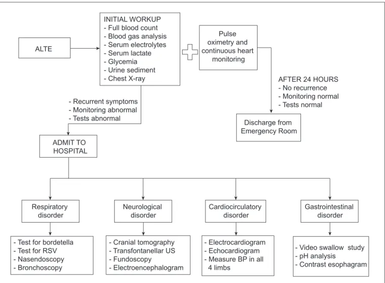
In view of this, tests are suggested that should at least be capable of identifying the most common and most serious causes. Initially, heart monitoring and continuous transcu-taneous oximetry can be set up in the emergency room(3,8,13). Initial laboratory tests should include a full blood count and venous blood gas analysis and serum lactate, glycemia and electrolytes should be assayed(3-5,9,13,20,21,27). Urine should be tested for signs of urinary infection, urine organic acids or reducing substances; and toxicology for drugs and psycho-active substances is also suggested(5,8,9,40). Chest X-rays can also be taken on admission(4,9).
In cases where infants have respiratory symptoms, consid-eration should be given to taking a nasopharynx swab to test for pertussis and respiratory syncytial virus. Where laryngeal stridor has been present for a prolonged period, cervical and thoracic X-rays, contrast esophagram, nasopharyngeal laryngoscopy and bronchoscopy can be considered, although the last two are not recommended for initial work up(3,41).
Cranial tomography, transfontanellar ultrasound and ophthalmoscopy to rule out bleeding in the CNS are rec-ommended by some specialists (especially if violence is suspected), while an electroencephalogram and polysom-nography can complement imaging exams for neurological investigation(3,9,13,28).
An electrocardiogram should be requested if arrhythmia is suspected, especially if peripheral perfusion is poor. If there is a possibility that the long QT syndrome is present then continuous monitoring with a Holter may be indicated. If there is a history of tiring while suckling and frequent lush-ing, a chest X-ray should be taken and arterial blood pressure and peripheral oxygen saturation should be measured in all four limbs to rule out aortic coarctation(3,5,9,21).
If the event occurred during feeding, the possibility of a disorder of deglutition should initially be investigated by video swallow study, followed by neurological assessment and tests for gastroesophageal relux disease (GERD) us-ing esophageal pH monitorus-ing, barium contrast swallow, esophageal manometry or scintigraphy with isotope-labeled milk (milk scan)(3,9,20).
Infants who are suspected of suffering from primary or secondary apnea should be put on cardiorespiratory moni-toring, which may reveal trigger factors such as sleeping/ feeding and the presence of arrhythmia(3).
Etiologies of ALTE
be digestive, neurological, cardiocirculatory, metabolic, endocrine or infectious(13,19,22). Other causes have also been described, including parental mental disease and negligence or violence(10,19,22).
Although a large number of diseases can manifest with ALTE, the causes of around a half of cases remain unidenti-ied, even after careful investigation of patients who have been admitted to hospital(2,7-9,15,20,27). When no clinical or surgical causes are found to explain the occurrence of an ALTE, the episode is deined as idiopathic, with percentages varying from 16 to 44% of cases(3,6,7,17,18,22,27).
Studies have demonstrated that a wide range of diagno-ses are associated with ALTE in apparently healthy infants, such upper airway infections, croup, choking, foreign body aspiration, whooping cough, acute viral bronchiolitis, bac-terial pneumonia, bacbac-terial or aseptic meningitis, apnea of prematurity, upper digestive hemorrhage, patent ductus ateriosus, double aortic arch, supraventricular tachycardia, viral or bacterial sepsis, epilepsy, febrile convulsions, birth injury, neonatal jaundice, inborn errors of metabolism, opioid intoxication, urinary tract infection, gastroesopha-geal relux disease, Münchausen syndrome by proxy, drug intoxication, ischemic cerebrovascular accident, ventriculo-peritoneal shunt dysfunction, CNS neoplasm, acute gastro-enteritis and laryngomalacia(6,7,9,18,20,22). Of these, the most commonly identiied causes are acute viral bronchiolitis and gastroesophageal relux disease(4,7,9,18,20,31).
Diseases of the gastrointestinal tract
Certain diseases of the gastrointestinal tract, such as intussusception, volvulus, infectious gastroenteritis and strangulated hernia can manifest with ALTE, but the most common is gastroesophageal relux disease – GERD, with incidence rates as high as 40% among infants with ALTE in some studies(4,20,31,42).
While GERD is often detected in infants who have suffered an ALTE, it is still necessary to consider whether it is merely a co-existing condition, since it may or may not be the primary cause of the event(8,27). It is believed that up to 89% of infants with ALTE have GERD that can by detected by esophageal pH monitoring and milk scans, but generally conirmed episodes of relux do not correlate with manifestations of ALTE(9,19,20). Furthermore, other causes of ALTE can be mistaken for GERD, such as overfeeding by volume, which causes gastric hyperdisten-sion and frequent regurgitations, leading to choking and episodes of aspiration(27).
Child abuse
It is believed that between 3 and 15.8% of ALTE episodes are related to child abuse(4,19,31,43). Abused infants generally do not exhibit clinical evidence of abuse on initial examination and so this does not rule out a hypothesis of abuse(3,8,13,19-22,31,43). Studies have demonstrated that cases of ALTE have been associated with intentional poisoning, intentional suffoca-tion, shaken baby syndrome and Münchausen syndrome by proxy(3,8,13,20-22,43). Therefore, signs of possible violence, such as bruising and hematoma, burns, fractures, retinal hemorrhage, subdural hematoma, diffuse axonal injury and cerebral edema must always be followed-up, although there are generally no signs of violence on initial examination(30,42-44).
When signs suggestive of violence are present, it is im-portant that the medical team takes care to correlate indings with the carers’ accounts of what happened and establish the sequence of events. For example, it should be determined whether the infant was shaken in response to the ALTE, in an attempt at resuscitation, or if the infant had been healthy, was shaken and then suffered an ALTE as a result of injuries provoked by this aggression(10,19,22). It is also important to inquire whether any medications had been administered to infants the active ingredients of which could be identii-able in urine tests(39,45,46).
The American Academy of Pediatrics’ Committee on Child Abuse and Neglect directs physicians to be alert to the possibility of abuse if ALTE are recurrent, if they always takes place under supervision by the same carer, if there is a history of other infants dying in the care of the same person or if the initial clinical examination inds blood from the infant’s mouth or nose, which is suggestive of an attempt at suffocation(3,13,14,27).
Infections and epilepsy
Infections should also be considered as possible causes of ALTE, and tests should be conducted to rule out infections even if the infant is apparently healthy on initial clinical ex-amination(37,47). Respiratory infections are the most common, in particular respiratory syncytial virus infections(17,26,47-50).
Disorders of a neurological nature are also found in infants after ALTE. Epilepsy is the principal etiology described, but central nervous system malformations have also been reported(21,27-28,51).
Home monitoring
as long as the infant’s carers understood how they worked and were able to take the correct action if the monitor’s alarm sounded(2). However, while such monitors are effective instruments for detecting episodes of oxygen desaturation and arrhythmia, it was demonstrated that they did not affect morbidity or mortality from ALTE or SIDS(3,13,27,52). Additionally, it was found that the monitors identified episodes of asymptomatic apnea not requiring intervention, which are common both among healthy infants and among those who have suffered an ALTE previously(26). It was also found that lay people using these monitors were susceptible to confusing true ALTE with false alarms caused by incorrect connections, thereby generating additional anxiety, particularly dur-ing the first months of use, and makdur-ing it necessary
to provide the families involved with psychological support(5,52-54). As a result, the American Academy of Pediatrics recommends that home-use cardiorespiratory monitors should not be used to try to prevent ALTE or SIDS and that their use be restricted to preterm new-borns with recurrent episodes of apnea, bradycardia and hypoxemia; infants with tracheostomies dependent on mechanical ventilation; and to infants with unstable airways, abnormal respiratory rhythms and symptomatic chronic lung disease(55).
Comments
The huge range of diseases that can be associated with ALTE cases means that there is no consensus on best
INITIAL WORKUP - Full blood count - Blood gas analysis - Serum electrolytes - Serum lactate - Glycemia - Urine sediment - Chest X-ray ALTE
Pulse oximetry and continuous heart
monitoring
AFTER 24 HOURS - No recurrence - Monitoring normal - Tests normal
Discharge from Emergency Room - Recurrent symptoms
- Monitoring abnormal - Tests abnormal
ADMIT TO HOSPITAL
Respiratory disorder
Neurological disorder
Cardiocirculatory disorder
Gastrointestinal disorder
- Test for bordetella - Test for RSV - Nasendoscopy - Bronchoscopy
- Cranial tomography - Transfontanellar US - Fundoscopy
- Electroencephalogram
- Electrocardiogram - Echocardiogram - Measure BP in all 4 limbs
- Video swallow study - pH analysis
- Contrast esophagram
Figure 1 - Suggested conduct for ALTE cases
References
1. Kahn A, Blum D, Hennart P, Samson-Dollfus D, Tayot J, Gilly R et al. A critical comparison of the history of sudden-death syndrome and infants hospitalised for near-miss for SIDS. Eur J Pediatr 1984;143:103-7.
2. Autoria não referida. National Institutes of Health. Consensus development conference on infantile apnea and home monitoring. Pediatrics 1987;79:292-9. 3. Dewolfe CC. Apparent life-threatening event: a review. Pediatr Clin North Am
2005;52:1127-46.
4. Shah S, Sharieff GQ. An update on the approach to apparent life-threatening events. Curr Opin Pediatr 2007;19:288-94.
5. Rivarola MR, Nunes ML; Comitê de Síndrome da Morte Súbita do Lactente da Associação Latinoamericana de Pediatria. Consensus document for the clinical evaluation and follow up of infants with an apparent life threatening event (ALTE) and its differential diagnosis with irst seizure. J Epilepsy Clin Neurophysiol 2007;13:51-7.
6. Anjos AM, Nunes ML. The epidemiological proile of children with Apparent Life Threatening Event (ALTE) and prospective evaluation of underlying etiological factors. Rev Bras Saude Mater Infant 2009;9:301-9.
7. Romaneli MT, Fraga AM, Morcillo AM, Tresoldi AT, Baracat EC. Factors associated with infant death after apparent life-threatening event (ALTE). J Pediatr (Rio J) 2010;86:515-9.
8. Gibb SM, Waite AJ. Symposium: Intensive care - the management of apparent life-threatening events. Curr Paediatr 1998;8:152-6.
9. Davies F, Gupta R. Apparent life-threatening events in infants presenting to an emergency department. Emerg Med J 2002;19:11-6.
10. Barnes PD, Galaznik J, Gardner H, Shunan M. Infant acute life-threatening event - dysfagic choking versus nonaccidental injury. Semin Pediatr Neurol 2010;17:7-11.
11. Forsyth WB, Allen JE, Brinkley JW, Chenoweth AD, Hunter G, Miller RE et al. Committee on Infant and Preschool child. The sudden infant death syndrome. Pediatrics 1972;50:964-5.
12. Bergman AB, Ray CG, Pomeroy MA, Wahl PW, Beckwith JB. Studies of the sudden infant death syndrome in King County, Washington. III. Epidemiology. Pediatrics 1972;49:860-70.
13. Farrell PA, Weiner GM, Lemons JA. SIDS, ALTE, apnea, and the use of home monitors. Pediatr Rev 2002;23:3-9.
14. Krugman RD, Bays JA, Chadwick DL, Kanda MB, Levitt CJ, McHugh MT. Distinguishing sudden infant death syndrome from child abuse fatalities. Pediatrics 1994;94:124-6.
15. Mitchell EA, Thompson JM. Parental reported apnoea, admissions to hospital and sudden infant death syndrome. Acta Paediatr 2001;90:417-22. 16. Edner A, Wennborg M, Alm B, Lagercrantz H. Why do ALTE infants not die in
SIDS? Acta Paediatr 2007;96:191-4.
17. Sánchez Etxaniz J, Santiago Burruchaga M, González Hermosa A, Rodríguez Serrano R, Astobiza Beobide E, Vega Martín MI. Epidemiological characteristics and risk factors for apparent life-threatening events. An Pediatr (Barc) 2009;71:412-8.
18. Kiechl-Kohlendorfer U, Hof D, Pupp P, Traweger-Ravanell B, Kiechl S. Epidemiology of apparent life threatening events. Arch Dis Child 2005;90:297-300.
19. Semmekrot BA, Van Sleuwen BE, Engelberts AC, Joosten KF, Mulder JC, Liem KD et al. Surveillance study of apparent life-threatening events (ALTE) in the Netherlands. Eur J Pediatr 2010;169:229-36.
20. McGovern MC, Smith MB. Causes of apparent life threatening events in infants: a systematic review. Arch Dis Child 2004;89:1043-8.
21. Samuels MP, Poets CF, Noyes JP, Hartmann H, Hewerston J, Southall DP. Diagnosis and management after life threatening events in infants and young children who received cardiopulmonary resuscitation. BMJ 1993;306:489-92. 22. Altman RL, Brand DA, Forman S, Kutscher ML, Lowenthal DB, Franke KA
et al. Abusive head injury as a cause of apparent life-threatening events in infancy. Arch Pediatr Adolesc Med 2003;157:1011-5.
23. Andres V, Garcia P, Rimet Y, Nicaise C, Simeoni U. Apparent life-threatening events in presumably healthy newborns during early skin-to-skin contact. Pediatrics 2011;127:e1073-6.
24. De Piero AD, Teach SJ, Chamberlain JM. ED evaluation of infants after an apparent life-threatening event. Am J Emerg Med 2004;22:83-6.
25. Brand DA, Altman RL, Purtill K, Edwards KS. Yield of diagnostic testing in infants who have had an apparent life-threatening event. Pediatrics 2005;115:885-93.
26. Ramanathan R, Corwin MJ, Hunt CE, Lister G, Tinsley LR, Baird T et al.
Cardiorespiratory events recorded on home monitors: comparison of healthy infants with those at increased risk for SIDS. JAMA 2001;285:2199-207. 27. Kahn A; European Sociaty for the Study and Prevention of Infant Death.
Recommended clinical evaluation of infants with an apparent life-threatening event. Eur J Pediatr 2004;163:108-15.
28. Franco P, Montemitro E, Scaillet S, Groswasser J, Kato I, Lin JS et al. Fewer spontaneous arousals in infants with apparent life-threatening event. Sleep 2011;34:733-43.
29. Al-Kindy HA, Gélinas JF, Hatzakis G, Côté A. Risk factors for extreme events in infants hospitalized for apparent life-threatening events. J Pediatr 2009;154:332-7.
30. Kahn A, Montauk L, Blum D. Diagnostic categories in infants referred for an acute event suggesting near-miss SIDS. Eur J Pediatr 1987;146:458-60. 31. Pinho AP, Nunes ML. Epidemiological proile and strategies for diagnosing
SIDS in a developing country. J Pediatr (Rio J) 2011;87:115-22.
32. Bonkowsky JL, Guenther E, Pilloux FM, Srivastava R. Death, child abuse, and adverse neurological outcome of infants after an apparent life-threatening event. Pediatrics 2008;122:125-31.
33. Steinschneider A, Richmond C, Ramaswamy V, Curns A. Clinical characteristics of an apparent life-threatening event (ALTE) and the subsequent occurrence of prolonged apnea or prolonged bradycardia. Clin Pediatr (Phila) 1998;37:223-9. 34. Poets A, Steinfeldt R, Poets CF. Sudden deaths and severe apparent
life-threatening events in term infants within 24 hours of birth. Pediatrics 2011;127:e869-73.
35. Hewertson J, Poets CF, Samuels MP, Boyd SG, Neville BG, Southall DP. Epileptic seizure-induced hypoxemia in infants with apparent life-threatening events. Pediatrics 1994;94:148-56.
36. Curcoy AI, Trenhchs V, Morales M, Serra A, Pou J. Retinal hemorrhages and apparent life-threatening events. Pediatr Emerg Care 2010;26:118-20. 37. Claudius I, Keens T. Do all infants with apparent life-threatening events need
to be admitted? Pediatrics 2007;119:679-83.
38. Zuckerbraun NS, Zomorrodi A, Pitetto RD. Occurrence of serious bacterial infection in infants aged 60 days or younger with an apparent life-threatening event. Pediatr Emerg Care 2009;25:19-25.
39. Hickson GB, Cooper WO, Campbell PW, Altemeier WA 3rd. Effects of
practice when the infant is seen for the irst time after suf-fering an event. The majority of authors agree that initial laboratory work up should be conducted and recommend heart monitoring and pulse oximetry for a minimum of 24 hours(1,3-5,8,20,25,26,29,30,32,33,37,38). A majority of them also recommend hospital admission for investigation of any
pediatrician characteristics on management decisions in simulated cases involving apparent life-threatening events. Arch Pediatr Adolesc Med 1998;152:383-7.
40. Perret C, Tabin R, Marcoz JP, Llor J, Chesaux JJ. Apparent life-threatening event in infants: think about star anise intoxication! Arch Pediatr 2011;18:750-3. 41. Willis MW, Bonkowsky JL, Srivastava R, Grimmer JF. Usefulness of airway
evaluation in infants initially seen with an apparent life-threatening event. Arch Otolaryngol Head Neck Surg 2011;137:359-62.
42. Tolia V, Vandenplas Y. Systematic review: the extra-esophageal symptoms of gastro-esophageal relux disease in children. Aliment Pharmacol Ther 2009;29:258-72.
43. Vellody K, Freeto JP, Gage SL, Collins N, Gershan WM. Clues that aid in the diagnosis of nonaccidental trauma presenting as an apparent life-threatening event. Clin Pediatr (Phila) 2008;47:912-8.
44. Guenther E, Powers A, Srivastava R, Bonkowsky JL. Abusive head trauma in children presenting with an apparent life-threatening event. J Pediatr 2010;157:821-5.
45. Pitetti RD, Whitman E, Zaylor A. Accidental and nonaccidental poisonings as a cause of apparent life-threatening events in infants. Pediatrics 2008;122:e359-62.
46. Aviner S, Berkovitch M, Dalkian H, Braunstein R, Lomnicky Y, Schlensinger M. Use of a homeopathic preparation for “infantile colic” and an apparent life-threatening event. Pediatrics 2010;125:e318-23.
47. Altman RL, Li KI, Brand DA. Infections and apparent life-threatening events.
Clin Pediatr 2008;47:372-8.
48. Estrada B, Carter M, Barik S, Vidal R, Herbert D, Ramsey KM. Severe human metapneumovirus infection in hospitalized children. Clin Pediatr (Phila) 2007;46:258-62.
49. Arms JL, Ortega H, Reid S. Chronological and clinical characteristics of apnea associated with respiratory syncytial virus infection: a retrospective case series. Clin Pediatr (Phila) 2008;47:953-8.
50. Stock C, Teyssier G, Pichot V, Goffaux P, Barthelemy JC, Patural H. Autonomic dysfunction with early respiratory syncytial virus-related infection. Auton Neurosci 2010;156:90-5.
51. Anjos AM, Nunes ML. Prevalence of epilepsy and seizure disorders as causes of apparent life-threatening event (ALTE) in children admitted to a tertiary hospital. Arq Neuropsiquiatr 2009;67:616-20.
52. Scollan-Koliopoulus M, Koliopoulus JS. Evaluation and management of apparent life-threatening events in infants. Pediatr Nurs 2010;36:77-84. 53. Desmarez C, Blum D, Montauk L, Kahn A. Impact of home monitoring for
sudden infant death syndrome on family life. A controlled study. Eur J Pediatr 1987;146:159-61.
54. Abendroth D, Moser DK, Dracup K, Doering LV. Do apnea monitors decrease emotional distress in parents of infants at high risk for cardiopulmonary arrest? J Pediatr Health Care 1999;13:50-7.