REVIEW ARTICLE
The link between cardiovascular risk, Alzheimer’s disease,
and mild cognitive impairment: support from recent
functional neuroimaging studies
Luiz K. Ferreira,
1,2Jaqueline H. Tamashiro-Duran,
1,2Paula Squarzoni,
1,2Fabio L. Duran,
1,2Tania C. Alves,
1,2Carlos A. Buchpiguel,
2,3Geraldo F. Busatto
1,21Laboratory of Psychiatric Neuroimaging (LIM-21), Department and Institute of Psychiatry, School of Medicine, Universidade de Sa˜o Paulo
(USP), Sa˜o Paulo, SP, Brazil.2Center for Interdisciplinary Research on Applied Neurosciences (NAPNA), USP, Sa˜o Paulo, SP, Brazil.
3Nuclear Medicine Division (LIM-43), Department of Radiology, School of Medicine, USP, Sa˜o Paulo, SP, Brazil.
Objective:To review functional neuroimaging studies about the relationship between cardiovascular risk factors (CVRFs), Alzheimer’s disease (AD), and mild cognitive impairment (MCI).
Methods:We performed a comprehensive literature search to identify articles in the neuroimaging field addressing CVRF in AD and MCI. We included studies that used positron emission tomography (PET), single photon emission computerized tomography (SPECT), or functional magnetic resonance imaging (fMRI).
Results:CVRFs have been considered risk factors for cognitive decline, MCI, and AD. Patterns of AD-like changes in brain function have been found in association with several CVRFs (both regarding individual risk factors and also composite CVRF measures). In vivo assessment of AD-related pathology with amyloid imaging techniques provided further evidence linking CVRFs and AD, but there is still limited information resulting from this new technology.
Conclusion:There is a large body of evidence from functional neuroimaging studies supporting the hypothesis that CVRFs may play a causal role in the pathophysiology of AD. A major limitation of most studies is their cross-sectional design; future longitudinal studies using multiple imaging modalities are expected to better document changes in CVRF-related brain function patterns and provide a clearer picture of the complex relationship between aging, CVRFs, and AD.
Keywords: PET; SPECT; fMRI; Alzheimer’s disease; cardiovascular risk factors
Introduction
The term functional neuroimaging refers to a group of radiological, nuclear, and molecular imaging techniques used to evaluate brain function. These methods have been increasingly applied to investigate brain activity abnormalities associated with neuropsychiatric disorders in vivo. Alzheimer’s disease (AD) –– the commonest cause of dementia –– has been extensively studied using neuro-functional techniques and these findings have provided important insights about its pathophysiology.1
In this review, we will highlight the following functional neuroimaging modalities: positron emission tomography (PET) and single photon emission computerized tomo-graphy (SPECT). The latter has been used to document regional cerebral blood flow (rCBF) abnormalities with perfusion tracers such as technetium-labeled hexam-ethylpropylene amine oxime (99mTc-HMPAO, exameta-zime) and the former has been applied to demonstrate
regional brain glucose metabolism using 18F-fluoro-[2]-deoxyglucose (FDG-PET). We also review recent results from functional magnetic resonance imaging (fMRI) that measures changes in neural activity by relying on the blood oxygenation level-dependent (BOLD) signal.2
In AD, consistent patterns of localized functional brain abnormalities associated with cognitive decline have been described using both PET and SPECT. Such brain changes are most significantly located in the precuneus and posterior cingulate gyrus,3-6 with some additional involvement of the hippocampus, amygdala, parahippo-campal gyrus, and the posterior parietal and temporal neocortices.7-9 Such AD-related functional changes can aid in the diagnosis of AD.10 Accordingly, the U.S. National Institute on Aging and Alzheimer’s Association diagnostic criteria for AD recommend the incorporation of FDG-PET as an imaging biomarker to help diagnose the condition.11,12 Moreover, longitudinal changes in FDG-PET might reflect disease progression and can be used as secondary surrogate markers of outcome in trials evaluating novel treatment strategies.13
Individuals with objective cognitive decline not severe enough to fulfill the criteria for dementia receive the diagnosis of mild cognitive impairment (MCI) and have a high risk of developing dementia.14 FDG-PET studies
Correspondence: Geraldo F. Busatto, LIM-21, Centro de Medicina Nuclear, 36andar, Rua Dr. Ovidio Pires de Campos, s/n6, CEP 05403-010, Sa˜o Paulo, SP, Brazil.
E-mail: [email protected]
Submitted Sep 30 2013, accepted Jan 03 2014. ß2014 Associac¸a˜o Brasileira de Psiquiatria
have identified regional deficits of glucose metabolism in the hippocampus as well as in the posterior cingulate gyrus of patients with MCI.15 Functional neuroimaging studies –– using fMRI, FDG-PET, and SPECT –– have been used to predict conversion from MCI to AD.16-18Because MCI is etiologically heterogeneous,19FDG-PET has been investigated as a tool to estimate which MCI patients have high likelihood of converting to AD.20 Finally, functional neuroimaging findings that correlate with cognitive changes in MCI have been described by a number of studies. One example is that increases in hippocampal activation during memory encoding and retrieval have been documented in MCI patients (com-pared with healthy controls), possibly suggesting an early compensatory strategy that disappears as the disease progresses to clinical dementia.21-23
Cardiovascular risk factors (CVRFs), such as hyper-tension, diabetes, dyslipidemia, obesity, and smoking, are highly prevalent in the population and have a significant impact on cognitive performance.24-26 Such conditions are now recognized as risk factors not only for vascular dementia (VaD) but also for AD.27-30 The present article aims to critically review functional neuroi-maging studies that have investigated the impact of CVRFs on brain functioning in individuals with AD and MCI, and to discuss how such findings have provided new insights about the pathophysiology of AD and MCI.
Methods
We carried out a comprehensive search using the MEDLINE database (http://www.ncbi.nlm.nih.gov/pubmed/) for neurofunctional studies investigating the impact of CVRFs on brain function. To identify relevant articles, we used the following keywords: 1) for functional neuroimaging: PET, ‘‘positron emission tomography,’’ SPECT, ‘‘single photon emission computerized tomography,’’ ‘‘functional magnetic,’’ ‘‘functional resonance,’’ FMRI, ‘‘blood oxygena-tion level-dependent response,’’ ‘‘blood oxygenaoxygena-tion level dependent response’’; 2) for CVRF: ‘‘diabetes mellitus,’’ hypertension, obesity, overweight, smoking, tobacco, dysli-pidemia, hypercholesterolemia, cholesterol, apolipoprotein, ‘‘physical fitness,’’ sedentar*, cardiovascular (sedentar*was used as a wildcard in the search strategy to retrieve keywords related to sedentary lifestyle, such as ‘‘sedentari-ness,’’ ‘‘sedentarism,’’ and ‘‘sedentary’’); and 3) for AD and MCI: ‘‘Alzheimer’s disease,’’ Alzheimer, Alzheimer’s, ‘‘mild cognitive impairment,’’ MCI.
The search strategy was not limited to a particular period of time or set of languages, and it retrieved a total of 1295 articles.
We included those studies that were considered important to summarize the current knowledge about the relationship between brain function, CVRFs, AD, and MCI. The references from the included articles were also examined and relevant cited studies were included. We selected recent articles that provided relevant information for a critic and broad overview of the current knowledge in this field.
Results
We organized the information resulting from this review into subsections. We first present findings regarding the relationship between CVRFs, cognitive decline, and dementia in ‘‘CVRFs and cognitive decline.’’ Next, we focus on brain metabolism and perfusion (‘‘CVRFs and reduced cerebral blood flow and glucose metabolism: PET and SPECT findings’’) and then on results from resting-state fMRI studies (‘‘the potential of resting-state fMRI studies’’). After that, because of the great potential of amyloid imaging to provide information about AD and risk factors, we dedicate a section to studies using amyloid markers (‘‘unraveling the relationship between AD and CVRFs with PET amyloid imaging’’). The im-portance of composite measures is highlighted in ‘‘the effects of combined CVRFs’’ and, finally, we provide a summary of hypothetical mechanisms that contribute to the association between CVRFs, AD, and MCI in ‘‘microstructural and molecular mechanisms underlying cardiovascular risk-related brain function deficits.’’
Cardiovascular risk factors and cognitive decline
The two leading causes of dementia are AD and VaD.31-33 While the symptoms of VaD are traditionally thought to be a direct consequence of cerebral infarcts, senile plaques and neurofibrillary tangles associated with neuronal death are the neuropathological hallmarks of AD.34 Postmortem investigations show that the AD neuropathology begins and is more severe in the hippocampal and entorhinal regions, spreading progres-sively to the temporal and parietal cortices and finally to frontal regions.35
A large body of epidemiological and clinical evidence in recent years has indicated that, rather than separate entities with distinct causes, VaD and AD share similar CVRFs.36 Thus, several studies have suggested that, even in the absence of stroke, the incidence of AD is significantly influenced by the presence and severity of elevated blood pressure,37-39 diabetes,30,40 smoking,41 and physical inactivity.42 High cholesterol levels have been associated with increased risk of AD in a number of studies, but there are also negative reports, and these conflicting results are thought to be caused by different mediating effects of age, gender, andAPOEgenotype.43 Moreover, amyloid-b deposition –– a pathological hall-mark of AD which can now be measured in vivo with amyloid imaging techniques –– has been associated with vascular risk factors such as physical inactivity, higher plasma concentrations of cortisol, and hypertension.44-46 The diagnosis of MCI has also been associated with several CVRFs; these include hypertension,47,48 diabetes,49,50and increased cholesterol levels.51,52
disease increases the risk of dementia in patients with AD pathology.
Clinical trials evaluating the effects of CVRF-controlling agents also reinforce the relationship between cogni-tive decline and CVRFs.54-57 There is evidence that antihypertensive treatment in midlife can reduce the sub-sequent risk of cognitive decline and dementia in old age,54,55 although negative results have also been reported (for a review, see Peters & Becket).54 Further-more, it is well established that CVRF-reducing lifestyle habits during early and mid-adulthood, including physical exercise and dieting, may reduce the risk of cognitive deficits and AD later in life.56-58Moreover, several studies have indicated that cognitive impairment may be present in individuals suffering from other cardiovascular condi-tions such as congestive heart failure (CHF),59-61 atrial fibrillation (AF),62,63 and coronary artery disease.64-66 Such cognitive deficits may be reduced by stabilization of the cardiac condition.67
The blurring of boundaries between AD and VaD is also supported by different lines of structural neuroimaging research. For instance, white matter hyperintensities (WMH), which are thought to reflect microvascular injuries,68-71 can be found in more than half of T2-weighted or fluid-attenuated inversion recovery (FLAIR) datasets of elderly individuals investigated with magnetic resonance imaging (MRI) 72,73; the presence of such lesions is related to CVRFs, such as hypertension, diabetes, smoking, and hypercholesterolemia, as well as with signs of endothelial dysfunction.68-71 WMH are seen in excess in samples of patients with either AD or VaD74,75; in elderly individuals with no overt signs of dementia, their presence may be related to a dysexecu-tive profile of cognidysexecu-tive deficits.76,77 Besides, WMH burden at baseline has been associated with subsequent brain amyloid deposition in a longitudinal study.78 More subtle WM changes, such as those identified on diffusion MRI, have been associated with cognitive dysfunction in AD79and brain amyloid load.80However, there has also been reports of WMH being associated with cognitive decline but not with brain amyloid deposition or long-itudinal change in AD-specific biomarkers, such as CSF Ab42.81,82Other brain lesions of vascular origin, such as silent infarcts, have also been associated with the diagnosis of AD.74,75 Finally, morphometric MRI studies have shown that CVRFs are associated with gray matter volume reductions in the medial temporal cortex as well as in other brain regions implicated in the pathophysiol-ogy of AD, such as the precuneus and posterior cingulate gyrus.83-86Two recent systematic reviews have provided further evidence linking the cardiovascular system and AD: one review found that high blood pressure is associated with hippocampal volume reduction,87 and the other described a correlation between medial temporal lobe volume and cardiorespiratory fitness.88
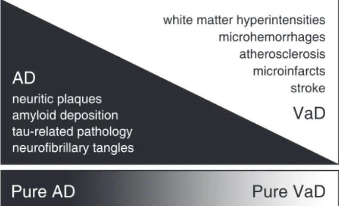
Based on the above epidemiological, clinical, and structural neuroimaging findings, it has been proposed recently that AD and VaD actually represent two extremes of a dementia spectrum ranging from patients with pure VaD to patients with pure AD, with a majority of
patients having contributions from both neuropathological pathways.53,74,89,90Figure 1 (adapted from Viswanathan et al.90) illustrates the concept of a spectrum ranging from pure AD to pure VaD. Such lines of evidence have also provided support to a more ambitious ‘‘vascular hypoth-esis’’ for AD, which proposes that the neuropathological changes that characterize AD would originate primarily from microvascular abnormalities.91
Cardiovascular risk factors and reduced cerebral blood flow and glucose metabolism: PET and SPECT findings
Comparing healthy controls and elderly patients with CHF, our group found significant rCBF reductions in patients, which were circumscribed to brain regions commonly affected in the early stages of AD –– namely, the precuneus and posterior cingulate gyrus. In addition, we found a significant direct association between lower cognitive test scores and rCBF reductions in the posterior cingulate gyrus.92This was, to the best of our knowledge, the first demonstration that a non-severe cardiac condi-tion could lead to circumscribed brain funccondi-tioning abnormalities similar to those considered typical of mild AD, even in individuals with no features of dementia or MCI. Similarly, subsequent rCBF investigations reported findings of AD-like hypoperfusion changes localized to regions such as the hippocampus and precuneus in association with other CVRFs, such as hypertension.93
More recently, a SPECT study of 18 overweight and obese participants showed extensive decreased prefron-tal rCBF associated with obesity.94This study highlights the association of prefrontal cortex abnormalities to obesity, but it is difficult to establish if these findings are the cause of overeating, reflect a vulnerability of this brain region to the metabolic and cardiovascular changes induced by obesity, or both. Moreover, the age range of the sample was very wide (20-82 years), and it is possible
that the effects of increased weight are dynamic and change throughout the lifespan.
Recently, there have also been large studies in this field using FDG-PET. Reiman et al.95 searched for significant associations between serum cholesterol levels and cerebral metabolic rates of glucose metabolism (CMRgl) in 117 cognitively normal middle-aged and elderly individuals (age 47-68 years). Higher serum total cholesterol levels were associated with lower CMRgl bilaterally in the precuneus, lateral parietal neocortex (encompassing the superior parietal lobule and angular and supramarginal gyri), lateral temporal neocortex (involving the superior temporal gyrus), and lateral prefrontal cortex (including the superior frontal gyrus), in a pattern that showed a substantial degree of overlap with the pattern of regional brain functional changes com-monly seen in subjects with mild AD.95
Investigating a subgroup of the same cohort, Langbaum et al.96detected lower frontotemporal glucose metabolism in proportion to elevated blood pressure indices.96 In additional studies with relatively modest samples, findings of CMRgl as assessed with FDG-PET have been described in association with CVRFs such as insulin resistance97and obesity98 as well as with WMH volume, which has been considered a marker of cerebrovascular burden.81,99Taken together, these stu-dies show a significant association between those risk factors and reduced CMRgl, variably implicating the precuneus, posterior cingulate gyrus, lateral parietal neocortex, and lateral temporal neocortex,96,97 as well as the lateral prefrontal cortex.96,98
Both type 1 and type 2 diabetes mellitus have also been associated with neurofunctional deficits.97,100It has been reported that the regional pattern of brain hypome-tabolism in cognitively normal patients with diabetes resembles, to some extent, the regional pattern of AD-related neurofunctional abnormalities, because both involve the posterior cingulate cortex, precuneus, and the inferior lateral parietal lobe.97 Moreover, higher fasting serum glucose levels in adults with no history of diabetes have also been correlated with lower CMRgl in the precuneus and posterior cingulate cortex, among other brain regions.101
In conclusion, FDG-PET and brain perfusion SPECT studies assessing non-demented individuals with a high CVRF burden have provided support to the ‘‘vascular hypothesis’’ of AD by showing patterns of brain functional abnormalities similar to those observed in AD.
A note of caution: it is still very challenging to unravel the relationships between AD and CVRF because they share several neuropathological features, such as brain atrophy and perfusion and metabolism deficits, which could hinder interpretation of neuroimaging findings obtained with current technologies.102Moreover, CVRFs may dynamically change neurofunctional findings in patients with AD. An interesting study showed that diabetic patients with AD had increased rCBF in the left inferior temporal gyrus on baseline SPECT when compared to non-diabetic patients with AD. In other
words, functional changes related to AD may differ depending on the presence of diabetes.103
To date, the majority of published studies have been cross-sectional, with limited information about the tem-poral dynamics of functional brain changes. A longitudinal study of patients with AD showed that participants with more severe CVRFs at baseline presented greater cognitive decline and more widespread rCBF reduc-tions.104Therefore, CVRFs can contribute to an acceler-ated progression of clinical and neurofunctional changes in patients with AD.
The APOE epsilon 4 (APOE e4) allele is not only an important risk factor for AD and cardiovascular dis-eases,27,105 but is also associated with functional brain changes106-108that can correlate with behavioral perfor-mance.109
Glucose hypometabolism has been associated with the presence of the APOE e4 allele in non-demented older adults.110-112In the FDG-PET study by Reiman et al.,95the sample of cognitively normal middle-aged and elderly individuals was subdivided into APOE e4 homozygous (n=24), heterozygous (n=38), and non-carriers (n=55). In some cortical regions, the relationship between hypome-tabolism and CVRFs had greater salience in e4 allele carriers than in non-carriers. The authors postulated that higher cholesterol levels, particularly in association with thee4 allele, would increase the risk of AD by accelerating some of the brain changes associated with normal aging.95 In our own FDG-PET study,113we aimed to investigate whether the associations between reduced CMRgl and elevated CVRF scores would be present regardless of
APOEgenotype. After controlling for the presence of the
e4 allele, the CVRF-related regional brain hypofunctional patterns retained statistical significance in the precuneus and posterior cingulate gyrus, suggesting that findings similar to those reported in AD subjects can be seen in association with the severity of CVRFs independently of
APOE status. On the other hand, findings involving the lateral temporoparietal neocortices lost their significance when the analysis was repeated after controlling for the effects of thee4 allele,113 indicating that metabolism in those latter regions may be influenced by APOE, as suggested by previous PET studies of non-elderly subjects.95,114,115
Although e4-related hypometabolism has been asso-ciated with the neuropathological processes of AD,112,116 such findings may not be necessarily pathological or specifically linked to AD.117For instance, APOEe4 is also a risk factor for vascular disease.118 Moreover, a study that used both FDG-PET and amyloid imaging to assess cognitively normal older adults showed that cerebral hypometabolism associated with e4 is not mediated by amyloid deposition.119Finally, posterior cingulate cortex hypometabolism has also been associated with vascular risk factors.113Thus, a vascular component –– in addition to other AD-related processes –– should be considered as part of the causal hypothesis of cerebral hypometabolism ine4 carriers.
factors.120In a study of middle-aged women,e4 carriers with higher cardiovascular fitness exhibited increased metabolism in the inferior temporal cortex and decreased metabolism in the middle and superior frontal gyri and right inferior parietal lobule when compared with low-fitness participants.121 Interestingly, such differences were not found among non-carriers of the e4 allele, suggesting that physical activity may have a greater impact on the population at risk for AD/vascular disease. This is in accordance with a study that reported higher cerebral amyloid burden in sedentarye4 carriers, while there were no significant differences between physically active and inactive subjects within the group ofe4 non-carriers.122
The potential of resting-state fMRI studies
PET and SPECT techniques rely on the use of radio-pharmaceuticals administered via the intravenous route. Another option is fMRI, which consists of multiple acquisitions of brain images measuring changes in the ratio of deoxygenated to oxygenated hemoglobin (the BOLD effect); these changes reflect regional neural activity.2
During fMRI acquisitions, the subject may be asked to perform cognitive tasks or, alternatively, to lie still while not engaging in any specific task in order to study the brain during an unconstrained state (resting-state fMRI [rs-fMRI]). Interregional correlations of BOLD signal time courses can provide estimates of functional connectivity, and it has been shown that functionally related brain regions (e.g., the bilateral primary motor cortices) exhibit high resting functional connectivity.123,124 Functionally discrete networks, such as the primary sensorimotor network, the frontoparietal attention network and the default mode network (DMN), can be identified on fMRI.125 The DMN comprises the posterior cingulate cortex, the ventromedial prefrontal cortex, and the inferior parietal lobule, has been implicated in episodic memory retrieval,125-127 and is one among a number of resting-state networks that have been characterized using fMRI (for a review, see van den Heuvel & Pol125).
The DMN is of special interest because: 1) age-related decreases in DMN functional connectivity have been the most consistent finding in rs-fMRI studies of the elderly population (for a review, see Ferreira & Busatto128); 2) patients with AD and MCI exhibit increased age-related changes in the DMN129-131; 3) hypoperfusion and hypometabolism (assessed by SPECT and PET) in the precuneus and posterior cingulate cortex –– a major DMN hub –– are frequently found in AD132,133; 4) functional connectivity within the DMN has been shown to correlate with behavioral performance in healthy older adults128 and in patients with AD129,130; and 5) baseline functional connectivity within the DMN has been associated with conversion from MCI to AD.134
A study of middle-aged subjects with type 2 diabetes focused on the functional connectivity of the posterior cingulate cortex found decreased connectivity in the bilateral middle temporal gyrus, the left medial and right
inferior frontal gyri, and the left thalamus in the diabetes group as compared with controls. Moreover, insulin resistance correlated negatively with the connectivity between the posterior cingulate cortex and the right inferior frontal gyrus and the right precuneus.135 It is interesting to note the existing overlap between these results and the regions presenting negative correlations between resting CMRgl and insulin resistance as found by another group97; one possibility is that insulin resistance impairs neural metabolism, thus leading to less efficient interregional network integration and to a number of pathophysiological processes related to AD.136,137
Hippocampal connectivity has been studied in older adults with type 2 diabetes using rs-fMRI. These patients exhibited decreased connectivity between the hippocam-pus and major hubs of the DMN (posterior cingulate cortex, medial prefrontal cortex and inferior parietal lobule).138The decreased hippocampal connectivity with the medial prefrontal cortex has also been recently reported in women with higher fasting insulin levels (a marker of insulin resistance).139It is relevant to note that the impact of diabetes in resting brain networks has not only been found in the DMN but also in language and attention networks, especially in patients with microvas-cular complications.140
A rs-fMRI study demonstrated that connectivity of the precuneus and the anterior cingulate cortex with the whole DMN were, respectively, positively and negatively correlated with body mass index, and connectivity of the left insula to a temporal lobe network showed negative correlation.141Perhaps some of these findings are related to the modulation of eating behavior, while others may reflect brain changes secondary to the metabolic abnormalities associated with obesity. The multiple possibilities of interpretation highlight how challenging it is to understand results from cross-sectional studies of conditions (such as obesity) that can both determine and/ or modify neural function patterns.
It is important to note that studying the impact of cardiovascular health in the brain using the BOLD signal can be problematic because it relies on hemodynamic response, which can be altered by cardiovascular disease142 and is modulated by cardiorespiratory fit-ness.143,144Therefore, although the study of resting brain networks using fMRI has provided interesting information, there are still considerable challenges to be overcome before it can be considered a clinically useful and reliable indicator of early brain abnormalities due to CVRFs.145
Unraveling the relationship between AD and
cardiovascular risk factors with PET amyloid imaging
In recent years, it has become possible to use nuclear medicine and molecular imaging techniques to study in vivo patterns of Ab deposition, one of the pathologi-cal hallmarks of AD.132,146 Amyloid imaging consists of injection of a radiolabeled ligand targeting Ab
developed for this purpose was carbon-11 labeled Pittsburgh Compound-B (PiB). Studies using this tech-nology have shown that amyloid deposition: 1) occurs years before clinical dementia147,148; 2) is not linearly related to cortical atrophy and cognitive decline147-149; 3) is more intense in patients with MCI who convert to AD than in nonconverters150,151; and 4) plateaus when clinical dementia is established (while other neurodegen-erative imaging biomarkers, such as brain atrophy, keep progressing).148,149
For the next few years, the amount of information provided by amyloid imaging studies is expected to increase sharply. For instance, this imaging modality is now being used in a sub-study of the Alzheimer’s Disease Neuroimaging Initiative, a large longitudinal multicenter project in the U.S. involving cohorts of elderly controls and subjects with MCI or AD. In this project, subjects have been investigated with multiple neuroimaging modalities and then followed up longitudinally, thus allowing measurements of change in these biomarkers over time.152 Furthermore, in 2012, the U.S. Food and Drug Administration approved florbetapir (18F) –– a PET radiopharmaceutical agent that binds to amyloid aggre-gates –– for clinical use in adults being evaluated for AD diagnosis.153
Regarding CVRFs, preliminary amyloid imaging stu-dies with PET have shown that engagement in physical exercise may be associated with decreased amyloid deposition in cognitively normal older adults154and that sedentary APOEe4 allele carriers exhibit greater amyloid deposition than physically active carriers.122Moreover, a study of late middle-aged to older adult subjects showed that systolic blood pressure correlated positively with 11
C-PiB accumulation in the posterior cingulate gyrus and precuneus, as well as in the frontal and temporal neocortices.96 On the other hand, a prospective cohort study of 53 older adults could not find significant differences in 11C-PiB retention between subjects with and without impaired glucose homeostasis.155 Each CVRF may present a particular association with AD pathophysiology.
A recent florbetapir-PET study found not only that hypertensive APOE e4 carriers showed higher amyloid deposition than subjects with just one of these risk factors (hypertension or APOE e4 genotype), but also that the subgroup of APOE e4+ individuals with unmedicated hypertension exhibited higher levels of amyloid burden than those with medicated hypertension.45These findings provide evidence that treatment of CVRFs may change the impact of APOE on amyloid deposition. The more widespread use of amyloid imaging is expected to foster longitudinal studies that use this technology to measure the effect of interventions. For instance, amyloid imaging findings have already been described as a secondary outcome in an AD clinical trial of liraglutide, a medication currently used for the treatment of diabetes,156as well as in a clinical trial of physical activity seeking to delay the progression of WMH to MCI.157
Advances in amyloid imaging have also provided interesting opportunities to unravel the relationship
between cerebrovascular disease (CVD) and AD. In a recent study, PiB-PET was performed a few days after stroke, and, in 20 out of 21 individuals, there was higher PiB retention in the ipsilateral peri-infarct brain region than in the contralateral side.158 In one recent long-itudinal study, baseline severity of white matter lesions correlated with increased PiB retention after a mean follow-up interval of 28 months.78 The authors sug-gested that the association between WMH –– a sign of CVD –– and progression of amyloid load might be mediated by impaired amyloid clearance due to vascular damage.
Subtle white matter changes as a suggestion of decreased axonal integrity (assessed by diffusion-weighted MRI) in the internal capsule and parahippo-campal region have been associated with amyloid deposition in older adults.80 Interestingly, such asso-ciations were no longer significant after controlling for
APOE genotype; one hypothesis is that APOE e4 increases amyloid deposition not only in the brain parenchyma but also in the blood vessels, thus leading to amyloid angiopathy, which is in turn associated with WMH.159
Finally, cerebral microhemorrhages (which are asso-ciated with several vascular risk factors) have been positively associated with amyloid deposition in the parieto-occipital region in a study of healthy older adults and patients with MCI or dementia.160
Overall, findings from in vivo amyloid imaging reinforce the relationship between AD and CVRFs. Further long-itudinal studies should address in greater depth the relationship between CVRFs and Ab deposition in the brain, using PET for amyloid imaging.122Such studies will be of key importance to demonstrate that the patterns of CVRF-related hypofunctioning reviewed in the present article may indeed be seen as correlates of AD-related neuropathology.
The effects of combined CVRFs
CVRFs rarely occur in isolation in elderly popula-tions161,162; therefore, the approach of investigating the impact of single risk factors on the brain may be limited. As multiple combinations of different CVRFs are present in the population, including information on multiple risk factors can provide a more accurate profile of each individual.163 The Framingham Coronary Heart Disease Risk (FCHDR) index is a widely used composite measure that takes into account multiple risk factors (age, sex, blood pressure, smoking status, total cholesterol and high-density lipoprotein cholesterol levels, and presence of diabetes) to assess the 10-year risk of coronary heart disease.163-165
prefrontal cortex) and medial (i.e., superior medial frontal, and superior orbital frontal gyri) prefrontal cortices.166
With the aim of extending the above findings to more cognitively preserved elderly subjects, our group recently acquired FDG-PET data from 59 cognitively intact older adults. The subgroup with high FCHDR scores exhibited reduced CMRgl in the precuneus, posterior cingulate gyrus, and lateral temporal and parietal neocortices when compared to those with low scores (Figure 2).113 This pattern of results provides further evidence of the substantial degree of overlap in regard to the location of foci of cerebral hypofunction across imaging studies of AD and CVRFs. Most of these results retained their statistical significance after correction for gray matter
atrophy (partial volume correction); thus, the findings represent true metabolic deficits, unrelated to the degree of atrophic changes.113
One other feature of our FDG-PET results is reminis-cent of findings reported in functional imaging studies of incipient AD: the lack of hypometabolism in frontal regions (Figure 2).113This stands in contrast both to the recently reported findings of cardiovascular risk-related prefrontal hypofunctioning in elderly subjects classified using the FCHDR index166 and to the results of other FDG-PET imaging studies that investigated the influence of single CVRFs on brain functioning.95-98One important difference between our PET study and the one by Kuczynski et al.166is that the authors of the latter study did not exclude subjects with lacunar infarcts. They
Figure 2 Reduced brain glucose metabolism in older adults with high cardiovascular risk. Areas of reduced cerebral glucose metabolism (as assessed with FDG-PET) in a group of cognitively intact older adults with high cardiovascular risk (according to FCHDR scores) compared to an age-matched group with low cardiovascular risk are highlighted in yellow, overlaid on axial slices of a reference MRI scan that approximates the Talairach & Tournoux stereotactic atlas167(for details, see
Tamashiro-Duran et al.113). Data were analyzed using voxel-based, statistical parametric mapping methods and findings reached
actually suggested that findings of frontal lobe hypome-tabolism in individuals with higher FCHDR scores could be determined by the greater incidence of lacunar infarcts in those subjects, leading to localized frontal metabolic changes.166This reasoning may explain the absence of frontal metabolic changes in our FDG-PET study, since we excluded subjects with vascular-related silent brain lesions as assessed by MRI, including lacunar infarcts. It is therefore plausible to argue that, in the absence of lacunar infarcts, the frontal lobe is not especially vulnerable to the damaging effects of CVRFs in cogni-tively intact individuals.
Beason-Held et al.168 used FCHDR scores to investi-gate the relationship between baseline CVRFs and subsequent changes in resting rCBF (as assessed with PET) in cognitively preserved older adults (n=97) who underwent repeated annual imaging assessments over a period of up to 8 years. The authors found a significant relationship between higher baseline FCHDR scores and greater progression of rCBF deficits in the precuneus, as well as in frontal regions, the insula, and the brain stem. This was the first longitudinal PET study to provide evidence of progressive functional brain deficits in individuals with a high cardiovascular risk burden.168
The impact of summarizing vascular risk factors in one index has also been shown in a study of 43 elderly subjects with variable cognitive deficits; FCHDR index and amyloid deposition were positively correlated, but no individual component of the FCHDR (including diabetes, hypertension, and elevated lipids) was significantly associated with brain amyloid burden.169
Microstructural and molecular mechanisms underlying cardiovascular risk-related brain function deficits
The continued influence of CVRFs and cardiac disorders that lead to chronically reduced rCBF is thought to lead to numerous local neuropathological consequences,170 including structural deformities of brain microvessels,171,172 microembolic events, decreased oxygen and nutrient supply, metabolic deficits, toxic disturbances,173-175 and endothelial dysfunction.176Such deficits at the microstruc-tural level are all likely to contribute to the findings of regional brain hypofunction detectable using FDG-PET, rCBF SPECT, and rs-fMRI in clinical studies.
It is also important to consider the possibility of direct links between microvascular changes secondary to chronic cerebral blood flow reductions and triggering of the accumulation of Ab-peptide that characterizes AD.34 Recently, molecular models have implicated circulatory defects (i.e., alterations in vascular smooth muscle cells of meningeal arterioles due to CVRF) as critical factors that impair the removal of Abfrom the brain across the blood brain barrier.171Chronic inflammatory processes, closely linked to endothelial dysfunction, have also been increasingly implicated as relevant to the development of the Ab-related molecular changes underlying the symp-toms of AD.177-181
Specific CVRFs are associated with non-vascular microstructural and molecular changes, which may also
contribute to the presence of cognitive deficits and related functional imaging deficits. In the case of smoking, for instance, nicotine self-administration has been shown to decrease neurogenesis and neuroplasticity and increase cell death in the hippocampus (dentate gyrus) of rats and mice.182,183 Diabetes entails specific inflammatory changes involving protein kinase C activation, excess production of reactive oxygen species, protein glycosyla-tion, and cellular activation of the receptor for advanced glycation endproducts.184,185Finally, recent research on glycogen synthase kinase-3 (GSK-3) provides one other interesting link between AD and diabetes. GSK-3 is a pivotal enzyme in glycogen synthesis and is thought to participate in the development of insulin resistance primarily by inhibiting glycogen synthase activity and, thus, decreasing the synthesis of glycogen.186GSK-3 has been implicated in the pathophysiology of AD because it promotes tau phosphorylation187,188 and its activity is regulated by the Abpeptide.189
There is evidence that impaired glucose metabolism plays an important role in the pathogenesis of AD (for a recent review, see Chen & Zhong190) and may represent a link between CVRFs/CVD and AD: persistently sub-optimal glucose and/or oxygen supply may trigger a series of downstream events, such as oxidative stress, mitochondrial dysfunction, inflammation, GSK-3 activa-tion, amyloid deposiactiva-tion, tau hyperphosphorylaactiva-tion, and neuronal death.190,191
In addition to the hypothesis that vascular impairments may lead or contribute to AD neuropathology, it is also plausible –– and supplementary –– to understand that AD and vascular burden can represent processes occurring simultaneously in the same individual that, when com-bined, lead to an increased risk of cognitive decline and dementia. In other words, brains suffering from this ‘‘double hit’’ may be more prone to declines in function, thus leading to clinical dementia.
Discussion
There is compelling evidence from neurofunctional studies to support that CVRFs are related to AD. Although some findings are conflicting and heteroge-neous, a large body of the literature supports the understanding that CVRFs and CVD contribute to brain changes related to cognitive decline and increased risk of dementia in AD, and may also play a role in the pathophysiology of AD.
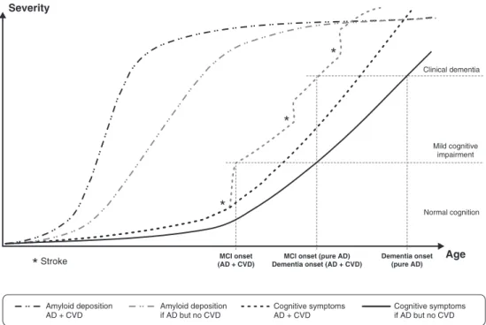
line), and the illustration only partially accounts for this heterogeneity. Nevertheless, two interesting aspects should be highlighted.
First, it would be very informative to test the accuracy of the model depicted by the dash-dot lines with longitudinal studies assessing amyloid deposition in middle-aged adults with normal cognition and different cardiovascular risk profiles. In other words, future research should provide more evidence to allow us to better answer the following questions: do baseline CVRFs increase subsequent amyloid deposition and/or tau-related pathology? Does treatment of CVRF modifies the longitudinal dynamics of AD-related neuropathology? Second, because CVRFs and CVD contribute to cognitive decline (three lines to the right), adequate treatment of cardiovascular conditions and prevention of CVD should delay the onset of clinical dementia even in patients with co-occurring AD-related pathology. This aspect is particularly relevant for public health, as AD + CVD comorbidity is very common and the impact of delaying clinical onset by just a few years is substantial.192
The present article sought to summarize the current relevant knowledge in this field, and, although compre-hensive, was not a systematic review. Instead, we provided a broad overview addressing a number of
relevant topics, some of which can be further explored by future original studies or systematic reviews.
In conclusion, CVRFs are important determinants of brain health in older adults. Functional neuroimaging studies have provided multiple levels of evidence that CVRFs are associated with neuronal changes even in cognitively normal adults. Overall, these results point toward the hypothesis that CVRFs may be causally related to AD. Thus, greater knowledge about how these factors influence brain function over time may provide important insights for the development of strategies aimed at delaying or preventing pathologic brain changes, with relevant public health implications regarding the prevention of AD. Combining currently available resources for CVRF prevention/treatment and recent multimodal techniques for monitoring of AD-related pathology and changes in brain function can result in an unprecedented impact on how we understand the aging brain and promote successful aging.
Acknowledgements
This work was supported by grants number 2012/11898-5 (LKF) and 2012/50329-6 (GFB) from Fundac¸a˜o de Amparo a` Pesquisa do Estado de Sa˜o Paulo (FAPESP). TCA and GFB are also supported by
Conselho Nacional de Desenvolvimento Cientı´fico e Tecnolo´gico (CNPq), Brazil.
Disclosure
The authors report no conflicts of interest.
References
1 Reiman EM, Jagust WJ. Brain imaging in the study of Alzheimer’s disease. Neuroimage. 2012;61:505-16.
2 Huettel SA, Song AW, McCarthy G. Functional magnetic resonance maging. 2nd ed. Sunderland: Sinauer Associates; 2009.
3 Che´telat G, Desgranges B, de la Sayette V, Viader F, Berkouk K, Landeau B, et al. Dissociating atrophy and hypometabolism impact on episodic memory in mild cognitive impairment. Brain. 2003;126:1955-67.
4 Kawachi T, Ishii K, Sakamoto S, Sasaki M, Mori T, Yamashita F, et al. Comparison of the diagnostic performance of FDG-PET and VBM-MRI in very mild Alzheimer’s disease. Eur J Nucl Med Mol Imaging. 2006;33:801-9.
5 Minoshima S, Giordani B, Berent S, Frey KA, Foster NL, Kuhl DE. Metabolic reduction in the posterior cingulate cortex in very early Alzheimer’s disease. Ann Neurol. 1997;42:85-94.
6 Mosconi L. Brain glucose metabolism in the early and specific diagnosis of Alzheimer’s disease. FDG-PET studies in MCI and AD. Eur J Nucl Med Mol Imaging. 2005;32:486-510.
7 Jagust W. Positron emission tomography and magnetic resonance imaging in the diagnosis and prediction of dementia. Alzheimers Dement. 2006;2:36-42.
8 Mosconi L, Tsui WH, Herholz K, Pupi A, Drzezga A, Lucignani G, et al. Multicenter standardized 18F-FDG PET diagnosis of mild cognitive impairment, Alzheimer’s disease, and other dementias. J Nucl Med. 2008;49:390-8.
9 Petrie EC, Cross DJ, Galasko D, Schellenberg GD, Raskind MA, Peskind ER, et al. Preclinical evidence of Alzheimer changes: convergent cerebrospinal fluid biomarker and fluorodeoxyglucose positron emission tomography findings. Arch Neurol. 2009;66:632-7.
10 Bloudek LM, Spackman DE, Blankenburg M, Sullivan SD. Review and meta-analysis of biomarkers and diagnostic imaging in Alzheimer’s disease. J Alzheimers Dis. 2011;26:627-45.
11 Jack CR Jr, Albert MS, Knopman DS, McKhann GM, Sperling RA, Carrillo MC, et al. Introduction to the recommendations from the National Institute on Aging-Alzheimer’s Association workgroups on diagnostic guidelines for Alzheimer’s disease. Alzheimer’s Dement. 2011;7:257-62.
12 McKhann GM, Knopman DS, Chertkow H, Hyman BT, Jack CR Jr, Kawas CH, et al. The diagnosis of dementia due to Alzheimer’s disease: recommendations from the National Institute on Aging-Alzheimer’s Association workgroups on diagnostic guidelines for Alzheimer’s disease. Alzheimers Dement. 2011;7:263-9.
13 Hampel H, Frank R, Broich K, Teipel SJ, Katz RG, Hardy J, et al. Biomarkers for Alzheimer’s disease: academic, industry and regulatory perspectives. Nat Rev Drug Discov. 2010;9:560-74. 14 Petersen RC. Mild cognitive impairment as a diagnostic entity.
J Intern Med. 2004;256:183-94.
15 Herholz K. Cerebral glucose metabolism in preclinical and prodromal Alzheimer’s disease. Expert Rev Neurother. 2010;10: 1667-73.
16 Bru¨ck A, Virta JR, Koivunen J, Koikkalainen J, Scheinin NM, Helenius H, et al. [11C]PIB, [18F]FDG and MR imaging in patients with mild cognitive impairment. Eur J Nucl Med Mol Imaging. 2013;40:1567-72.
17 Miller SL, Fenstermacher E, Bates J, Blacker D, Sperling RA, Dickerson BC. Hippocampal activation in adults with mild cognitive impairment predicts subsequent cognitive decline. J Neurol Neurosurg Psychiatry. 2008;79:630-5.
18 Yuan Y, Gu ZX, Wei WS. Fluorodeoxyglucose-Positron-Emission Tomography, Single-Photon Emission Tomography, and Structural MR Imaging for Prediction of Rapid Conversion to Alzheimer
Disease in Patients with Mild Cognitive Impairment: A Meta-Analysis. AJNR Am J Neuroradiol. 2009;30:404-10.
19 Albert MS, DeKosky ST, Dickson D, Dubois B, Feldman HH, Fox NC, et al. The diagnosis of mild cognitive impairment due to Alzheimer’s disease: recommendations from the National Institute on Aging-Alzheimer’s Association workgroups on diagnostic guide-lines for Alzheimer’s disease. Alzheimers Dement. 2011;7:270-9. 20 Alexopoulos P, Guo LH, Jiang M, Bujo H, Grimmer T, Fo¨rster S,
et al. Amyloid cascade and tau pathology cerebrospinal fluid markers in mild cognitive impairment with regards to Alzheimer’s disease cerebral metabolic signature. J Alzheimers Dis. 2013;36:401-8.
21 Kircher TT, Weis S, Freymann K, Erb M, Jessen F, Grodd W, et al. Hippocampal activation in patients with mild cognitive impairment is necessary for successful memory encoding. J Neurol Neurosurg Psychiatry. 2007;78:812-8.
22 Parra MA, Pattan V, Wong D, Beaglehole A, Lonie J, Wan HI, et al. Medial temporal lobe function during emotional memory in early Alzheimer’s disease, mild cognitive impairment and healthy ageing: an fMRI study. BMC Psychiatry. 2013;13:76.
23 Zamboni G, Wilcock GK, Douaud G, Drazich E, McCulloch E, Filippini N, et al. Resting functional connectivity reveals residual functional activity in Alzheimer’s disease. Biol Psychiatry. 2013; 74:375-83.
24 Fitzpatrick AL, Kuller LH, Lopez OL, Diehr P, O’Meara ES, Longstreth WT Jr, et al. Midlife and late-life obesity and the risk of dementia: cardiovascular health study. Arch Neurol. 2009; 66:336-42.
25 Li J, Wang YJ, Zhang M, Xu ZQ, Gao CY, Fang CQ, et al. Vascular risk factors promote conversion from mild cognitive impairment to Alzheimer disease. Neurology. 2011;76:1485-91.
26 Obisesan TO, Obisesan OA, Martins S, Alamgir L, Bond V, Maxwell C, et al. High blood pressure, hypertension, and high pulse pressure are associated with poorer cognitive function in persons aged 60 and older: the Third National Health and Nutrition Examination Survey. J Am Geriatr Soc. 2008;56:501-9.
27 Irie F, Fitzpatrick AL, Lopez OL, Kuller LH, Peila R, Newman AB, et al. Enhanced risk for Alzheimer disease in persons with type 2 diabetes and APOE epsilon4: the Cardiovascular Health Study Cognition Study. Arch Neurol. 2008;65:89-93.
28 Rosendorff C, Beeri MS, Silverman JM. Cardiovascular risk factors for Alzheimer’s disease. Am J Geriatr Cardiol. 2007;16:143-9. 29 Stuerenburg HJ, Ganzer S, Arlt S, Mu¨ller-Thomsen T. The influence
of smoking on plasma folate and lipoproteins in Alzheimer disease, mild cognitive impairment and depression. Neuro Endocrinol Lett. 2005;26:261-3.
30 Xu W, Qiu C, Gatz M, Pedersen NL, Johansson B, Fratiglioni L. Mid- and late-life diabetes in relation to the risk of dementia: a population-based twin study. Diabetes. 2009;58:71-7.
31 Bottino CM, Azevedo D Jr, Tatsch M, Hototian SR, Moscoso MA, Folquitto J, et al. Estimate of dementia prevalence in a community sample from Sa˜o Paulo, Brazil. Dement Geriatr Cogn Disord. 2008;26:291-9.
32 Plassman BL, Langa KM, Fisher GG, Heeringa SG, Weir DR, Ofstedal MB, et al. Prevalence of dementia in the United States: the aging, demographics, and memory study. Neuroepidemiology. 2007;29:125-32.
33 Scazufca M, Menezes PR, Vallada HP, Crepaldi AL, Pastor-Valero M, Coutinho LM, et al. High prevalence of dementia among older adults from poor socioeconomic backgrounds in Sa˜o Paulo, Brazil. Int Psychogeriatr. 2008;20:394-405.
34 Hardy J, Selkoe DJ. The amyloid hypothesis of Alzheimer’s disease: progress and problems on the road to therapeutics. Science. 2002;297:353-6.
35 Braak E, Griffing K, Arai K, Bohl J, Bratzke H, Braak H. Neuropathology of Alzheimer’s disease: what is new since A. Alzheimer? Eur Arch Psychiatry Clin Neurosci. 1999;249: 14-22.
36 Akinyemi RO, Mukaetova-Ladinska EB, Attems J, Ihara M, Kalaria RN. Vascular risk factors and neurodegeneration in ageing related dementias: Alzheimer’s disease and vascular dementia. Curr Alzheimer Res. 2013;10:642-53.
Framingham heart study. Int J Obes Relat Metab Disord. 2003;27:260-8.
38 Havlik RJ, Foley DJ, Sayer B, Masaki K, White L, Launer LJ. Variability in midlife systolic blood pressure is related to late-life brain white matter lesions: the Honolulu-Asia Aging study. Stroke. 2002;33:26-30.
39 Skoog I, Lernfelt B, Landahl S, Palmertz B, Andreasson LA, Nilsson L, et al. 15-year longitudinal study of blood pressure and dementia. Lancet. 1996;347:1141-5.
40 Luchsinger JA, Reitz C, Honig LS, Tang MX, Shea S, Mayeux R. Aggregation of vascular risk factors and risk of incident Alzheimer disease. Neurology. 2005;65:545-51.
41 Anstey KJ, von Sanden C, Salim A, O’Kearney R. Smoking as a risk factor for dementia and cognitive decline: a meta-analysis of prospective studies. Am J Epidemiol. 2007;166:367-78.
42 Erickson KI, Weinstein AM, Lopez OL. Physical activity, brain plasticity, and Alzheimer’s disease. Arch Med Res. 2012;43:615-21. 43 Solfrizzi V, D’Introno A, Colacicco AM, Capurso C, Todarello O, Pellicani V, et al. Circulating biomarkers of cognitive decline and dementia. Clin Chim Acta. 2006;364:91-112.
44 Brown BM, Peiffer JJ, Taddei K, Lui JK, Laws SM, Gupta VB, et al. Physical activity and amyloid-b plasma and brain levels: results from the Australian Imaging, Biomarkers and Lifestyle Study of Ageing. Mol Psychiatry. 2013;18:875-81.
45 Rodrigue KM, Rieck JR, Kennedy KM, Devous MD Sr, Diaz-Arrastia R, Park DC. Risk factors forb-amyloid deposition in healthy aging: vascular and genetic effects. JAMA Neurol. 2013;70:600-6. 46 Toledo JB, Toledo E, Weiner MW, Jack CR Jr, Jagust W, Lee VM, et al. Cardiovascular risk factors, cortisol, and amyloid-bdeposition in Alzheimer’s Disease Neuroimaging Initiative. Alzheimers Dement. 2012;8:483-9.
47 Das SK, Bose P, Biswas A, Dutt A, Banerjee TK, Hazra AM, et al. An epidemiologic study of mild cognitive impairment in Kolkata, India. Neurology. 2007;68:2019-26.
48 Reitz C, Tang MX, Manly J, Mayeux R, Luchsinger JA. Hypertension and the risk of mild cognitive impairment. Arch Neurol. 2007;64:1734-40.
49 Lopez OL, Jagust WJ, Dulberg C, Becker JT, DeKosky ST, Fitzpatrick A, et al. Risk factors for mild cognitive impairment in the Cardiovascular Health Study Cognition Study: part 2. Arch Neurol. 2003;60:1394-9.
50 Luchsinger JA, Reitz C, Patel B, Tang MX, Manly JJ, Mayeux R. Relation of diabetes to mild cognitive impairment. Arch Neurol. 2007;64:570-5.
51 Kivipelto M, Helkala EL, Ha¨nninen T, Laakso MP, Hallikainen M, Alhainen K, et al. Midlife vascular risk factors and late-life mild cognitive impairment: A population-based study. Neurology. 2001; 56:1683-9.
52 Solomon A, Ka˚reholt I, Ngandu T, Winblad B, Nissinen A, Tuomilehto J, et al. Serum cholesterol changes after midlife and late-life cognition: twenty-one-year follow-up study. Neurology. 2007;68:751-6.
53 Toledo JB, Arnold SE, Raible K, Brettschneider J, Xie SX, Grossman M, et al. Contribution of cerebrovascular disease in autopsy confirmed neurodegenerative disease cases in the National Alzheimer’s Coordinating Centre. Brain. 2013;136:2697-706.
54 Peters R, Beckett N. Hypertension, dementia, and antihypertensive treatment: implications for the very elderly. Curr Hypertens Rep. 2009;11:277-82.
55 Purandare N. Preventing dementia: role of vascular risk factors and cerebral emboli. Br Med Bull. 2009;91:49-59.
56 Scarmeas N, Luchsinger JA, Schupf N, Brickman AM, Cosentino S, Tang MX, et al. Physical activity, diet, and risk of Alzheimer disease. JAMA. 2009;302:627-37.
57 Scarmeas N, Stern Y, Mayeux R, Manly JJ, Schupf N, Luchsinger JA. Mediterranean diet and mild cognitive impairment. Arch Neurol. 2009;66:216-25.
58 Buchman AS, Boyle PA, Yu L, Shah RC, Wilson RS, Bennett DA. Total daily physical activity and the risk of AD and cognitive decline in older adults. Neurology. 2012;78:1323-9.
59 Antonelli Incalzi R, Trojano L, Acanfora D, Crisci C, Tarantino F, Abete P, et al. Verbal memory impairment in congestive heart failure. J Clin Exp Neuropsychol. 2003;25:14-23.
60 Bennett SJ, Sauve´ MJ. Cognitive deficits in patients with heart failure: a review of the literature. J Cardiovasc Nurs. 2003;18:219-42.
61 Glynn LM, Christenfeld N, Gerin W. The role of rumination in recovery from reactivity: cardiovascular consequences of emotional states. Psychosom Med. 2002;64:714-26.
62 Bellomo A, Mancinella M, Troisi G, Marigliano V. [Relationship between atrial fibrillation and other cardiovascular risk factors in Alzheimer’s disease. New prevention opportunities]. Recent Prog Med. 2009;100:124-6.
63 Purnell C, Gao S, Callahan CM, Hendrie HC. Cardiovascular risk factors and incident Alzheimer disease: a systematic review of the literature. Alzheimer Dis Assoc Disord. 2009;23:1-10.
64 Breteler MM, Claus JJ, Grobbee DE, Hofman A. Cardiovascular disease and distribution of cognitive function in elderly people: the Rotterdam Study. BMJ. 1994;308:1604-8.
65 Launer LJ, Masaki K, Petrovitch H, Foley D, Havlik RJ. The association between midlife blood pressure levels and late-life cognitive function. The Honolulu-Asia Aging Study. JAMA. 1995;274:1846-51.
66 Rosengart TK, Sweet J, Finnin EB, Wolfe P, Cashy J, Hahn E, et al. Neurocognitive functioning in patients undergoing coronary artery bypass graft surgery or percutaneous coronary intervention: evidence of impairment before intervention compared with normal controls. Ann Thorac Surg. 2005;80:1327-34.
67 Almeida OP, Tamai S. Congestive heart failure and cognitive functioning amongst older adults. Arq Neuropsiquiatr. 2001;59:324-9.
68 Debette S, Beiser A, DeCarli C, Au R, Himali JJ, Kelly-Hayes M, et al. Association of MRI markers of vascular brain injury with incident stroke, mild cognitive impairment, dementia, and mortality: the Framingham Offspring Study. Stroke. 2010;41:600-6. 69 Kearney-Schwartz A, Rossignol P, Bracard S, Felblinger J, Fay R,
Boivin JM, et al. Vascular structure and function is correlated to cognitive performance and white matter hyperintensities in older hypertensive patients with subjective memory complaints. Stroke. 2009;40:1229-36.
70 Markus HS, Hunt B, Palmer K, Enzinger C, Schmidt H, Schmidt R. Markers of endothelial and hemostatic activation and progression of cerebral white matter hyperintensities: longitudinal results of the Austrian Stroke Prevention Study. Stroke. 2005;36:1410-4. 71 Rostrup E, Gouw AA, Vrenken H, van Straaten EC, Ropele S,
Pantoni L, et al. The spatial distribution of age-related white matter changes as a function of vascular risk factors––results from the LADIS study. Neuroimage. 2012;60:1597-607.
72 Gouw AA, Seewann A, van der Flier WM, Barkhof F, Rozemuller AM, Scheltens P, et al. Heterogeneity of small vessel disease: a systematic review of MRI and histopathology correlations. J Neurol Neurosurg Psychiatry. 2011;82:126-35.
73 So¨derlund H, Nyberg L, Adolfsson R, Nilsson LG, Launer LJ. High prevalence of white matter hyperintensities in normal aging: relation to blood pressure and cognition. Cortex. 2003;39:1093-105. 74 Erkinjuntti T, Gauthier S. The concept of vascular cognitive
impairment. In: Giannakopoulos P, Hof PR. Dementia in clinical practice. Basel: Karger Publishers; 2009. p. 79-85.
75 Teipel SJ, Meindl T, Wagner M, Kohl T, Bu¨rger K, Reiser MF, et al. White matter microstructure in relation to education in aging and Alzheimer’s disease. J Alzheimers Dis. 2009;17:571-83.
76 Ko¨hler S, Thomas AJ, Lloyd A, Barber R, Almeida OP, O’Brien JT. White matter hyperintensities, cortisol levels, brain atrophy and continuing cognitive deficits in late-life depression. Br J Psychiatry. 2010;196:143-9.
77 Longstreth WT Jr, Arnold AM, Beauchamp NJ Jr, Manolio TA, Lefkowitz D, Jungreis C, et al. Incidence, manifestations, and predictors of worsening white matter on serial cranial magnetic resonance imaging in the elderly: the Cardiovascular Health Study. Stroke. 2005;36:56-61.
78 Grimmer T, Faust M, Auer F, Alexopoulos P, Fo¨rstl H, Henriksen G, et al. White matter hyperintensities predict amyloid increase in Alzheimer’s disease. Neurobiol Aging. 2012;33:2766-73.
80 Chao LL, Decarli C, Kriger S, Truran D, Zhang Y, Laxamana J, et al. Associations between white matter hyperintensities andbamyloid on integrity of projection, association, and limbic fiber tracts measured with diffusion tensor MRI. PLoS One. 2013;8:e65175. 81 Lo RY, Jagust WJ, Alzheimer’s Disease Neuroimaging Initiative.
Vascular burden and Alzheimer disease pathologic progression. Neurology. 2012;79:1349-55.
82 Marchand WR, Lee JN, Suchy Y, Garn C, Johnson S, Wood N, et al. Age-related changes of the functional architecture of the cortico-basal ganglia circuitry during motor task execution. Neuroimage. 2011;55:194-203.
83 Almeida OP, Garrido GJ, Lautenschlager NT, Hulse GK, Jamrozik K, Flicker L. Smoking is associated with reduced cortical regional gray matter density in brain regions associated with incipient Alzheimer disease. Am J Geriatr Psychiatry. 2008;16:92-8. 84 Chen Z, Li L, Sun J, Ma L. Mapping the brain in type II diabetes:
Voxel-based morphometry using DARTEL. Eur J Radiol. 2012; 81:1870-6.
85 Korf ES, van Straaten ECW, de Leeuw FE, van der Flier WM, Barkhof F, Pantoni L, et al. Diabetes mellitus, hypertension and medial temporal lobe atrophy: the LADIS study. Diabet Med. 2007;24:166-71.
86 de Toledo Ferraz Alves TC, Scazufca M, Squarzoni P, de Souza Duran FL, Tamashiro-Duran JH, Vallada HP, et al. Subtle gray matter changes in temporo-parietal cortex associated with cardio-vascular risk factors. J Alzheimers Dis. 2011;27:575-89.
87 Beauchet O, Celle S, Roche F, Bartha R, Montero-Odasso M, Allali G, et al. Blood pressure levels and brain volume reduction: a systematic review and meta-analysis. J Hypertens. 2013;31: 1502-16.
88 Hayes SM, Hayes JP, Cadden M, Verfaellie M. A review of cardiorespiratory fitness-related neuroplasticity in the aging brain. Front Aging Neurosci. 2013;5:31.
89 Rocchi A, Orsucci D, Tognoni G, Ceravolo R, Siciliano G. The role of vascular factors in late-onset sporadic Alzheimer’s disease. Genetic and molecular aspects. Curr Alzheimer Res. 2009;6: 224-37.
90 Viswanathan A, Rocca WA, Tzourio C. Vascular risk factors and dementia: how to move forward? Neurology. 2009;72:368-74. 91 de la Torre JC. Three postulates to help identify the cause of
Alzheimer’s disease. J Alzheimers Dis. 2011;24:657-68.
92 Alves TC, Rays J, Fra´guas R Jr, Wajngarten M, Meneghetti JC, Prando S, et al. Localized cerebral blood flow reductions in patients with heart failure: a study using 99mTc-HMPAO SPECT. J Neuroimaging. 2005;15:150-6.
93 Dai W, Lopez OL, Carmichael OT, Becker JT, Kuller LH, Gach HM. Abnormal regional cerebral blood flow in cognitively normal elderly subjects with hypertension. Stroke. 2008;39:349-54.
94 Willeumier KC, Taylor DV, Amen DG. Elevated BMI Is associated With Decreased Blood Flow in the Prefrontal Cortex Using SPECT Imaging in Healthy adults. Obesity (Silver Spring). 2011;19:1095-7. 95 Reiman EM, Chen K, Langbaum JB, Lee W, Reschke C, Bandy D, et al. Higher serum total cholesterol levels in late middle age are associated with glucose hypometabolism in brain regions affected by Alzheimer’s disease and normal aging. Neuroimage. 2010;49:169-76.
96 Langbaum JB, Chen K, Launer LJ, Fleisher AS, Lee W, Liu X, et al. Blood pressure is associated with higher brain amyloid burden and lower glucose metabolism in healthy late middle-age persons. Neurobiol Aging. 2012;33:827.e11-9.
97 Baker LD, Cross DJ, Minoshima S, Belongia D, Watson GS, Craft S. Insulin resistance and Alzheimer-like reductions in regional cerebral glucose metabolism for cognitively normal adults with prediabetes or early type 2 diabetes. Arch Neurol. 2011;68:51-7. 98 Volkow ND, Wang GJ, Telang F, Fowler JS, Goldstein RZ,
Alia-Klein N, et al. Inverse association between BMI and prefrontal metabolic activity in healthy adults. Obesity (Silver Spring). 2009;17:60-5.
99 Erten-Lyons D, Woltjer R, Kaye J, Mattek N, Dodge HH, Green S, et al. Neuropathologic basis of white matter hyperintensity accumulation with advanced age. Neurology. 2013;81:977-83. 100 van Golen LW, Huisman MC, Ijzerman RG, Hoetjes NJ, Schwarte
LA, Lammertsma AA, et al. Cerebral blood flow and glucose
metabolism measured with positron emission tomography are decreased in human type 1 diabetes. Diabetes. 2013;62:2898-904. 101 Burns CM, Chen K, Kaszniak AW, Lee W, Alexander GE, Bandy D, et al. Higher serum glucose levels are associated with cerebral hypometabolism in Alzheimer regions. Neurology. 2013;80:1557-64.
102 Austin BP, Nair VA, Meier TB, Xu G, Rowley HA, Carlsson CM, et al. Effects of hypoperfusion in Alzheimer’s disease. J Alzheimers Dis. 2011;26:123-33.
103 Hirao K, Hanyu H, Sato T, Kanetaka H, Shimizu S, Sakurai H, et al. A longitudinal SPECT study of different patterns of regional cerebral blood flow in Alzheimer’s disease with or without diabetes. Dement Geriatr Cogn Dis Extra. 2011;1:62-74.
104 Kume K, Hanyu H, Sato T, Hirao K, Shimizu S, Kanetaka H, et al. Vascular risk factors are associated with faster decline of Alzheimer disease: a longitudinal SPECT study. J Neurol. 2011;258:1295-303.
105 Kivipelto M, Rovio S, Ngandu T, Ka˚reholt I, Eskelinen M, Winblad B, et al. Apolipoprotein E epsilon4 magnifies lifestyle risks for dementia: a population-based study. J Cell Mol Med. 2008; 12:2762-71.
106 Goveas JS, Xie C, Chen G, Li W, Ward BD, Franczak MB, et al. Functional network endophenotypes unravel the effects of apoli-poprotein E epsilon 4 in middle-aged adults. PLoS One. 2013;8:e55902.
107 Nichols LM, Masdeu JC, Mattay VS, Kohn P, Emery M, Sambataro F, et al. Interactive effect of apolipoprotein e genotype and age on hippocampal activation during memory processing in healthy adults. Arch Gen Psychiatry. 2012;69:804-13.
108 Trachtenberg AJ, Filippini N, Ebmeier KP, Smith SM, Karpe F, Mackay CE. The effects of APOE on the functional architecture of the resting brain. Neuroimage. 2012;59:565-72.
109 Westlye ET, Lundervold A, Rootwelt H, Lundervold AJ, Westlye LT. Increased hippocampal default mode synchronization during rest in middle-aged and elderly APOE 4 carriers: relationships with memory performance. J Neurosci. 2011;31:7775-83.
110 Drzezga A, Grimmer T, Riemenschneider M, Lautenschlager N, Siebner H, Alexopoulus P, et al. Prediction of individual clinical outcome in MCI by means of genetic assessment and (18)F-FDG PET. J Nucl Med. 2005;46:1625-32.
111 Mosconi L, De Santi S, Brys M, Tsui WH, Pirraglia E, Glodzik-Sobanska L, et al. Hypometabolism and altered cerebrospinal fluid markers in normal apolipoprotein E E4 carriers with subjective memory complaints. Biol Psychiatry. 2008;63:609-18.
112 Protas HD, Chen K, Langbaum JB, Fleisher AS, Alexander GE, Lee W, et al. Posterior cingulate glucose metabolism, hippocampal glucose metabolism, and hippocampal volume in cognitively normal, late-middle-aged persons at 3 levels of genetic risk for Alzheimer disease. JAMA Neurol. 2013;70:320-5.
113 Tamashiro-Duran JH, Squarzoni P, de Souza Duran FL, Curiati PK, Vallada HP, Buchpiguel CA, et al. Cardiovascular risk in cognitively preserved elderlies is associated with glucose hypometabolism in the posterior cingulate cortex and precuneus regardless of brain atrophy and apolipoprotein gene variations. Age (Dordr). 2013;35:777-92.
114 Langbaum JBS, Chen K, Caselli RJ, Lee W, Reschke C, Bandy D, et al. Hypometabolism in Alzheimer-affected brain regions in cognitively healthy Latino individuals carrying the apolipoprotein E epsilon4 allele. Arch Neurol. 2010;67:462-8.
115 Reiman EM, Chen K, Alexander GE, Caselli RJ, Bandy D, Osborne D, et al. Functional brain abnormalities in young adults at genetic risk for late-onset Alzheimer’s dementia. Proc Natl Acad Sci U S A. 2004;101:284-9.
116 Frank G, Hennig-Fast K, Klu¨nemann HH, Schmitz G, Greenlee MW. Differential impact of ApoE e4 on cortical activation during famous face recognition in cognitively intact individuals and patients with amnestic mild cognitive impairment. Alzheimer Dis Assoc Disord. 2011;25:250-61.
117 Trachtenberg AJ, Filippini N, Cheeseman J, Duff EP, Neville MJ, Ebmeier KP, et al. The effects of APOE on brain activity do not simply reflect the risk of Alzheimer’s disease. Neurobiology Aging. 2012;33:618.e1-618.e13.