Capsular block syndrome – report of two cases
Síndrome do bloqueio capsular tardio – relato de dois casos
Karina Ameno Cautela
1, Angelo Ferreira Passos
2, Abraão Garcia Mendes
313rd-year resident at the Department of Ophthalmology, University Hospital Cassiano Antônio de Moraes (HUCAM), Federal University of Espírito Santo (UFES), Vitória/ES, Brazil.
2Associate Professor, Federal University of Espírito Santo (UFES), Vitória/ES, Brazil.
3Ophthalmologist, Head of the Anterior Segment Unit of University Hospital Cassiano Antonio de Moraes (HUCAM), Vitória/ES, Brazil.
University Hospital Cassiano Antonio de Moraes (HUCAM), Vitória/ES, Brazil.
The authors declare no conflicts of interest
Received for publication: 10/6/2011 - Accepted for publication: 11/12/2011
A
BSTRACTTwo cases of late capsular block syndrome are reported. They were observed nine and five years after facoemulsification, in one eye with pseudoexfoliation syndrome and the other operated on for glaucoma with a functioning bleb. There was a large amount of milky material behind the intraocular lens, and reduction of the visual acuity (four lines of the Snellen chart in both cases). The condition was promptly resolved through minimal YAG laser posterior capsulotomy.
Keywords: Lens; capsule; Lens/surgery; Capsulorhexis/complication; Cataract extraction ; Postoperative complications; Case reports
R
ESUMORelato de dois casos de síndrome do bloqueio capsular, de ocorrência tardia, nove e cinco anos após facoemulsificação, respectiva-mente, em um olho com pseudoexfoliação capsular e outro operado de glaucoma, com bolha funcionante. A condição se constituiu na retenção de grande quantidade de líquido esbranquiçado, atrás da lente intraocular, o que levou à redução da AV, de quatro linhas de Snellen, nos dois casos. Houve pronta resolução do quadro com mínima capsulotomia posterior.
I
NTRODUCTIONC
apsular block syndrome occurs in the intra- or postoperative (PO) period; the PO form can be classified as early or late. Among the PO forms, the early type was the first to be described by Davison(1) in 1990. Some years later,in 1998, the late type was recognised as a variant of the same syndrome by Miyake et al.(2). It is also known as capsular bag
hyperdistention, capsulorhexis block syndrome, capsular bag distension syndrome, or viscoelastic retention syndrome.(3)
Masket was the first to use the term capsular block syndrome (CBS) in 1993(4). It involves a continuous capsulorhexis and occurs
due to adhesion of the anterior capsule (AC) to the intraocular lens (IOL)(1-3,5), preventing exchange between the inner capsular
bag and the anterior chamber and leading to fluid retention behind the IOL. Although the process is generally related to IOL implantation in the capsular bag, it can also occur after IOL implantation in the sulcus. (6-9)
The early type is not rare. However, there are few reports of the late type.(1,5-8,10-16) Both types can impair visual acuity (VA)
and lead to further complications that should be readily recognised and managed.
This paper reports two cases of CBS detected several years after cataract surgery, making considerations about its pathogenesis and treatment.
Description of cases Case 1
The patient was a 78-year-old white male seen on January 4, 2005. He had undergone cataract surgery in the left eye (LE) five years earlier; there was no information about the surgical procedure or the type of IOL.
The patient has bilateral Fuchs dystrophy, with diffuse corneal oedema in the pseudophakic LE and compensated dystrophy in the phakic right eye (RE). The RE had cataract and pseudoexfoliation of the lens capsule. He underwent needle micropuncture in the LE, with significant relief of symptoms and improvement of VA to 20/70.
The remainder of the ophthalmic examination revealed no significant findings in both eyes (BE), and he was later submitted to triple surgery in the RE, without complications and with a good visual outcome. His condition remained unchanged until October 7, 2010.
On November 30, 2009 he returned complaining of worsening vision in the LE, starting less than two months earlier. His corrected VA was 20/150, but refraction could not be assessed because of media opacity. IOP was 14 mmHg. On examination under slight mydriasis (the pupil would not dilate much) the IOL
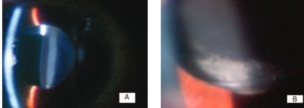
was centred in the capsular bag and there was a large amount of white fluid occupying the space between the IOL and the poste-rior capsule (Figure 1A). The capsulorhexis rim was normal in the superior and temporal regions (the parts that could be observed with the mydriasis obtained). The anterior capsule was fibrotic in this area and was apparently closely adhered to the IOL. The anterior chamber was deep. There were also posterior subcapsular opacities similar to Elschnig spots (Figura1B).
The patient received the diagnosis of CBS and underwent posterior capsulotomy. The fluid disappeared completely, rapidly dispersing through the anterior vitreous, with improvement of visual acuity to 20/70. The condition remained unchanged and this eye underwent corneal transplantation on May 14, 2010. On October 7, 2010 this eye had a corrected VA of 20/40 despite the graft sutures.
Case 2
The patient was a 76-year-old mixed-race female with nu-clear cataract and glaucoma in both eyes; she was using travaprost. On March 26, 2004 she underwent combined phacoemulsification with IOL implantation and trabeculectomy in the LE, without complications. The IOL implanted in the capsular bag was an AR 40 from Advanced Medical Optics made of hydrophobic acrylic and PMMA haptics, with a biconvex 6-mm optic, a total diameter of 13 6-mm, and +22.00 D. The type of viscoelastic used in the procedure or the diameter of the capsulorhexis were not recorded.
In April 2006 the patient underwent needling with subconjunctival injection of mitomycin to recover a failed fistula, with successful restoration of drainage.
No abnormalities were noted until an evaluation on May 25, 2009, but in November 18, 2009, with a corrected VA of 20/ 30, an opacity was noted behind the IOL. This should have been clarified in an immediate follow-up visit for papillary dilation, but that was only done in June 2010, when the VA was already 20/50 with a refraction of -2.00 -1.75, 75°; the patient was using a -1.00 -1.50, 80° lens. The posterior capsule (PC) was distended, with an accumulation of white fluid between the PC and the IOL (Figure 2A); the anterior chamber depth was apparently normal for a pseudophakic eye.
The IOP was 11 mmHg, and there was a well-formed filtration bubble.
A white mass was observed through the iridectomy in the superior periphery of the capsular bag (Figure 2B), as well as a posterior-inferior subcapsular spot-like opacity outside the dynamic pupillary area (Figure 2C). The edge of the anterior capsule had a significant degree of fibrosis and was apparently closely adhered to the IOL; the capsulorhexis diameter was 4.5 mm (Figure 2C).
Three days after YAG capsulotomy the corrected visual
Figure 1:A. Large amount of white fluid trapped behind the intraocular lens. The tortuous aspect of the anterior and posterior line of the fluid is due to the irregular corneal surface; thickened fibrotic anterior capsule; B.
acuity was 20/20 with the old lens.
Similar to the LE, the RE had undergone combined surgery followed by needling, more or less at the same time, but had no particular changes; it also had a filtration bubble and controlled IOP without medication. However, in July 2007 it had undergone posterior capsulotomy.
D
ISCUSSIONMiyake et al.(2) proposed a classification for CBS in three
types, depending on the time of onset: a) Intraoperative CBS: occurring in eyes with posterior capsule rupture and core dislocation blocking the anterior capsular opening after hydrodissection. It occurs more frequently in polar and mature cataracts and eyes with a greater visual axis;(17,18) b) Early
postoperative CBS: usually present on the first day,(4,6,7,9,13,14,18)
but may occur within the first two PO weeks. The seven cases described by Davison(1) were not observed on the first PO day,
only when they returned for a follow-up visit two weeks later; c) Late postoperative CBS: tends to be a chronic process which manifests on average 3.8 years after surgery.
This classification, proposed by Miyake et al.(2) in 1998,
was based on a review of 7 eyes with core dislocation after hydrodissection described by Hashimoto et al (17); 13 early cases
reported by Davison(1) and Holtz(10); and 44 late cases described
by Ota et al.(18), Eifrig(19) and, most importantly, by Miyake et
al.(5). Until a few years before that classification, only the early
type had been recognised; then, in 1996, Robert Drews reported the intraoperative type in the Video Journal of Cataract and Refractive Surgery (cited in Miyake et al.(2)). The late type used
to be considered as a different entity and was called secondary fluid cataract(5,18) or capsulorhexis-related lacteocrumenasia(19).
Many years later, in 2008, Kim et al.(15) proposed a new
classification for PO CBS with three groups, taking into account its pathogenesis, based on the analysis of 8 cases: a) acellular, occurring early on the 1st PO day due to retention of viscoelastic, with little cell reaction or fibrosis between the IOL and the ante-rior capsule; b) inflammatory: cases of early CBS with inflammatory reaction and fibrin; c) fibrotic: cases of late CBS due to other mechanisms, such as metaplasia and proliferation of lens epithelial cells (LECs).
The early type seems to be much more frequent than the late type. In 10 articles published on the syndrome from 1992 to 2001 there were 41 reports of the early type in 7 articles (6,7,9-11,13,14) vs. 4 reports of the late type in 3 articles.(8,12,14) However,
the classification proposed by Miyake et al.(2) was based on a
significant sample of 44 eyes with late CBS reported in only 3
papers.(5,18,19) Of these, 41 had been reported by Miyake et al.(5)
In early CBS, despite 6 reports of IOLs implanted in the capsular bag(1,10) and two reports of IOLs implanted in the
sulcus(7,9) with an apparently normal anterior chamber, the
ante-rior chamber depth is usually reduced.(1,2,5,8,10,11,14) In early CBS
with IOL in the capsular bag, since there is no retraction of the capsular bag and because it has a much larger volume than the IOL, the lens tends to be pushed forward by the fluid that collects behind it, which can lead to myopisation as described by Davison(1)
and Holtz.(10) Therefore, any case of suspected myopic biometric
error in the immediate PO period should be examined with pupil dilation to exclude CBS.(16) The same mechanism can also cause
angle closure with increased IOP.(1,2,5,8,10,14)
By contrast, in the late type this does not usually occur(2,5),
but there tends to backward distension of the PC, as suggested by Theng et al.(14) and verified in the two cases reported here.
Both eyes had a fibrotic ring around the capsulorhexis rim, and although there was a large accumulation of fluid behind the IOL, there was no significant IOL protrusion and the anterior chamber retained a depth compatible with pseudophakia. In only one case was it possible to assess refraction; a myopisation of 1 D was found, and as expected the IOP was normal. This generally occurs because in the late type capsular fibrosis tends to prevent the IOL from being pushed forward, probably not only due to the fibrotic ring which forms around the capsulorhexis rim but also to contraction of the capsular bag as a whole, with consequent tightening of the zonule.
This is generally expected in late CBS with IOL implantation in the capsular bag. However, in cases of IOL implantation in the sulcus, protrusion with myopisation and angle-closure glaucoma has been observed.(8) For the early type, Kim
et al.(15) reported that increased axial length is a risk factor,
mentioning a diameter greater than 25 mm. Although these ca-ses have been reported with different lens types(1,2,6,15,16,20), the
early type is more common in lenses with flexible haptics, as they are more easily pushed forward.(5) In particular, the condition
has been associated with a specific type of IOL, the Akreus Adapt by Bausch & Lomb. It has four points of support, a 6-mm optic made of hydrophilic acrylic, and no haptic angulation, which not only increases contact with the anterior capsule but also interfe-res with viscoelastic aspiration(15). Kim et al.(15) observed six
ca-ses in 206 caca-ses (2.9%) with this IOL, while with other IOLs, according to various authors, the frequency ranges from 0.3 to 1.6%(1,10,15,21,22). Marback et al.(16) reported an even lower
prevalence of 0.02%.
In most reports of the early type the IOL was a single-piece Acrysof MA60BM made hydrophobic acrylic(3,6,11,14,15), but
according to Durack et al. CBS is independent of the type of material. However, such a relationship is unclear in the case of lenses with a biconvex design, which seems to facilitate closure between the capsulorhexis rim and the IOL.(6) According to
Davison(1) the Acry Soft IOL can cause CBS because of its shape,
while PMMA IOLs can cause it because they produce a stronger reaction, with increased fibrosis and adhesion between the ante-rior capsule and the IOL. According to Holtz(10), CBS usually
does not occur with lenses that have positioning holes, as well as smaller lenses and those with firmer angled haptics.
Other factors are the size of the capsulorhexis (small), the type of viscoelastic, and the amount retained in the eye.(2,4,6,13,15)
All reports involved the use of sodium hyaluronate (Healon™ or
Healon GV™), and in four cases an association of sodium
hyaluronate with chondroitin sulphate (Viscoat™) was used.(3,14)
Sodium hyaluronate retained in the capsular bag behind the IOL increases the osmotic gradient, attracting fluid that passes through the lens capsule, which acts as a semipermeable membrane.(10)
This pushes the IOL forward toward the capsulorhexis rim, causing the blockade. This could not happen with viscoelastic of lower molecular weight, which would cause a lower oncotic pressure and would also be able to pass through the lens capsule, not being retained in the capsular bag(10).
However, even in early cases, Davison(1) and Masket(4)
minimise the role of the viscoelastic, emphasising the importance of retained lens materials such as subepithelial cells and their protein products, and possibly cortical remnants. This hypothesis is also cited in the early work of Miyake et al.(2,5)
Due to its pathogenesis, early CBS usually involves clear fluid without inflammatory reaction. However, Mardelli & Mehanna(20) described a case of early CBS with white fluid,
classifying it as a sterile endophthalmitis due to cortex retention behind the IOL, but a with clean vitreous. This case could be included in the inflammatory group in the classification by Kim et al.(15), who reported two cases related to important anterior
chamber inflammation and resolved with anti-inflammatory treatment.
As for late CBS, the accumulated fluid is white and milky, and Miyake et al.(5) suggest it is formed by LEC proliferation
and metaplasia, with production of numerous types of collagen and extracellular matrix which accumulate in the capsular bag. Secondarily, the process could be due to a difference in osmolality between the aqueous humour and the materials accumulated in the “closed chamber”, including residual LECs and their products and cortical remnants, which would attract aqueous humour.(5)
Another possibility described by Nishi et al.(12) is that residual
proliferating LECs become apoptotic and/or necrotic.
Eifrig(19) studied the fluid by electrophoresis and found a
large amount of alpha crystallin a and small amount of albumin, but no gamma globulin, which reinforces the hypothesis that the material is derived from the lens itself and is not due to an antigen-antibody reaction. A similar opaque fluid appears by liquefaction of the lens matrix in hypermature cataracts.(12)
LECs have a key role in the synthesis of lens protein matrix(23) and are found in the anterior capsule remnants in
cataract surgery.(1) They must retain their synthesis ability, at
least in part, in pseudophakic eyes, assuming that their products are constantly exchanged with the aqueous humour.(12) Thus,
according to Davison(1) and Holtz(10), AC adhesion with the IOL,
which under normal circumstances occurs approximately two weeks after surgery, must not be complete, as if that happened the vast majority of cases would suffer capsular block with retention of fluid in the capsular bag. In both eyes of this report there were inferior subcapsular spot-like opacities, and in one of
them a peripheral white mass could be seen through the superi-or iridectomy, resulting from LEC proliferation. These accumulations of proliferating cells could explain further production of the proteins mentioned above. However, in the large series of 41 eyes studied by Miyake et al.(5) capsular spot-like
opacities were described in only 11 eyes, while 12 eyes had fibrotic opacities and one had a Soemmering ring.
The two eyes reported here had IOLs in the capsular bag, which is the condition of most reported cases, both in early and late CBS.(1,2,6,11-15) However, there is one report of late CBS(8)
and three reports of early CBS(6,7,9) with IOLs in the sulcus. Thus,
regardless of the type of IOL implantation, the basic condition for CBS is occlusion of the anterior capsule opening preventing the exchange of fluid between the anterior chamber and the capsular bag.
In case 1 of this report, the phakic contralateral eye had capsular pseudoexfoliation, a condition which has not been associated with CBS. In case 2, the eye with CBS had undergone combined surgery (cataract + IOL implantation + trabeculectomy), similar to the contralateral eye.
The interval between the procedures in both eyes was a few months. Both underwent needling to recover their fistulas, which had suffered failure. However, the eye without CBS had undergone posterior capsulotomy more than two years earlier. All procedures were uneventful. The association of CBS with glaucoma surgery was observed and supported by Muñoz-Negrete & Rebolleda(3), who reported four cases (two
trabeculectomies and two Ahmed valve implants). The authors suggest that these procedures, combined with cataract surgery, must be associated with CBS because the relative reduction of pressure in the anterior chamber can cause IOL displacement toward the anterior capsule. The cases were observed one month, five months, two years and three years after surgery. The authors point to the fact that the cases detected five months and two years after surgery probably had CBS since the immediate postoperative period, stating that the syndrome should always be kept in mind and should be included in the differential diagnosis of shallow anterior chamber after combined cataract and glaucoma surgery. Two other cases of cataract + glaucoma surgery were reported by Sorenson et al.(24) and McQuenn &
Margo(25): one with trabeculectomy and the other with Ahmed
valve implantation. In their large series with 41 eyes, Miyake et al.(5) make a correlation between CBS and glaucoma (four
ca-ses), but also with other conditions that alter the blood-ocular barrier, such as diabetic retinopathy and uveitis (14 cases and 2 cases, respectively).
As to the time of onset of late CBS, it occurs on average 3.8 years(2,5) after cataract surgery, with reports ranging from 2
months to 6 years.(3,5,8,12,14,15) According to Nishi et al.(12), fluid
accumulation in late CBS slowly causes visual blurring after 5-7 years. Theng et al.(14) also believe that late CBS is subclinical in
its early stages and is only detected later when PC opacification and impaired VA occur.
In this report, one case manifested more five years and the other nine years after cataract surgery. However, contrary to what is said by Theng et al.(14), in both cases VA reduction
could not be explained by capsular opacity, because it was outside the visual axis. Therefore, the VA impairment of four lines in Snellen’s chart was related to the accumulation of large amounts of white fluid, which is consistent with the statement by Miyake et al.(5) that significant reductions in visual acuity related solely
Regarding the therapeutic approach, in early CBS expectant management can be adopted at first, since spontaneous resolution is relatively common, being reported within 2 weeks to 2 months(1,6,7,9,10). In an early case followed by Durack et al.(6)
that received no treatment for 11 months, the patient remained stable throughout the period and was then treated by posterior capsulotomy. When spontaneous resolution does not occur, ante-rior capsulotomy can be used(1,6). When this is not effective or
cannot be done because the pupil does not dilate, posterior capsulotomy is an alternative.(6,10,11) Invasive treatments have
been performed such as slit lamp needling(20) and aspiration of
material retained behind the IOL by irrigation/aspiration.(16,20)
For late CBS, there are only 3 reports of spontaneous resolution.(5) The generally indicated procedure is posterior
capsulotomy(6,8,12,14) because it promotes greater IOL stability in
the capsular bag and has a lower risk of complications. There is only one report of a case that did not resolve with the posterior YAG capsulotomy, where the patient was submitted to surgical capsulectomy with posterior vitrectomy.(3)
In both cases reported here, a minimal opening was done in the PC, because there was no capsular opacity corresponding to the dynamic pupil area. It was also carried out with a very low power, aiming only to release the fluid and trying to prevent this initial opening from pushing the PC due to the unknown pressure of the large amount of fluid, causing a large rupture and jeopardising IOL support and maintenance of the vitreous in its place. Contrary to what was pointed out by Muñoz-Negrete & Rebolleda,(3) despite the large amount of white fluid trapped
behind the IOL we had no difficulty reaching the PC, which was achieved with a single laser shot. Also, as expected,(5) there was
no inflammatory reaction in the eye as a result of fluid extravasation into the anterior vitreous.
R
EFERENCES1- Davison JA. Capsular bag distension after endophacoemulsification and posterior chamber intraocular lens implantation. J Cataract Re-fract Surg. 1990;16(1):99-108.
2- Miyake K, Otta I, Ichihashi S, Miyake S, Tanaka Y, Terasaki H. New classification of capsular block syndrome. J Cataract Refract Surg. 1998;24(9):1230-4. Comment in J Cataract Refract Surg. 2001;27(7):966.
3- Munõz-Negrete FJ, Rebolleda G. Capsular bag distension syndrome after combined cataract and glaucoma surgery. Acta Opthalmol Scand. 2005;83(2):252-5.
4- Masket S. Postoperative complications of capsulorhexis. J Cataract Refract Surg. 1993;19(6):721-4. Review.
5- Miyake K, Ota I, Miyake S, Horigushi M. Liquefied after cataract: a complication of continuous curvilinear capsulorhexis and intraocular lens implantation in the lens capsule. Am J Ophthalmol. 1998;125(4):429-35.
6- Durack I, Ozbeck Z, Ferliel ST, Oner FH, Söylev M. Early postoperative capsular block syndrome. J Cataract Refract Surg. 2001;27(4):555-9. 7- Basti S, Nayak H, Mathur U. Capsular bag distension after optic
cap-ture of a sulcus-fixated intraocular lens. J Cataract Refract Surg. 1999;25(2):293-5.
8- Liu TY, Chou PI. Capsular block syndrome associated with secondary angle-closure glaucoma. J Cataract Refract Surg 2001;27(9):1503-5. 9- Miyake K, Ota I, Miyake S, Terasaki H. Capsular block syndrome with external blockage of the capsular opening by a ciliary sulcus fixated posterior chamber lens. Am J Ophthalmol. 1999;127(5):605-7. 10- Holtz SJ. Postoperative capsular bag distension. J Cataract Refract
Surg. 1992;18(3):310-7. Comment in J Cataract Refract Surg. 1992;18(5):537.
11- Omar O, Eng CT, Chang A, Durcan FJ, Liss RP, Stark BI. Capsular bag distension with an acrylic intraocular lens. J Cataract Refract Surg. 1996;22 Suppl 2:1365-7.
12- Nishi O, Nishi K, Takahashi E. Capsular bag distension syndrome noted 5 years after intraocular lens implantation. Am J Ophthalmol. 1998;125(4):545-7. Comment in Am J Ophthalmol. 1999;127(2):244-5. 13- Sugiura T, Miyauchi S, Eguchi S, Obata H, Nanba H, Fujino Y, et al. Analy-sis of liquid accumulated in the distended capsular bag in early postop-erative capsular block syndrome. J Cataract Refract Surg. 2000;26(3):420-5. Comment in J Cataract Refract Surg. 2001;27(2):177-8.
14- Theng JT, Jap A, Chee SP. Capsular block syndrome: a case series. J Cataract Refract Surg. 2000;26(3):462-7. Comment in J Cataract Re-fract Surg. 2001;27(7):966. J Cataract ReRe-fract Surg. 2001;27(2):177-8. 15- Kim HK, Shin JP. Capsular block syndrome after cataract surgery: clinical analysis and classification. J Cataract Refract Surg. 2008;34(3):357-63.
16- Marback EF, Araújo RCA, Freitas MS. Síndrome do bloqueio capsular precoce relato de caso e especulação sobre o tamanho da parte óptica da lente como fator de risco. Rev Bras Oftalmol. 2009;68(6):344-7. 17- Hashimoto T, Izutani M, Tanaka Y, Tanaka Y. [Two cases of posterior
capsular rupture and dislocation of lens nucleus following hydrodissection]. Folia Ophthalmol Jpn. 1994;45:973-6.
18- Ota I, Miyake S, Miyake K. Dislocation of the lens nucleus into the vitreous cavity after standard hydrodissection. Am J Ophthalmol. 1996;121(6):706-8.
19- Eifrig DE. Capsulorhexis-related lacteocrumenasia. J Cataract Re-fract Surg. 1997;23(3):450-4.
20- Mardelli PG, Mehanna CJ. Phacoanaphylactic endophthalmitis sec-ondary to capsular block syndrome. J Catarct Refract Surg. 2007;33(5):921-2.
21- Agrawal S, Agrawal J, Agrawal TP. Complete capsular bag distension syndrome. J Cataract Refract Surg. 2000;26(9):1417-8.
22- Yepez JB, de Yepez JC, Arevalo JF. Intraoperative peripheral ante-rior capsulotomy to prevent early postoperative capsular block syn-drome. J Cataract Refract Surg. 2001;27(2):177-8.
23- Dantas AM. Fisiologia do cristalino e zônula. In: Conselho Brasileiro de Oftalmologia. Bases da oftalmologia. Rio de Janeiro: Guanabara-Koogan; 2008. Vol II. p. 827-69.
24- Sorenson AL, Holladay JT, Kim T, Kendall CJ, Carlson AN. Ultrasonographic measurement of induced myopia associated with capsular bag distention syndrome. Ophthalmology. 2000;107(5):902-8. Comment in Ophthalmology. 2001;108(9):1517.
25. McQueen BR, Margo CE. Capsular bag distention syndrome after combined cataract-lens implant surgery and Ahmed valve implanta-tion. Am J Ophthalm
Corresponding author:
Karina Ameno Cautela
Rua Dom Pedro II, 60, apto. 601 - Praia do Canto CEP 29055-600, Vitória/ES, Brazil