232
Therapeutical evaluation of bevacizumab
application in relapsed pterygium
Avaliação terapêutica da aplicação de bevacizumabe
em pterígio recidivado
Mayara Martins Abrahão¹, Leonardo Pinheiro Teixeira¹, Denise Borges de Andrade Mendanha¹, Luana Miranda Campos¹,
Mateus Martins Cortez Vilar¹, João Jorge Nassaralla Junior²
A
BSTRACTObjective: Therapeutic evaluation of Bevacizumab application in relapsed pterygium concerning visual acuity, keratometry, refrac-tion, symptomatology. Methods: Group 1 (4 patients) received 0.1 ml of Bevacizumab (avastin), being evaluated posteriorly on the tenth and thirtieth days after the application, seeking to compare with the exam previously made, being it realized with the other two groups, in which Group 2 (4 patients) received 0.2 ml of Bevacizumab and the Group 3 (3 patients) received 1 ml of the placebo injection. Results: In this study, eleven eyes of eleven patients were evaluated. Among these patients, 7 were women (63.6%) and 4 men (36.4%). There was a variation in the cylindrical diopter after the treatment with a dose of 0.1 ml of bevaciumab during the evaluation on the thirtieth day. Whereas the cylindrical shaft had a significantly larger modification after the application of 0.2 ml. Regarding the spherical diopter variation, there were modifications in the 3 groups. The keratometry varied in the 3 groups, mostly after the thirtieth day of evaluation. In relation to symptomatology, it was observed a reduction in the subjective evaluation of the eye burning sensation, the prurience mentioned by the patient and a reduction of the hyperemia biomicroscopy evaluation. Conclusion:
In bevacizumab application in the recurrent pterygium treatment, there is modification of the spherical and cylindrical parameters of refraction, besides the changes in keratometry and the reduction of the symptomatology.
Keywords: Pterygium/drug therapy; Bevacizumab/therapeutic use; Visual acuity; Refraction
R
ESUMOObjetivo: Avaliação terapêutica da aplicação de bevacizumabe em pterígio recidivado com relação a acuidade visual, ceratometria, refração, sintomatologia. Métodos: O Grupo 1 (4 pacientes) recebeu 0,1ml de bevacizumabe (avastin) sendo avaliado posteriormente, nos dias 10 e 30 após a aplicação, buscando-se comparar com o exame previamente realizado, sendo o mesmo realizado com os outros dois grupos, em que o Grupo 2 (04 pacientes) recebeu 0,2ml de bevacizumabe e o grupo 3 (3 pacientes) recebeu 0,1 de injeção placebo. Resultados: Neste estudo foram avaliados 11 olhos de 11 pacientes. Dentre esses pacientes, 7 (63,6%) eram mulheres e 4 (36,4%) ho-mens. Houve a variação na dioptria cilíndrica após o tratamento com dose de 0,1ml de bevacizumabe, durante a avaliação no trigésimo dia. Já o eixo cilíndrico teve uma modificação significativamente maior após a aplicação de 0,2ml. Em relação a variação dióptrica esférica, houve modificações nos três grupos. A ceratometria variou nos três grupos, principalmente no trigésimo dia de avaliação. Em relação a sintomatologia, observou-se uma redução na avaliação subjetiva da ardência, do prurido referida pelo paciente, e uma redução na avaliação biomicroscópica da hiperemia. Conclusão: Na aplicação do bevacizumabe no tratamento de pterígio recorren-te, há modificação dos parâmetros esféricos e cilíndricos da refração, além da mudança ceratométrica e redução da sintomatologia.
Descritores: Pterígio/tratamento farmacológico; Bevacizumabe/uso terapêutico; Acuidade visual; Refração.
1Instituto de Olhos de Goiânia, Goiânia, GO, Brazil.
2Setor de Retina e Vítreo, Instituto de Olhos de Goiânia, Goiânia, GO, Brazil.
Recebido para publicação em 01/02/2017 - Aceito para publicação em 20/09/2017.
Os autores declaram não haver conflito de interesses.
A
RTIGOO
RIGINALRev Bras Oftalmol. 2017;76 (5): 232-4
233
I
NTRODUCTIONP
terygium is a growth in the cornea, usually nasal, of con-tinuous fibrovascular tissue to the conjuctiva. It occurs in the area of palpebral fissure, more frequently in the nasal quadrant than in the temporal quadrant, although either one or the other, or both (“double” Pterygium) occur. High whitish opacities (“Vogt Islands”) and an iron deposit line (“Stocker”) can delineate the pterygium’s head in the córnea. (1)Pterygium consists of degenerative collagenous changes in the vascularized subepithelial stroma. (2) It is associated with
ultra-violet exposure. It is more prevalent and more severe in tropical áreas near the equator and less frequent and milder in cooler climates. Outdoor work and situations with high light reflectivity, including from sand and water, intensify the pterygium develop-ment, and using sunglasses and hats is a protective measure. (1-11)
Although pyterigium is classified as a degenerative desease, nowadays there are diferent scientific thinking strands that consider it a proliferative disease.(3) These diferences have been appearing due
to recent discoveries of several factors related to its pathogenesis, such as: cornea invasion and invasion of the subconjunctival tissue by fibroblast, conjuctival expression of tumor suppressor gene p53 and presence of angiogenic factors and immunosuppresion. Thus, its exact etiopathogeny remains uncertain.
Many studies have shown that VEGF in pterygium pathogene-sis is increasing. There is a variety of options for the pterygium treat-ment. The VEGF expression in the pterygium’s tissue lead us to think that antiangiogenic / anti-VEGF therapy could induce the blood vassels regression and, consequently, slow pterygium progression. (4)
Bevacizumab is a whole humanized monoclonal antibody that binds and inhibits all biologically active forms of VEGF-A. FDA aproved bevacizumab in 2004 for the metastic colorectal cancer systemic treatment (intravenous). Studies showed that intravenous administration could reduce the leakage by the NVC, improve vision significantly and reduce the retina center thikness. (1-12)
Hence, in this study, the bevacizumab application in relapsed pterygium was realized seeking to observe the signal and symp-toms modification, since only surgical treatments have a reduced efficiency. Such study, for being innovator, serves as basis for the achievement of bigger and more complex studies.
M
ETHODSThis study is randomized clinical case, realized in the (Goiania Eye Institute – Instituto de Olhos de Goiânia), between August 2015 and November 2015, in patients with relapsed pterygium, considering these the fibrovascular tissue growing above the limbo in an area where the excision was previously realized.
Initially, patients with relapsed pterygium were randomly selected, they were attended in the Goiania Eye Institute, in wich the surgical excision was indicated. Initially, they were collected visual accuity, keratometry, refraction, and subjective symptoma-tology, looking for lacrimation, eye burning, itching, photophobia, among others. These patients were allocated, posteriorly, into three groups with the same amount of patients, that received random and different medications.
Group 1 (4 patients) received 0.1 ml of bevacizumab (avas-tin), being evaluated posteriorly on the tenth and thirtieth days after the application, seeking to compare with the exam previously made, being it realized with the other two groups, in which Group 2 (4 patients) received 0.2 ml of Bevacizumab and the Group 3 (3 patients) received 1 ml of the placebo injection.
The injection applications were realized in the prelimbal area, next to the subconjuctival vessels of greater caliber, and all of the patients were treated with tobramycin every four hours.
Data were observed and translated into numbers cataloged in Microsoft Excel 2010 spreadsheets in wich the researchers themselves realized the statistical study. For the statistical analysis of the variables in this study, the statistical program Statistical Package for the Social Sciences (SPSS) 18.0 version was used, wich has resources to analyze data, create graphics and make fast and reliable predictions. In data obtaining, it was considered statistically significnt p less than 0.5 to greater reliability of results. The research was based on the Terms of Resolution 196/96 of the Brazilian National Health Council and it started only after the by the Research Ethics Committe of the Goiania Eye Institute, to ensure all the involved basic principles of bioethics, ie autonomy, non-maleficence, benevolence and justice.
R
ESULTSIn this study, eleven eyes of eleven patients diagnosed with recurrent pterygium attended in Goiania Eye Institute were evaluated. Among these patients, 7 (63.6%) were women and 4 (36.4%) were men, with ages ranging 55-72 years, the average age was 64 years, all coming from Goiania (Brazil).
Regarding the observed signs, first we highlight the varia-tion in the cylindrical diopter, in which we can contrast a larger change (0.1875 diopters, after treatment with 0.1 ml bevacizumab dose during the evaluation on the thirtieth day, since neither the dose of 0.2 ml of the anti-VEGF nor the placebo didn’t show any diopter change (Figure 1).
The cylindrical axis had a significantly larger modification after the application of 0.2 ml (17.5 degrees) compared with the evaluation after the first 10 days of the groups that received 0.1 ml of bevacizumab and 0.1 ml of placebo (Figure 2).
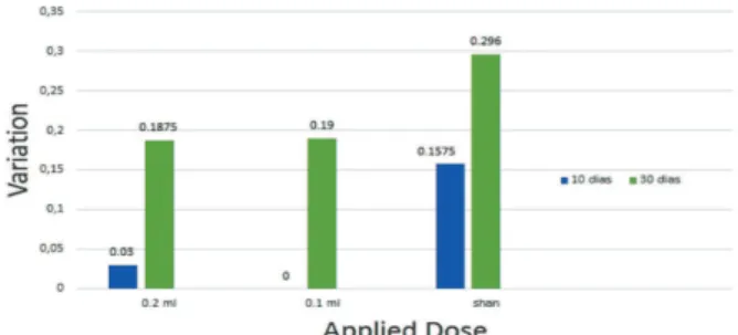
Regarding spherical diopter change, it was possible to ob-serve that there were changes in the three groups (0.6875, 0.083 and 0.1875 for Groups 1, 2 and 3, respectively), with no variation between the tenth day and the thirtieth day evaluations, highlighting a significantly larger alteration after applying 0.2 ml of anti-VEGF. Given the many evaluations, it was found that even though the keratometry varied in the three groups, mainly on the thirtieth day of evaluation,with modifications that vary between 0.1875 and 0.296, and the placebo group had a tendency to greater variation (Figure 3).
Rev Bras Oftalmol. 2017; 76 (5): 232-4 Therapeutical evaluation of bevacizumab application in relapsed pterygium
Figure 1: Variation of Cylindrical Diopter After Application of Bevacizumab
234
In relation to symptomatology, among the most mentio-ned signals, we have eye burning, itching and hyperemia. There were reductions in the subjective eye burning evaluation, itching reported by the patient and in the biomicroscopy evaluation of hyperemia. Since no hyperemia was observed after the first ten days in eight out of the eight patients undergoing anti-VEGF injection, and, on the other hand, all the three patients submitted to placebo continued with hyperemia.
D
ISCUSSIONThis study sought to prospectively demonstrate the effect of the treatment of relapsed pterygium with bevacizumab through a randomized clinical essay. Several references metion discove-ries that stood out in the last two decades, with the unveiling of a veriety of angiogenic modulators that may be related to the pathogenesis of pterygium, since it is formed predominantly by fibrovascular tissue. Thus, it becomes important to carry out studies demonstrating the anti-VEGF action not only in reducing the size of pterygium, but also in the modificaition of the signs and symptoms outlined in the ophtalmologic examination.
The most prevalent topographical change is symmetrical with-the-rule astigmatism, caused by the flattening of the cornea towards the lesion(6,7,8). In figures 1 and 2, we see a change in
corneal topography through the diopters and cylinder axis mo-dification, in wich after evaluation on the subsequent tenth and thirtieth days after the application, was observed a cylindrical axis alteration of about 17.5 degrees, after tem days of application, with a further reduction of this modification to 6.65 degrees, showing that bevacizumab application leads to a change of this axis. Such fact could be related to the size reduction (grade) of pterygium after applying this medication that showed that there was a re-duction of about 2 mm in the pterygium size after two months of anti-VEGF application.(9) The modifications in those parameters,
however, were not able to interfere with visual accuity of patients who proved to be equal in post-implementation evaluations.
Several studies show that pterygium manipulation may be responsible for modifying the refractive power of the cornea with astigmatism induction and modification of topographical regula-rity of it. (12-15) Figure 3 shows a marked change in keratometry
with placebo application. This change may have occured due to this manipulation, making it dubious, if keratometric alterations found after the medication application occured because of the manipulation or if there was the action of anti-VEGF with the reduction of the existing irregularities. This fact opens the door to studies that relate to the size of pterygium reduction with the modification of clinical parameters. However, it does not invali-date our study that was able to demonstrate significant variations in signs and symptoms related to this disease, therefore, leaving it to further correlation.
The application of bevacizumab also proved to be effective in reducing irritative symptoms (72.72%), corroborating with existing data in the literature that show a reduction of over 90% in irritative symptoms after treatment.
C
ONCLUSIONGiven the above, we can state that in the application of bevacizumab in the treatment of recurrent pterygium there is modification in the spherical and cylindrical parameters of re-fraction, besides keratometry alteration. However, such changes require studies correlating not only changes, but also quantifying the consequent improvement or worsening of this therapy.
Regarding biomicroscopic signs, not with standing visual acuity that showed no changes, there was a reduction of hype-remia, itching and photophobia within thirty days of treatment.
R
EFERENCES1. Yanoff M, Duker JS. Oftalmologia. 3a ed. Rio de Janeiro: Elsevier; 2011. 2. Kanski JJ. Oftalmologia clínica: uma abordagem sistemática. 6a ed.
Rio de Janeiro: Elsevier; 2008. p. 242-4.
3. Bradley JC, Yang W, Bradley RH, Reid TW, Schwab IR. The science of pterygia. Br J Ophthalmol. 2010;94(7):815-20.
4. Mauro J, Foster CS. Pterygia: pathogenesis and the role of subconjunc-tival bevacizumab in treatment. Semin Ophthalmol. 2009;24(3):130-4. 5. Hosseini H, Nejabat M, Khalili MR. Bevacizumab (Avastin) as a
potentialnovel adjunct in the management of pterygia. Med Hypo-theses. 2007;69(4):925-7.
6. Stern GA, Lin A. Effect of pterygium excision on induced corneal topographic abnormalities. Cornea. 1998; 17(1):23-7.
7. Tranjan Neto A, Alves MR, José NK. Alterações topográficas cornea-nas desencadeadas pelo pterígio. Arq Bras Oftalmol. 1996;59(5):443-8. 8. Almeida Jr GC, Ribeiro CS, Xavier JS, Paiva GP, Kashiwabuchi LK. Videoceratografia antes e após a cirurgia de pterígio. Arq Bras Oftalmol. 1999;62(3):244-52.
9. Stival LS, Lago AM, Figueiredo MN, Bittar RH, Machado ML, Nassa-ralla Junior JJ.Efficacy and safety of subconjunctival bevacizumab for recurrent pterygium. Arq Bras Oftalmol. 2014; 77(1):4-7.
10. Hurwitz H, Fehrenbacher L, Novotny W, Cartwright T, Hainsworth J, Heim W, et al. Bevacizumab plus irinotecan, fluorouracil, and leucovorin for metastatic colorectal cancer. N Engl J Med. 2004;350(23):2335-42. Comment in: N Engl J Med. 2004;350(23):2406-8; Nat Clin Pract Gas-troenterol Hepatol. 2004;1(2):72-3; Cancer Treat Rev. 2004;30(8):715-7; N Engl J Med. 2004;351(16):1690-1; author reply 1690-1.
11. Hill JC, Maske R. Pathogenesis of pterygium. Eye (Lond). 1989;3(Pt 2):218-26. 12. Morrow GL, Stein RM. Evaluation of corneal topography: past, present and future trends. Can J Ophthalmol.1992; 27(5):213-25. 13. Vadas MG, Monteiro MR, Gomes JÁ. Mudanças na refração após
cirurgia de correção de esotropia. Arq Bras Oftalmol. 2011;64(4):315-23. 14. Budak K, Khater TT, Friedman NJ, Koch DD. Corneal topographic changes induced by excision of perilimbal lesions. Ophthalmic Surg Lasers. 1999;30(6):458-64.
15. Adamis AP, Starck T, Kenyon KR. The management of pterygium. Ophthalmol Clin North Am. 1990;3(4):611-23.
16. Tomidokoro A, Oshika T, Amano S, Eguchi K, Eguchi S. Quantitative analysis of regular and irregular astigmatism induced by pterygium. Cornea. 1999;18(4):412-5.
17. Tomidokoro A, Miyata K, Sakaguchi Y, Samejima T, Tokunaga T, Oshika T. Effects of pterygium on corneal spherical power and astigmatism. Ophthalmology. 2000;107(8):1568-71.
Rev Bras Oftalmol. 2017; 76 (5): 232-4
Abrahão MM, Teixeira LP, Mendanha DBA, Campos LM, Vilar MMC, Nassaralla Junior JJ.
Figure 3: Variation of Keratometry After the Application of Beva-cizumab
Corresponding Author:
Mayara Martins Abrahão
Instituto de Olhos de Goiânia, Goiânia, GO, Brazil. Rua 9B, nº 48, St. Oeste, Goiânia–GO, 74110-120