Abstract
Submitted: November 28, 2016 0RGL¿FDWLRQ$SULO Accepted: April 19, 2017
casein phosphopeptide-amorphous
calcium phosphate cream with and
demineralization in a pH cyclic study
Casein phosphopeptide-amorphous calcium phosphate (CPP-ACP) complexes are anticariogenic and capable of remineralizing the early
To evaluate the effect of CPP-ACP and CPP-ACPF creams associated with a
Material and Methods: Previously selected by surface microhardness (SH) analysis, human enamel blocks (n=56) were submitted to daily treatment with dentifrice in a pH-cycling model. The enamel blocks were divided into four groups; G1: CrestTM Cavity Protection – Procter & Gamble (1,100 ppmF of NaF); G2: CrestTM +MI Paste (MP) – RecaldentTM GC Corporation Tokyo, Japan); G3: CrestTM + MI Paste Plus (MPP) – RecaldentTM 900 ppm as NaF, GC Corporation Tokyo, Japan), and G4: control, saliva. Specimens were soaked
60 s after the remineralization period. The undiluted MP and MPP creams were applied for 3 m/d. After cycling, SH was re-measured and cross section microhardness measurements were taken. Results: The SH values observed for the groups G3 (257±70), G1 (205±70), and G2 (208±84) differed from the G4 group (98±110) (one-way ANOVA and Tukey’s post hoc test). There were no differences between the groups G1xG2, G2xG3, and G1xG3 for demineralization inhibition. The percentage of volume mineral showed
(Kruskal-Wallis and Mann Whitney p<0.05). Conclusion: Fluoride dentifrice associated with CPP-ACPF inhibited subsurface enamel demineralization.
Ke yw or ds:
Priscila de Pinto SINFITELI1
Thereza Christina Lopes COUTINHO2
Patrícia Regina Almeida de OLIVEIRA1
Wesley Felisberto VASQUES1
Leandra Matos AZEVEDO1
André Maues Brabo PEREIRA3
Monica Almeida TOSTES2
1Universidade Federal Fluminense, Faculdade de Odontologia, Niterói, RJ, Brasil.
2Universidade Federal Fluminense, Faculdade de Odontologia, Departamento de Odontopediatria,
Niterói, RJ, Brasil.
3Universidade Federal Fluminense, Faculdade de Engenharia, Niterói, RJ, Brasil.
Corresponding address: Monica Almeida Tostes Universidade Federal Fluminense, Faculdade de Odontologia. Rua Mário Santos Braga, no 30 - Campus Valonguinho, Centro - Niterói - RJ - Brazil - 24040-110
Introduction
The complex casein phospho-peptide-amorphous calcium phosphate (CPP-ACP) comes from a milk protein called casein. CPP-ACP represents an alternative-remineralizing agent, capable of stabilizing calcium phosphate, maintaining the supersaturation of these ions in the oral environment. Amorphous calcium phosphate (ACP) favors the proximity between calcium and phosphate ions in an amorphous phase. They can enhance remineralization, decrease demineralization or even both in an acid challenge to teeth surfaces4,17,18.
I n v it r o1,5,7,10,12-14,16,17,23 and in v iv o3,19,20 studies
have been carried out to understand the association
is little knowledge on whether this combination improves remineralization. Furthermore, some studies have shown that there was no additional
caries when used as a supplement to the regular
10,16. Meyer-Lueckel, et al.11 (2015)
compared the remineralizing effects induced by the application of casein phosphopeptide-stabilized amorphous calcium phosphate complexes (CPP-ACP
in sit u model. They concluded that the additional use of a CPP-ACP containing cream
toothpaste. In contrast, Reynolds, et al.18 (2008),
also using an in sit u caries model, showed that the
F dentifrice when compared to that containing 1,100 ppm F alone. In addition, other authors have shown that there was a higher increase in remineralization and a decrease in lesion depth when combining the two12.
Considering there are few reports on the
dentifrice, it seems relevant to investigate its performance regarding the enamel surface and remineralization throughout thelesion.
The surface hardness test (SH) used in some
studies has shown to be sensitive enough to detect the early stages of enamel demineralization, occurring at the enamel surface, but not able to measure lesion depth1,8,14,22,23. Thus, we used cross section hardness
(CSH) to evaluate lesion depth. The technique is well
established for enamel and can be used with enamel
block lesions2,6,9.
The aim of this study is to evaluate the effect of
remineralization, using SH and CSH. To do so, we tested the null hypothesis that, in a pH cycling model
dentifrice, MI Paste (MP), and MI Paste Plus (MPP),
Material and Methods
Sample preparation
In this study, 20 human third molars, which had been extracted for surgical reasons, were used. The study was approved by the Ethical Committee of Medical Science, Fluminense Federal University (Protocol number 393000/13). The sample size was performed assuming 80% of power to detect the
level (http://www.openepi.com). A minimum of 12 was
or hypomineralized lesions and any other visible defects. The teeth were stored in thymol 0.1% during the sample preparation process. A total of 80 enamel blocks were obtained from the buccal and lingual surfaces of 20 third human molars. After embedding the blocks in acrylic resin, the buccal surfaces of the enamel specimens (2x2x2 mm) were ground with SiC paper (400, 600 and 1,200 grits) (Struers
the specimens were polished using a 1 μm diamond polishing suspension with a polishing cloth (Arotec Ind. & Com., Cotia, SP, Brazil). The baseline surface microhardness (SH) of all specimens was measured using a microhardness tester (Micromet 2001, Buehler, IL, USA) with a Knoop diamond indenter under a 50 g
were made and the mean values of enamel surface hardness were calculated (SH baseline=SH0). After SH measurements, 56 enamel blocks were selected with a Knoop hardness number (KHN) ranging from 275.72 to 369.72. The enamel blocks were distributed into 4 groups of 14 blocks each: Group 1 - Crest TM
RecaldentTM (GC Corporation Tokyo, Japan); Group
3 - CrestTM + MI Paste Plus (MPP) - RecaldentTM 900
ppm as NaF (GC Corporation Tokyo, Japan); and Group 4 - saliva, used for negative control.
pH cycling and treatment with dentifrices
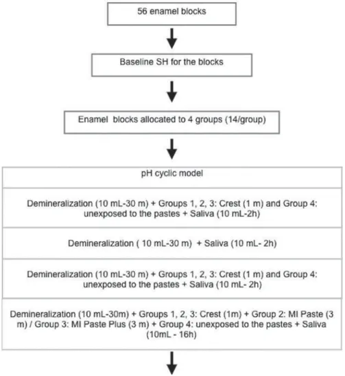
The enamel blocks were submitted to pH cycles
for 5 d, at 37°C. After sonication and the rinsing with distilled water, the specimens were immersed separately in 10 mL of demineralizing solution [2 Mm
Ca (Ca (NO3)2, 2 Mm PO4 (KH2 PO4) and 75 Mm of
acetate at 4.3 pH]22 according to the protocol in Figure
0.67 g/L NaCl; 0.1168 g/L CaCl2; 8 g/L CMC; 0.0408
g/L MgCl2; 0.96 g/KCL; 1 g/L C8H8O3; 24 g/L C6H14O6;
964,938 ml/L H2O; 0.274 g/L KH2 PO414.
At each transfer between different solutions, the enamel specimens were rinsed in distilled water for 1 m at 37°C. The treatment was applied in slurry in a proportion of 1:3 of deionized water for 60 s, three times a day, in the groups. A standardized volume (0.15 mL) was applied in each sample. The
MP (Group 2) and MPP (Group 3) formulas were used undiluted (0.03 g) for 3 m/d. Negative control (Group 4) remained unexposed to the pastes. The
second and the third demineralization cycles. After the last demineralization challenge, the enamel specimens were rinsed in distilled water for 1 m, and then
were changed every day (Figure 1).
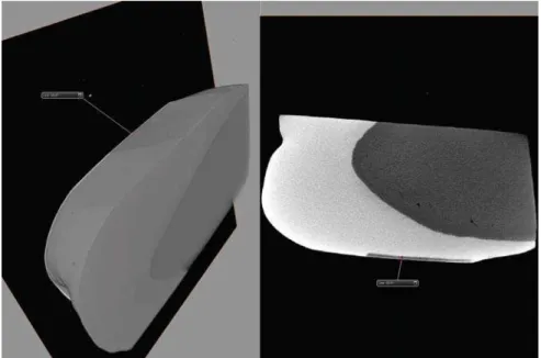
The lesion depth value formed with this protocol was tested previously. The three enamel blocks were submitted to pH cycles as described for control group.
After, the blocks were scanned with micro-CT(ZEISS Xradia 510 Versa). Three-D image analysis software (AVIZO) was used for visualization. The formed lesion depth was of 59.81 (Figure 2).
Surface hardness
(SH)
Pre and post-treatment measurements (SH0 and SH1, respectively) were conducted with the same static load and time used in baseline measurements. Five indentations with a space of 100μm from the baseline
indentations were made with a Knoop diamond
indenter under a 50 g load for 15 s. The percentage change of SH (%SH) was calculated [% SH = 100 (SH1 – SH0)/SH1].
Cross section hardness (CSH)
To perform CSH tests, the samples were sectioned
perpendicularly to the surface through the center. One
half of each sample was embedded in acrylic resin
indentations each were made, one at the center of the
exposed dental enamel and another one at a distance of 100 μm from the central row of indentations using a 25-gram load for 15 s. The indentations were made
at 25, 50, 75, 90, and 150 μm from the outer enamel surface. The mean values of the two measuring points were calculated at each distance from the surface. The Knoop microhardness number was converted to % volume mineral vs. depth, following the methods described by Featherstone, et al.6 (1983). Thus, this
formula was used in this study
to evaluate the change depth of enamel6.
Statistical analysis
The data were analyzed using Statgraphics Centurion XVI software (STATPOINT Technologies, Inc, USA). Initially, all the data (SH0, SH1) were checked by Shapiro-Wilk’s test and Levene’s test. Based on these preliminary analyses, the SH0 and SH1 data were submitted to the one-way analysis of variance and the Tukey’s HSD post hoc test. The % volume mineral data were analyzed by Kruskal Wallis and Mann-Whitney
by Graphpad
prism 6.0 (GraphPad Software, Inc, USA).
Results
From the initial 56 sections in this study, after pH-cycling we rejected three of them because of extensive loss of the external surface of the lesion, which made
surface microhardness analysis of all groups is shown descriptively in Table 1. The specimens showed a
(p<0.05). One-way ANOVA and Tukey’s HSD post hoc
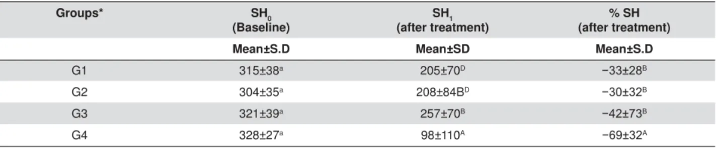
mean of enamel SH between the groups (p<0.05). The SH values observed for G3 (257±70), G2 (208±84) and G1 (205±70) groups differed from G4 (98±110). When comparing the G1 experimental group, the combined G2 and G3 treatment groups, we found
(p<0.05). On the other hand, the combined treatment groups, both containing CPP-ACP, were statistically
There were no differences between G1xG2, G2xG3 and G1xG3 for demineralization inhibition (p>0.05).
and distance factors, which indicates that the effect of the treatments was different depending on the depth of the enamel surface.
The percentage of volume mineral vs. depth of the
surface for all groups is shown in Figure 3. Percentage
compared to the groups G1 and G4 (only at 25 and
μ
other distances from the surface (p>0.05).
Discussion
The objective of this study was to evaluate the
when associated with regular fluoride dentifrice on demineralized enamel using surface hardness and cross section hardness in a pH-cycling model, simulating oral cavity conditions. When carried out this way, the hardness measurement is very sensitive to changes in mineral density2. We converted the
indentation lengths to % volume mineral according to Featherstone, et al.6 (1983).
In the cyclic model used in this study, we changed demineralization and remineralization solutions after each cycle, so that the concentration of calcium and phosphate ions in the solutions would not affect the
results. The sub-saturation condition can lead to the dissolution of hydroxyapatite and diffusion of calcium and phosphate ions towards the enamel surface, reducing the SH after pH cycling24. In addition,
we rinsed enamel surfaces after treatment with dentifrices, so that any treatment effect would be due to the binding of active ingredients to enamel22.
under these conditions and showed a significant difference when treatment was employed (Table 1).
When comparing G1 (FD 1,100 ppm F) to the combined groups (G2 and G3), we found no statistically
p<0.05). On the other hand, the combined treatment group and the
saliva group (p<0.05). Presumably, the concentration
enamel during exposure to acid challenge. Additionally, the specimens of the control group were exposed to
could decrease the remineralizing potential in the specimens of this group. Thus, this may explain the dose response of all groups.
Groups* SH0
(Baseline)
SH1 (after treatment)
% SH (after treatment)
Mean±S.D Mean±SD Mean±S.D
G1 315±38a 205±70D íB
G2 304±35a 208±84BD íB
G3 321±39a 257±70B íB
G4 328±27a 98±110A íA
'LIIHUHQWXSSHUDQGORZHUFDVHVXSHUVFULSWOHWWHUVLQGLFDWHVLJQL¿FDQWGLIIHUHQFHEHWZHHQWKHWHVWHGJURXSVDWS:HXVHORZHU
case superscript letters to compare the values of the same row and upper case letters to compare the values of each column.
*Q DQG**DQG*Q
Table 1- Surface microhardness results (mean±SD) of human enamel specimens according to different groups
We expected that the MPP cream comprising 900
show a superior effect. However, the effect of treatment with MPP and FD was similar to the one only with FD. It is worth to highlight that we applied MPP and MP as a top coating, which could show a lower penetration to enamel surface compared to the NaF solution7. In
reactivity are larger. These two aspects could have contributed to the results. Despite that, the G3 showed higher SH than the other groups (p<0.05). According to Pignatelli, et al.15 (2016), the application of NaF
+ CPP-ACP treatments can be synergistic, because while the former precipitates quickly, the latter forms a protective layer that involves the soluble NaF deposit
and fluoride dentifrice associated with CPP-ACP products differed from those observed with saliva (G4 -negative control). The regular dentifrice associated with MPP showed better, but not statistically different results compared to MP. Our results suggest that both
were effective in preventing mineral loss. The results corroborate with other previous in vit r o studies10,16. In
NaF 1,100 ppm and the application of combined groups10,16. Pulido, et al.16 (2008) suggested that a
longer CPP-ACP application time can be necessary to remineralization. In another study, the CPP-ACP showed higher remineralizing potential when used with
authors reported that the MP was superior compared
10. Here, the
time and application and the pH cyclic model were different when compared to the studies cited above, but relatively were similar.
Although there are few in vit r o study evaluating the association of dentifrice with MP and MPP, our data corroborates a clinical report indicating that brushing
demineralization compared to the group undergoing no treatment, while CPP-ACP containing cream
to remineralization11. Clinically, Sitthisettapong, et
al.20,21 (2015, 2012)did not detect any difference
between the daily application of CPP-ACP containing
alone in nursery schools for a study period of one year. According to these authors, this might be due to the
observed in this study.
We aimed at evaluating the association between
alone. In a recent publication by our research group using the same methodology, the microhardness values obtained after using MP and MPP formulations did not differ from each other, however, we obtained a response dose with MPP and FD14.
Microhardness values obtained after using MP (G2) and MPP (G3) and FD formulations did not differ from
difference in the enamel surface microhardness, based
dentifrice during cariogenic challenges, which can
ACPF interacting with the ACP of the casein complex, rendering both inorganic components ineffective, or due to medium saturation7,8. The outcome measures
for MI Paste Plus compared to MI Paste group
remineralization14.
The surface hardness test (SH) is sensitive to detect the early stages of enamel demineralization at the enamel surface, but is not able to measure lesion depth6. Thus, we used cross section hardness (CSH) to
evaluate the lesion depth6. Regarding the percentage
of volume mineral vs depth, we observed that the
d of pH cycling. In this model, G1 produced a minor inhibitory effect on dental enamel lesion formation;
from the control group (G4). This study used 1,100
and the application of CPP-ACP cream once a day for 3 m. Figure 3 shows greater percentage of volume
Pulido, et al.16 (2008) and Kumar, et al.10 (2008)
observed, although not statistically different, the combined treatment group had a minor increase in
ppm F) was used. In the current study, the MP and MPP creams containing 10% (w/w) CPP–ACP had the same concentration used in other studies5,7,12-14,16,19,22,23.
Clinically, Shen, et al.19 (2011) demonstrated
that there is need for an additional remineralization
increase the natural remineralization of saliva. These authors showed that MI Paste and MI Paste Plus provided high levels of salivary calcium and phosphate and enhanced remineralization of enamel lesions more than other products with high concentrations of
18 (2008) observed
that the dentifrice containing 2% CPP-ACP and 1,100 ppm F was better than all other formulations. Microradiography of the remineralized lesions
and 2800 ppm F) remineralized predominantly at the surface, whereas the 2% CPP-ACP dentifrice and the 2% CPP-ACP with 1,100 ppm F dentifrice produced a more homogenous remineralization throughout the lesion body. Thus, the exposure of the specimen to intra-oral conditions leads to key factors such as product dilution with saliva, product adsorption into soft and hard surfaces, and clearance. Thus, the Tooth Mousse Plus product provided high concentrations of stabilized and bioavailable calcium, phosphate, and
of in sit u enamel subsurface lesions compared to
19.
toothpaste, under laboratorial conditions, were able to inhibit carious lesions in enamel and showed no
alone. However, prevention of lesion depth was better
Despite the limitations of this in vit r o study, the use of
to control carious lesions.
Conclusion
The results obtained in this study showed similar remineralizing effects for MI Paste and MI PASTE Plus
PASTE
reduction in lesion depth.
Acknowledgements
The authors would like to thank José Maria Suhett de Azevedo (Labiom-R) for his laboratorial assistance. They also thank the Brazilian Federal Agency for Support and Evaluation of Higher Education for the
The Foundation for Research Support of the State of Rio de Janeiro for the concession of scholarships to
responsible for the design of this study, data collection and analysis, and also for the decision to publish, or the preparation of the manuscript.
References
casein phosphopeptide/amorphous calcium phosphate nanocomplexes on enamel remineralization in vit r o. Caries Res. 2010;44:364-71.
2- Arends J, ten Bosch JJ. Demineralization and remineralization evaluation techniques. J Dent Res. 1992;71:924-8.
3- Bröchner A, Christensen C, Kristensen B, Tranæus S, Karlsson L, Sonnesen L, et al. Treatment of post-orthodontic white spot lesions
with casein phosphopeptide-stabilised amorphous calcium phosphate. Clin Oral Investig. 2011;15:369-73.
4- Cochrane NJ, Cai F, Huq NL, Burrow MF, Reynolds EC. New approaches to enhanced remineralization of tooth enamel. J Dent Res.
2010;89:1187-97.
enamel lesions. J Dent. 2014;42:466-74.
6- Featherstone JD, Ten Cate JM, Shariati M, Arends J. Comparison
7- Hamba H, Nikaido T, Inoue G, Sadr A, Tagami J. Effects of CPP-ACP
a quantitative assessment using micro-computed tomography. J Dent.
2011;39:405-13.
8- Karlinsey RL, Mackey AC, Stookey GK, Pfarrer AM. I n v it r o
assessments of experimental NaF dentifrices containing a prospective calcium phosphate technology. Am J Dent. 2009;22:180-4.
9- Kielbassa AM, Wrbas K-Th, Schulte-Mönting J, Hellwig E. Correlation of transversal microradiography and microhardness on in sit u-induced
demineralization in irradiated and nonirradiated human dental enamel. Arch Oral Biol. 1999;44:243-51.
10- Kumar VL, Itthagarun A, King NM. The effect of casein phosphopeptide-amorphous calcium phosphate on remineralization of
in vit ro study. Aust Dent J.
11- Meyer-Lueckel H; Wierichs RJ, Schellwien T, Paris S. Remineralizing
in sit u. Caries Res. 2015;49:56-62.
12- Mielczarek A, Gedrange T, Michalik J. An in vit r o evaluation of the
J Dent. 2015;28:51-6.
13- Oliveira GM, Ritter AV, Heymann HO, Swift E Jr, Donovan T, Brock
lesions in vitro. J Dent. 2014;42:1592-602.
14- Oliveira PR, Fonseca AB, Silva EM, Coutinho TC, Tostes MA.
15- Pignatelli I, Kumar A, Shah K, Balonis M, Bauchy M, Wu B, et
al. Vertical scanning interferometry: a new method to quantify re-/ de-mineralization dynamics of dental enamel. Dent Mater. 2016;32:e
251-61.
16- Pulido MT, Wefel JS, Hernandez MM, Denehy GE,
Guzman-Armstrong S, Chalmers JM, et al. The inhibitory effect of MI paste,
caries-like lesions in enamel. Oper Dent. 2008;33:550-5.
17- Reynolds EC. Remineralization of enamel subsurface lesions by
casein phosphopeptide-stabilized calcium phosphate solutions. J Dent Res. 1997;76:1587-95.
18- Reynolds EC, Cai F, Cochrane NJ, Shen P, Walker G, Morgan MV, et
al. Fluoride and casein phosphopeptide-amorphous calcium phosphate. J Dent Res. 2008;87:344-8.
19- Shen P, Manton DJ, Cochrane NJ, Walker GD, Yuan Y, Reynolds C, et al. Effect of added calcium phosphate on enamel remineralization by
in sit u trial. J Dent. 2011;39:518-25
20- Sitthisettapong T, Doi T, Nishida Y, Kambara M, Phantumvanit P.
Effect of CPP-ACP paste on enamel carious lesion of primary upper
one-year clinical trial. Caries Res. 2015;49:434-41.
21- Sitthisettapong T, Phantumvanit P, Huebner CT, Derouen D. Effect
of CPP-ACP paste on dental caries in primary teeth: a randomized trial. J Dent Res. 2012;91:847-52.
22- Souza CC, Cury JL, Coutinho TC, Da Silva EM, Tostes MA. Effect of
demineralized enamel: a laboratory study. Am J Dent. 2014;27:215-9. 23- Vyavhare S, Sharma DS, Kulkarni VK. Effect of three different pastes on remineralization of initial enamel lesion: an in vit r o study. J
Clin Pediatr Dent. 2015;39:149-60.
24- White DJ. The application of in v it r o models to research on