Clinical evaluation of desensitizing
treatments for cervical dentin
hypersensitivity
Abstract: The aim of this study was to compare different treatments for dentin hypersensitivity in a 6-month follow-up. One hundred and one teeth exhibiting non carious cervical lesions were selected. The assess-ment method used to quantify sensitivity was the cold air syringe, re-corded by the visual analogue scale (VAS), prior to treatment (baseline), immediately after topical treatment, after 1 week, 1, 3 and 6 months. Teeth were randomly assigned to ive groups (n = 20): G1: Gluma De-sensitizer (GD); G2: Seal&Protect (SP); G3: Oxa-gel (OG); G4: Fluoride (F); G5: Low intensity laser-LILT (660 nm/3.8 J/cm²/15 mW). Analysis was based on the non-parametric Kruskal-Wallis test that demonstrated statistical differences immediately after the treatment (p = 0.0165). To observe the individual effects of each treatment, data was submitted to Friedman test. It was observed that GD and SP showed immediate effect after application. Reduction in the pain level throughout the six-month follow-up was also observed. In contrast, LILT presented a gradual reduc-tion of hypersensitivity. OG and F showed effects as of the irst and third month respectively. It can be concluded that, after the 6-month clinical evaluation, all therapies showed lower VAS sensitivity values compared with baseline, independently of their different modes of action.
Descriptors: Dentin hypersensitivity; Gels; Laser therapy, low-level; Patient outcome assessment.
Ana Cecilia Corrêa Aranha(a) Luiz André Freire Pimenta(b) Giselle Maria Marchi(c)
(a) Assistant Professor, PhD, Department of
Restorative Dentistry, School of Dentistry, University of São Paulo (USP), São Paulo, SP, Brazil.
(b) Clinical Professor, PhD, Department
of Dental Ecology, School of Dentistry, University of North Carolina at Chapel Hill, Chapel Hill, North Carolina, USA.
(c) Assistant Professor, PhD, Department
of Restorative Dentistry, Piracicaba Dental School, University of Campinas (UNICAMP), Piracicaba, SP, Brazil.
Corresponding author:
Ana Cecilia Corrêa Aranha Depto. de Dentística Restauradora, Faculdade de Odontologia da USP Av. Prof. Lineu Prestes, 2227, Cid. Universitária
São Paulo - SP - Brazil CEP: 05508-900 E-mail: [email protected]
Introduction
Dentin hypersensitivity is a common complaint and it is one of the most painful and least success-fully resolved problems of the teeth.1,2 It is deined as a short and sharp pain arising from exposed dentin, in response to chemical, thermal, tactile or osmotic stimuli, that cannot be explained as arising from other forms of old dental defect or pathology.1-3
There are many and varied etiological and pre-disposing factors related to dentin hypersensitivity. Removal of enamel, as a result of attrition, abrasion and erosion, or denudation of the root surface by loss of the overlying cementum and periodontal tis-sues is commonly cited.2
As exposure of the root area may be multifacto-rial, chronic trauma from tooth brushing, tooth lex-ure due to abnormal occlusal loading forces, para-functional habits, acute and chronic inlammatory gingival and periodontal diseases, acute trauma, periodontal surgery, and acidic dietary components, are commonly cited as major causes of cervical le-sions and dentin hypersensitivity.4
Microscopically, the features that determine the degree of hypersensitivity in subjects include the number, diameter and size of the open dentinal tu-bules. In sensitive teeth, the number of tubules per unit area is about eight times greater than the num-ber found in non-sensitive teeth, and the tubular di-ameter is two times greater.5
Numerous desensitizing agents have been clini-cally tested over several decades in an effort to al-leviate discomfort from cervical dentin hypersensi-tivity.3,4,6-8 Results have been variable and to some extent inconclusive, due to the different methodolo-gies employed, variability of the subjective response and the inluence of a placebo effect.
Thus, the purpose of this study was to investi-gate the clinical eficacy of some desensitizer agents in reducing cervical dentin hypersensitivity over a 6-month period.
Materials and Methods
The research protocol was initially submitted to the Ethics Committee of the School of Dentistry at Piracicaba, Brazil. Patients who gave their oral and voluntary written informed consent and were aware
of the study inclusion and exclusion criteria were examined prior to entry into the study. A detailed medical and dental history was recorded by exam-iner 1 to rule out certain participants. Patients were considered suitable for the study if they had sensitive teeth showing abrasion, erosion or recession with exposure of cervical dentin. Teeth with evidence of pulpitis, carious lesions, defective restorations, fac-ets of attrition, premature contact, cracked enamel, active periodontal disease, daily doses of medica-tions or any factor that could be responsible for sen-sitivity complaints, were also excluded. Other exclu-sion criteria were profesexclu-sional desensitizing therapy during the previous 3 months; neither pregnant nor lactating women were recruited.
After clinical examination, thirty-nine patients (101 teeth) were selected. Of these teeth, 68.3% were premolars, 14.8% were canines, 9.9% were incisors and 6.9% were molars. All lesions were lo-cated in the facial surface of the teeth.
If the patient had two lesions side-by-side in the same quadrant, just one of the lesions would receive the treatment at that moment. So, all patients would have at least one lesion per quadrant to be treated. Comparisons were made between the treatments, as it was dificult to subject the patients to the ive de-sensitizing methods provided.
In the irst screening visit, non-luoride tooth-paste Phillips (SmithKline Beecham, Brentford, UK) and soft toothbrush (Colgate-Palmolive, São Paulo, SP, Brazil) were dispensed for home use during the period of the study. Dietary counseling and oral hy-giene instructions techniques were also provided.
long, anchored at the left end by the descriptor “no pain” and at the other end by “unbearable pain.” The patients were asked to rate their pain according to the scale in order to mark the severity of their hy-persensitivity. The distance of this point in millime-ters from the left end of the scale was recorded and used as the VAS score. Patients were accepted entry into the study with a VAS score ≥ 40 mm.9
Five minutes after the irst measurement, patients received the treatment according to the manufactur-ers’ instructions (Table 1), by examiner 2. No nega-tive control group was allowed by the Ethics Com-mittee.
Prior to the topical application of the desensitiz-ing agents, the area received oral prophylaxis with pumice and was isolated with cotton rolls.
Group 1 - Gluma Desensitizer
A few drops of Gluma Desensitizer (Heraeus Kul-zer, Armonk, NY, USA) were applied with a cotton pellet using a gentle but irm rubbing motion. After 30 seconds, the area was dried thoroughly until the luid disappeared and the surface was not shinny.
Group 2 - Seal&Protect
A few drops of Seal&Protect (Dentsply, Petrópo-lis, RJ, Brazil) were applied to the dentin surface with an applicator tip. The surface was left undis-turbed for 20 seconds and the excess solvent re-moved by gently airing for a few seconds and cured for 10 seconds. With a cotton pellet, the oxygen-in-hibited layer was removed and the excess checked with a periodontal probe.
Group 3 - Oxa-Gel
The 3% potassium oxalate gel (Oxa Gel, Art Dent, Araraquara, SP, Brazil) was applied to the dentin surface with a cotton pellet and left undis-turbed for 2 minutes. After that, only the excess was removed.
Group 4 - Acidulated Phosphate Fluoride The gel form of Acidulated Phosphate Fluoride (Nuprogel, Dentsply, Petrópolis, RJ, Brazil) was ap-plied for 1 minute. Excess gel was removed with a cotton pellet and the patients were advised not to drink or eat for the next hour after the application of the product.
Group 5 - Low Intensity Laser Therapy (LILT)
The equipment used was a low power laser of GaAlAs semiconductor laser diode (MMOptics, São Carlos, SP, Brazil) operating at a continuous wave-length of 660 nm and power of 15 mW at an energy level of 3.8 J/cm², following the protocol of the Spe-cial Laboratory of Lasers in Dentistry (LELO) at the University of São Paulo.
The irradiation method involved three different irradiation points in the exposed dentin (mesial, dis-tal and central surfaces of the lesion) and one point in the tooth apex, each one lasting 10 seconds, in contact mode. This procedure was repeated three times, with intervals of 3 days between them.
Five minutes after inishing the procedure, the level of hypersensitivity was quantiied and the data was described as POST-1. For group 5, after the
Table 1 - Manufacturers and composition of the desensitizing products.
Group Material Manufacturer Active ingredients
1 Gluma Desensitizer Heraeus Kulzer Inc., Armonk, NY, USA 5% glutaraldehyde and 35% hydroxyethyl methacrylate (HEMA)
2 Seal&Protect Dentsply, Petrópolis, RJ, Brazil Di- and trimethacrylate resins, PENTA, silica, triclosan, cetylamine hydrofluoride and acetone
3 Oxa-gel Art Dent, Araraquara, SP, Brazil 3% potassium oxalate, pH 4
4 Acidulated Phosphate
Fluoride (Nuprogel) Dentsply, Petrópolis, RJ, Brazil 2.59% sodium fluoride, 1.16% phosphoric acid
third irradiation section, the sensitivity was quanti-ied as POST-1.
The effectiveness of the therapies was assessed by examiner 1 at the ive examination periods: immedi-ately after the application of the desensitizing agent 1), after 1 week 2), 1 month (POST-3), 3 months (POST-4) and 6 months (POST-5).
Results
For comparing the effectiveness of the treat-ments, teeth were used as a statistical unit rather than a subject. Data was submitted to the non-para-metric Kruskal-Wallis analysis of variance and mul-tiple comparison tests with the level of signiicance of 5%. Comparing the desensitizing treatments, the statistical analysis revealed signiicant differ-ences between the periods of examination. Statisti-cal differences were observed immediately after the treatment (p = 0.0165). Oxa-Gel and LILT showed the higher scores of sensitivity when compared to Gluma Desensitizer and Seal&Protect. Acidulated Phosphate Fluoride presented an intermediary level of sensitivity.
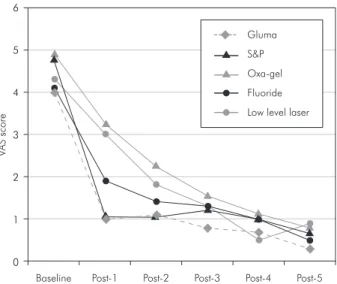
To observe the individual effect of each treat-ment, data was submitted to Friedman Analysis of Variance Test (p = 0.000). Reduction of sensitivity was signiicant for all treatments. Table 2 and Graph 1 indicate the mean scores for treatment at different time intervals.
It was observed that Gluma Desensitizer and Seal&Protect showed immediate effect after ap-plication and no statistically signiicant differenc-es were observed between the two therapidifferenc-es. The sensitivity level was kept the same until the end of the study. Regarding irradiation with LILT, the
ef-fectiveness was not immediate. The sensitivity level dropped in the irst week of evaluation, remaining constant until the end. The desensitizer agents Oxa-Gel and APF gel showed effects as of the irst and third months respectively.
Discussion
In the present study, it was noted that indepen-dently of the treatment, after six months of clinical follow up, all desensitizing agents were capable of reducing dentin hypersensitivity, presenting no sta-tistically signiicant differences on the Post-5 scale.
Considerable evidence has been accumulated to support the hydrodynamic theory.10,11 This theory proposes that stimulus on the exposed dentin sur-face causes a displacement of the luid inside the tu-bules that activates the nerve terminals in the dentin
Graph 1 - Illustrative representation of mean scores for treatment at different time intervals.
0 1 2 3 4 5 6
Baseline Post-1 Post-2 Post-3 Post-4 Post-5 S&P
Fluoride Oxa-gel Gluma
Low level laser
V
A
S
sco
re
Gluma S&P Oxa-Gel APF Laser
Baseline 4.0 A 4.75 A 4.90 A 4.1 A 4.3 A
Post-1 (5 min) 1.0 B 1.05 B 3.25 A 1.9 A 3.0 A
Post-2 (1 week) 1.1 A 1.05 A 2.25 A 1.4 A 1.8 A
Post-3 (1 month) 0.8 A 1.20 A 1.55 A 1.3 A 1.3 A
Post-4 (3 months) 0.7 A 1.00 A 1.15 A 1.0 A 0.5 A
Post-5 (6 months) 0.3 A 0.65 A 0.80 A 0.5 A 0.9 A
*Similar letters in a horizontal line imply no statistical significant differences. APF: Acidulated phosphate fluo-ride.
Table 2 - Baseline and post-treatment mean visual analogue
and pulp, causing pain. Taking into consideration that the application of desensitizing agents is a non-invasive treatment and also its potential in reducing the luid movement through the narrowing or occlu-sion of tubule openings, its use is strongly recom-mended as observed in the literature.6-8
However, the advent of dental lasers has raised another option for the treatment of dentinal hyper-sensitivity and has become a research interest in the last decades.12-15 In the present study, laser therapy provided a considerable decrease in sensitivity level; however, the response was slower when compared to the effect of desensitizing agents.
Although information on the neurophysiologic mechanism is not conclusive, it is postulated that a low intensity laser mediates an analgesic effect re-lated to the depolarization of C-iber afferents. This interference in the polarity of cell membranes by increasing the amplitude of the action potential of cell membranes can block the transmission of pain stimuli in hypersensitive dentin.14
Due to the lack of information related to the ir-radiation protocol used and the subjectivity of the evaluation of dentin hypersensitivity, contradictory results are reported in the literature.12
It is worth emphasizing, furthermore, that al-though the mechanisms of low intensity laser activity are still not clear, the results obtained in this study may have occurred due to the biomodulation effect of the irradiation. Histological studies have reported that hard tissue formation is enhanced as a reaction of dental pulp to laser light.16,17 In the present study, the non-immediate effect of Low Intensity Laser, but gradual reduction in sensitivity over a period of 6 months can explain the biomodulation effect.
Although speculative, the mechanisms proposed for the effects of low intensity laser require serious considerations and new experiments. It can be stat-ed that the diode laser is an effective method for the treatment of dentin hypersensitivity, considering the treatment to be predictable, reliable and simple.
With regard to Gluma Desensitizer and Seal&Protect, both desensitizers showed an imme-diate effect after application and the level of sensi-tivity remained the same until the 6-month period.
The Gluma Desensitizer product contains 5%
glutaraldehyde and 35% hydroxyethyl methacry-late (HEMA). The hypothesis for the immediate occlusion of the dentin tubules is an effect of glu-taraldehyde on the proteins of the dentinal luid. In the reaction of glutaraldehyde with dentin, the two groups of aldehydes present in glutaraldehyde in-terlace themselves with the amino groups of dentin collagen, leading to a ixing of proteins, forming a barrier.18,19 The positive results of Gluma Desensi-tizer presented in this study are in agreement with the literature.6,7,20,21
The desensitizing agent Seal&Protect showed similar results to those shown by Gluma. The agent Seal&Protect is derived from the adhesive system Prime&Bond NT that has an anti-microbial charac-teristic, resulting from the incorporation of triclosan, and acid monomers, which are self-conditioning.8,22
Considering the desensitizing effect of Oxa-Gel, it did not differ statistically in relation to the base-line up to the irst month after application. As of the irst month, a gradual reduction in sensitivity lev-els was noted until the six-month evaluation term. In the literature, the solution of 3% monohydroge-nated-monopotassium (pH 2) acts as a weak dentin acid conditioner, increasing the concentration of ionized calcium to extremely high levels, resulting in an accelerated formation of crystals. However, in spite of the satisfactory results found in literature 23-26, it is reported that water spray can remove the
oxalate crystals on the dentin surface, because the desensitizer agent is short-lived.27,28
The desensitizing effect of potassium is also relat-ed to the inactivation of nerve ibers. This double ac-tion of potassium oxalate may increase its possibility of combining therapies, both physical by tubular oc-clusion and neural by depolarizing the membrane.29
The non-immediate effect of potassium oxalate could be compared to the APF gel agent. Statisti-cally signiicant differences in the level of sensitivity were detected as of the third month, suggesting an interference of the placebo effect.
applica-tion, so that the anti-hyperesthesia effect is of short duration. In spite of luoride being recognized as an effective anti-caries agent, its use as a desensitizing agent is still reported as unsuccessful when com-pared to therapeutic agents such as Gluma or Seal& Protect, despite its distinct modes of action.
When evaluating the results of this study, it should be mentioned that the teeth were used as the unit of analysis. This might not be the most appro-priate way to analyze the data considering the po-tential effects of study participation, especially in a pain-related study; however, it enables the research to assess as many different products with a smaller number of patients.
It should be considered that the evaluation of treatments for dentin hypersensitivity is not a simple procedure due to the interference of the placebo ef-fect, the natural desensitization of the dentin, and the mechanical occlusion of the dentin tubules by smear layer or secondary dentin.6,8,12,20,30 Because it is a painful and subjective phenomenon, the pain from the cervical lesion may be modiied by the sub-ject’s emotional components.23,24
Taking into consideration the effectiveness of the agents used, it is observed that each agent has advantages and disadvantages, in relation to cost and time consumption. Opting for a desensitizing agent, the factors that lead to dentin exposure and hypersensitivity should be controlled by means of guidance on diet and brushing, and also occlusal adjustment, in order for an eficient treatment to be carried out.
Conclusion
After the 6-month follow-up period, it was pos-sible to conclude that hypersensitive dentin is a chal-lenging condition that involves speciic approaches and a multidisciplinary treatment. All therapies studied in the present study showed lower VAS sen-sitivity values compared with baseline, independent-ly of their different modes of action.
Acknowledgments
The authors would like to thank Prof. Gláucia Ambrosano for her support in the statistical analy-sis.
References
1. Addy M, Urquhart E. Dentine hypersensitivity: its preva-lence, aetiology and clinical management. Dent Update. 1992;19(10):407-8, 410-2.
2. Walters PA. Dentinal hypersensitivity: a review. J Contemp Dent Pract. 2005;6(2):107-17.
3. Canadian Advisory Board on Dentin Hypersensitivity. Consensus-based recommendations for the diagnosis and management of dentin hypersensitivity. J Can Dent Assoc. 2003;69(4):221-8.
4. Chabanski MB, Gillam DG. Etiology, prevalence and clini-cal features of cerviclini-cal dentin sensitivity. J Oral Rehabil. 1997;24(9):15-9.
5. Yoshiyama AM, Suge T, Kawasaki A, Ebisu S. Morphological characterization of tubule-like structures in hypersensitive human radicular dentin. J Dent. 1996;24(1-2):57-63. 6. Kakaboura A, Rahiotis C, Thomaidis S, Doukoudakis S.
Clinical effectiveness of two agents on the treatment of tooth cervical hypersensitivity. Am J Dent. 2005;18(4):291-5. 7. Olusile AO, Bamise CT, Oginni AO, Dosumu OO. Short-term
clinical evaluation of four desensitizing agents. J Contemp Dent Pract. 2008;9(1):22-9.
8. Pamir T, Dalgar H, Onal B. Clinical evaluation of three de-sensitizing agents in relieving dentin hypersensitivity. Oper Dent. 2007;32(6):544-8.
9. Clarck GE, Troullos ES. Designing hypersensitivity clinical studies. Dent Clin North Am. 1990;34(3):531-44.
10. Brännström M. Sensitivity of dentin. Oral Surg Oral Med Oral Pathol. 1966;21(4):517-27.
11. Gillam DG. Mechanisms of stimulus transmission across dentin – a review. J West Soc Periodontol Periodontal. 1995;43(2):53-65.
12. Benetti AR, Franco EB, Franco EJ, Pereira JC. Laser Therapy for dentin hypersensitivity: a critical appraisal. J Oral Laser Appl. 2004;4:271-8.
13. Gerschman JA, Ruben J, Gebart-Eaglamont J. Low level la-ser therapy for dentinal tooth hypersensitivity. Aust Dent J. 1994;39(6):353-7.
14. Kimura Y, Wilder-Smith P, Yonaga K, Matsumoto K. Treat-ment of dentin hypersensitivity by lasers: a review. J Clin Periodontol. 2000;27(10):715-21.
16. Ferreira AN, Silveira L, Genovese WJ, de Araújo VC, Frigo L, de Mesquita RA et al. Effect of GaAIAs laser on reactional dentinogenesis induction in human teeth. Photomed Laser Surg. 2006;24(3):358-65.
17. Matsui S, Tsujimoto Y, Matsushima K. Stimulatory effects of hydroxyl radical generation by Ga-Al-As laser irradiation on mineralization ability of human dental pulp cells. Biol Pharm Bull. 2007;30(1):27-31.
18. Dijkman GEHM, Jonebloed WL, de Vries J, Ogaard B, Ar-ends J. Closing of dentinal tubules by Glutaraldeyde treat-ment, a scanning electron microscopy study. Scand Dent Res. 1994;102(3):144-50.
19. Schüpbach P, Lutz F, Finger WJ. Closing of dentinal tubules by Gluma Desensitizer. Eur J Oral Sci. 1997;105(5):414-21. 20. Camps J, Pizzanti S, Dejou J, Franquin JC. Effects of
desen-sitizing agents on human dentin permeability. Am J Dent. 1998;1(6):286-90.
21. Davidson DF, Suzuki M. The Gluma bonding System: a clini-cal evaluation of its various components for the treatment of hypersensitive root dentin. J Can Dent Assoc. 1997;63(1):38-41.
22. Azzopardi A, Bartlett DW, Watson TF, Sherriff M. The meas-urement and prevention of erosion and abrasion. J Dent. 2001;29(6):395-400.
23. Cooley RL , Sandoval VA. Effectiveness of potassium oxalate treatment on dentin hypersensitivity. Gen Dent. 1989;37(4):330-3.
24. Gillam DG, Mordan NJ, Sinodinou AD, Tang JY, Knowles JC. The effects of oxalate-containing products on the ex-posed dentin surface: an SEM investigation. J Oral Rehabil. 2001;28(11):1037-44.
25. Muzzin KB, Johnson R. Effects of potassium oxalate on dentin hypersensitivity in vivo. J Periodontol. 1989;60(3):151-8. 26. Pashley DH, Galloway SE. The effects of oxalate treatment
on the smear layer of ground surfaces of human dentin. Arch Oral Biol. 1985;30(10):731.
27. Kerns DG, Scheidt MJ, Pashley DH, Horner JA, Strong SL, Van Dyke TE. Dentinal tubule occlusion and root hypersen-sitivity. J Periodontol. 1991;62(7):421-8.
28. Pereira JC, Segala AD, Gillam DG. Effect of desensitizing agents on the hydraulic conductance of human dentin sub-jected to different surface pre-treatments – an in vitro study. Dent Mater. 2005;21(2):129-38.
29. Orchardson R, Gillam DG. The efficacy of potassium salts as agents for treating dentin hypersensitivity. J Orofac Pain. 2000;14(1):9-19.