Agda Rísia David Pinto Coelho(a) Orlando Tanaka(b)
Jucienne Salgado Ribeiro(a) Maria Ângela Naval Machado(c) Elisa Souza Camargo(b)
(a) MSc in Orthodontics; (b)PhD, Professor of Orthodontics – Department of
Orthodontics, Pontifical Catholic University of Paraná, Curitiba, PR, Brazil.
(c) PhD, Professor of Pathology, Department of Pathology, Pontifical Catholic University of Paraná, Curitiba, PR, Brazil.
Corresponding author:
Elisa Souza Camargo Rua Fernando Simas, 327 Curitiba - PR - Brazil CEP: 80430-190
E-mail: [email protected]
Received for publication on Apr 23, 2009 Accepted for publication on Nov 16, 2009
Transverse craniofacial dimensions in
Angle Class II, Division 1 malocclusion
according to breathing mode
Abstract: The aim of this longitudinal study was to assess the relation between the transverse craniofacial dimensions of subjects with Class II, Division 1 malocclusion and the breathing mode presented by them. Forty Angle Class II, Division 1 malocclusion subjects of both genders participated in the study, 23 of which were predominantly nose breath-ers and 17 were predominantly mouth breathbreath-ers. The mean age ranged from 10 years and 9 months to 14 years – Age range 1; and from 13 years and 4 months to 16 years and 6 months – Age range 2. Measure-ments of six transverse craniofacial dimensions were performed in P-A teleradiographs: Total Sphenoid, Total Zygomatic, Total Nasal Cavity, Total Maxilla, Total Mastoid and Total Antegonion. The transversal craniofacial dimensions were measured and compared in both groups at age ranges 1 and 2. The longitudinal assessment of age ranges 1 and 2 showed that there was no statistically signiicant inluence of the breath-ing mode on the craniofacial dimensions evaluated, or on the alteration of these dimensions. Breathing mode had no inluence on craniofacial development in the sample studied.
Descriptors: Mouth breathing; Cephalometry; Diagnosis.
Introduction
The clinical characteristics associated with mouth breathing include ir-regularities in the maxillary dental arches, proclined maxillary and man-dibular incisors, tendency toward posterior crossbite and toward anterior open bite.1,2 These individuals also present the long face or adenoid facies syndrome, showing an elongation of the inferior facial height, slightly open lips, narrow maxillary arch, deep palate and Class II malocclusion.3,4
However, these clinical signs have been queried, since not all individ-uals who presented them were predominantly mouth breathers, and this facial type could also be a congenital trait, not necessarily related to the breathing mode.5,6 Muscle patterns and skeletal growth are inluenced only slightly by the breathing mode, since they are genetically transmit-ted and the facial type could be a congenital trait.7 Profit and Fields8 (2000) have also emphasized the strong correlation between breathing mode and craniofacial dimensions.
Division 1 malocclusion in individuals with adenoid facies.1,2,6,9,10
Leech11 (1958) analyzed the relation between breathing mode and the development of maloc-clusion and concluded that predominantly mouth breathing did not seem to affect the skeletal and dental patterns. He also observed that the width of the bones was not altered in subjects with this breathing mode.
On the other hand, Linder-Aronson12 (1963) found a positive correlation between the breathing mode and craniofacial development, having ob-served that the facial width of mouth breathers was smaller than that of nose breathers. Moreover, Gross
et al.13 (1994) found a constriction of the maxillary
arches in subjects with open mouth posture.
Due to the controversial results and the few lon-gitudinal studies found in the related literature, the aim of this study was to conduct a longitudinal as-sessment of the transverse craniofacial dimensions of boys and girls with Class II, Division 1 malocclu-sion, and assess the relation between these measure-ments and the breathing mode presented by them.
Material and Methods
This research project was submitted to and ap-proved by the Research Ethics Committee, Health and Biological Science Department, Pontiical Cath-olic University of Paraná.
A quantitative study was conducted in a sample of 40 white Brazilian boys and girls, with Class II, Division 1 malocclusion, 23 of which were predomi-nantly nose breathers and 17 were predomipredomi-nantly mouth breathers. The irst age range observed was from 10 years and 9 months to 14 years (Age range 1), and the second was from 13 years and 4 months to 16 years and 6 months (Age range 2).
The cases were considered subjects with Class II, Division 1 malocclusion when, at maximum habitu-al intercuspation, they presented the irst permanent mandibular molar distally located in relation to the irst permanent maxillary molar, unilaterally or bi-laterally (with or without subdivision), and maxil-lary incisors in labioversion. The subjects chosen had undergone no previous orthodontic treatment, and did not show premature loss of teeth, large
den-tal cavities, or deleterious oral habits.
Anamnesis was taken, and the subjects under-went clinical extra- and intraoral exams and pos-teroanterior cephalometric radiography (P-A). The breathing mode was assessed by a multidisciplinary team, formed by professionals in the ields of Otolar-yngology, Orthodontics and Speech and Language Pathology.14 Additionally, a questionnaire was sent to the subjects’ parents.
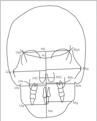
Based on the P-A cephalometric radiographs, ar-eas of interest were traced and the following cepha-lometric points (Figure 1) were marked: Sphenoid (Sph), Zygomatic (Zyg), Nasal Cavity (NC), Maxilla (Mx), Mastoid (Ms), Antegonion (Ag), and crista
galli (Nc). Except for crista galli, all points were
marked at both sides, right (R) and left (L). Next, the horizontal sphenoid line (HSL) joining LSph and RSph was traced. The True Median Line (TML) was then drawn from the midpoint of the HSL to the mentum, passing through the Nc point. The fol-lowing linear measurements were then obtained:
To-LMs
HSL
Nc
RSph
RZyg
RNC RMx
RMs
RAg LZyg
LNC
TML LMx
LAg LSph
tal Sphenoid (TSph), Total Zygomatic (TZyg), Total Nasal Cavity (TNC), Total Maxilla (TMx), Total Mastoid (TMs), and Total Antegonion (TAg). Each total measurement was obtained by adding the mea-surements of the right and left sides, which, in turn, were taken from the cephalometric point on each side, perpendicularly to the True Median Line. Each of the 6 transverse dimensions was measured for Age range 1 and Age range 2, and the behavior of these measurements between age ranges 1 and 2 was also determined, for a total of 18 measurements obtained in this study. All the procedures were performed by a single operator, with the help of a digital caliper.
The transverse craniofacial dimensions were measured and compared in the following groups: predominantly nose breathers at age ranges 1 and 2, and predominantly mouth breathers at age ranges 1 and 2.
Results
For each sample measurement, the following data were obtained, among others: mean, conidence in-terval for the mean, minimum value and maximum value of the sample, standard deviation and variance coeficient. The data were stratiied according to the breathing mode (predominantly nose breathers, NB, and predominantly mouth breathers, MB).
The Kolmogorov-Smirnov normality test and Levene’s test for equality of variances showed a nor-mal distribution of the variables, with a conidence level over 95%, and the tests indicated equality of variance among the NB and MB subjects.
The tracings and measurements performed on the P-A cephalometric radiographs were repeated and submitted to Dahlberg’s error test, which con-irmed the validity of the measurements obtained.
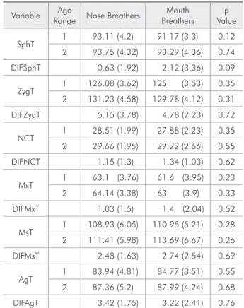
Student’s t test was applied to each of the 18 vari-ables (Table 1) in order to determine whether there were relevant differences in:
Transverse craniofacial measurements among NB and MB at Age range 1;
transverse craniofacial measurements among NB and MB at Age range 2;
alteration in the transverse craniofacial measure-ments from Age range 1 to Age range 2 among NB and MB.
• • •
No signiicant difference was detected in any of the measurements, thus showing that there was no relation between breathing mode and transverse craniofacial dimensions at age ranges 1 and 2, and between breathing mode and the development of the transverse craniofacial dimensions from Age range 1 to Age range 2.
Correlation tests and Student’s t tests (Table 2) were also performed for paired samples, for the pur-pose of analyzing whether there were signiicant dif-ferences in the transverse craniofacial measurements between age ranges 1 and 2, for NB subjects, MB subjects, and for all individuals.
Alteration of all the transverse craniofacial mea-surements between age ranges 1 and 2 proved sta-tistically signiicant. Only the TSph (Total Sphenoid) variable showed no signiicant alteration between age ranges 1 and 2, in the predominantly nose breathers.
Table 1 - Means and standard deviations for the variables in both age ranges according to breathing mode, and Stu-dent’s t test for independent samples.
Variable Age
Range Nose Breathers
Mouth Breathers
p Value
SphT 1 93.11 (4.2) 91.17 (3.3) 0.12 2 93.75 (4.32) 93.29 (4.36) 0.74
DIFSphT 0.63 (1.92) 2.12 (3.36) 0.09
ZygT 1 126.08 (3.62) 125 (3.53) 0.35 2 131.23 (4.58) 129.78 (4.12) 0.31
DIFZygT 5.15 (3.78) 4.78 (2.23) 0.72
NCT 1 28.51 (1.99) 27.88 (2.23) 0.35 2 29.66 (1.95) 29.22 (2.66) 0.55
DIFNCT 1.15 (1.3) 1.34 (1.03) 0.62
MxT 1 63.1 (3.76) 61.6 (3.95) 0.23 2 64.14 (3.38) 63 (3.9) 0.33
DIFMxT 1.03 (1.5) 1.4 (2.04) 0.52
MsT 1 108.93 (6.05) 110.95 (5.21) 0.28 2 111.41 (5.98) 113.69 (6.67) 0.26
DIFMsT 2.48 (1.63) 2.74 (2.54) 0.69
AgT 1 83.94 (4.81) 84.77 (3.51) 0.55 2 87.36 (5.2) 87.99 (4.24) 0.68
DIFAgT 3.42 (1.75) 3.22 (2.41) 0.76
Discussion
The main goal of orthodontics is to achieve a bal-ance between esthetics and functionality of the den-tocraniofacial complex by means of a precise diagno-sis and correction of any anomalies in this complex.
In addition to being commonly adopted for eval-uating symmetries, the P-A teleradiographs used in this research allow an analysis of the transverse cra-niofacial dimensions.15-18
The research described in this article pointed out that growth of transverse craniofacial dimensions continues even after puberty; this was also observed by Athanasiou et al.16 (1992) and by Lux et al.18 (2004). An increase in these measurements in the sample studied was detected in a time period of two and a half years, starting at the age of 10 years and 9 months up to age 14. The exception noted referred to an increase in the Total Sphenoid measurement (TSph), which showed no statistically signiicant difference in individuals who were predominantly
nose breathers. Therefore, bone development in this region did not occur in a way similar to the increase in the other measurements analyzed. This was prob-ably associated with the fact that the sphenoid bone is located in the skull, where growth ceases at an early age when compared with facial growth.8,18
The study described in this paper did not identify a correlation between the transverse craniofacial di-mensions and the breathing mode in individuals with Class II, Division 1 malocclusion (Table 1). These results indicate that the transverse craniofacial mea-surements are determined mainly by genetic factors, and are not inluenced by the breathing mode. In this respect, Brodie19 (1941) stated that the morpho-genetic pattern of the head is established when the individual is approximately 3 months old, and that facial growth follows a consistent vector and does not register individual variations. For Gwynne-Ev-ans and Ballard7 (1958), muscle patterns and skel-etal growth are genetically transmitted and are in-luenced only slightly by the breathing mode; there is no morphological type associated with mouth breathing. The high relation of heredity in the cra-niofacial dimensions and low relation of heredity in the variation of dental arches have already been es-tablished. However, the impact of these on the etio-logic factors of malocclusions, which present dental as well as skeletal components, is still unknown.8
As regards identifying the breathing mode, there is no consensus about characterizing it as predomi-nantly mouth breathing or nose breathing. Several diagnostic methods have been used. In this study, a multidisciplinary approach was chosen because of the conidence level and precision.14
It was not the aim of the present study to establish a causal relationship between the breathing mode and malocclusion, but, rather, to analyze the inluence of the breathing mode on the development of trans-verse craniofacial measurements in individuals with Class II, Division 1 malocclusion. As was shown, no such inluence was detected; however, mouth breath-ing cannot be considered the only etiologic factor of Class II, Division 1 malocclusion, since the sample in this research included 23 individuals with this mal-occlusion, who were predominantly nose breathers. Other studies1, 9, 10 have tried to establish a causal Table 2 - Student’s t test for paired samples.
Breathing
Mode Pair
Average
Difference D.F. t p value
Nose Breathers
(NB)
SphT1-SphT2 – 0.64 22 – 1.58 0.127
ZygT1-ZyhT2 – 5.15 22 – 6.54 0.000*
NCT1-NCT2 – 1.15 22 – 4.24 0.000*
MxT1-MxT2 – 1.04 22 – 3.31 0.003*
MsT1-MsT2 – 2.48 22 – 7.3 0.000*
AgT1-AgT2 – 3.42 22 – 9.37 0.000*
Mouth Breathers
(MB)
SphT1-SphT2 – 2.12 16 – 2.6 0.019*
ZygT1-ZyhT2 – 4.78 16 – 8.84 0.000*
NCT1-NCT2 – 1.34 16 – 5.36 0.000*
MxT1-MxT2 – 1.4 16 – 2.81 0.012*
MsT1-MsT2 – 2.74 16 – 4.44 0.000*
AgT1-AgT2 – 3.22 16 – 5.51 0.000*
Total (NB+MB)
SphT1-SphT2 – 1.26 39 – 2.97 0.005*
ZygT1-ZyhT2 – 4.99 39 – 9.92 0.000*
NCT1-NCT2 – 1.23 39 – 6.58 0.000*
MxT1-MxT2 – 1.19 39 – 4.33 0.000*
MsT1-MsT2 – 2.59 39 – 8.03 0.000*
AgT1-AgT2 – 3.33 39 – 10.39 0.000*
relationship between the breathing mode and the development of malocclusions. Hawkins9 (1969) ob-served that Class II, Division 1 malocclusions with adenoid facies could be attributed to predominantly mouth breathing, which would produce muscular imbalance, resulting in the narrowing of the maxil-lary arch, protrusion of the maxilmaxil-lary anterior teeth, shortening of the upper lip and lower lip in a hypo-tonic condition. Linder-Aronson1 (1979) associated characteristics such as narrow maxilla, proclined maxillary and mandibular incisors, and a tendency toward posterior cross bite and anterior open bite with mouth breathing. Melsen et al.10 (1987) noticed that children who were mouth breathers showed a greater incidence of distocclusion, anterior open bite, posterior cross bite and crowding.
Studies with objectives similar to those set for this research have endeavored to identify whether there was a correlation between the breathing mode and the development of dentocraniofacial dimen-sions. The results of the current research agree with the results found by Leech11 (1958), who analyzed boys and girls with ages between 2 and 13 years, 56% with Angle Class I, 36% with Class II, and 8% with Class III, and concluded that mouth breath-ing seemed to affect neither the skeletal and dental patterns nor the bone width. According to Shaugh-nessy20 (1983), although discussed at length in the literature, the long face or adenoid face syndrome does not characterize a pathognomonic sign of mouth breathing, and the alterations attributed to mouth breathing would indeed depend on individu-al adaptation of the muscular function.
On the other hand, the results obtained by the re-search described in this paper differ from those ob-tained by Linder-Aronson12 (1963), who compared the differences between nose breathers and mouth breathers in a 2-year longitudinal study that began with subjects who were 10 years old. In addition to the breathing mode, the presence of labial sealing at
rest was assessed and complementary exams, such as P-A cephalometric radiographs, were performed, with measurements of the maximum height of the face and nose. It was observed that children who were mouth breathers had a signiicantly larger fa-cial index than children who were nose breathers, that is, smaller facial width. Gross et al.13 (1994) performed a longitudinal assessment in 348 children in order to establish the inluence of an open mouth in dentofacial development. They observed that chil-dren with an open mouth posture showed a greater constriction of the maxillary arches than children with labial sealing. However, they did not perform measurements in the skeletal structures, and this has made it impossible to compare their study with the present study.
From the results presented in this paper, one may conclude with a high level of conidence that the breathing mode has no inluence on the transverse craniofacial measurements in individuals with Angle Class II, Division 1, malocclusion between the ages of 10 years and 9 months and 14 years (Age range 1), and between the ages of 13 years and 4 months and 16 years and 6 months (Age range 2). It was also observed that there was a signiicant develop-ment of the transverse dimensions of the craniofa-cial complex between the two age ranges analyzed, considering the predominantly nose breathers and the predominantly mouth breathers that comprised the sample, except for the Total Sphenoid measure-ment (TSph) in predominantly nose breathers.
Further research should be conducted with larg-er samples, stratiied according to gendlarg-er and type of occlusion, facial pattern and with longitudinal analyses ranging from childhood to adult life.
Conclusions
This study concluded that the breathing mode had no inluence on the transverse craniofacial de-velopment.
References
1. Linder-Aronson S. Respiratory function in relation to facial morphology and the dentition. Br J Orthod. 1979 Apr;6(2):59-71.
changes during growth. Am J Orthod Dentofacial Orthop. 2006 Dec;130(6):721-31.
3. Vig KW. Nasal obstruction and facial growth: the strength of evidence for clinical assumptions. Am J Orthod Dentofac Orthop. 1998 Jun;113(6):603-11.
4. Peltomäki T. The effect of mode of breathing on craniofacial growth -- revisited. Eur J Orthod. 2007 Oct; 29(5):426-9. 5. O’Ryan FS, Gallagher DM, LaBanc JP, Epker BN. The relation
between nasorespiratory function and dentofacial morphol-ogy: a review. Am J Orthod. 1982 Nov;82(5):403-10. 6. Lessa FC, Enoki C, Feres MF, Valera FC, Lima WT,
Matsu-moto MA. Breathing mode influence in craniofacial develop-ment. Braz J Otorhinolaryngol. 2005 Mar-Apr;71(2):156-60. Epub 2005 Aug 2.
7. Gwynne-Evans E, Ballard CF. The mouth breather. Am J Orthod. 1958 Jul;44(7):559.
8. Proffit W, Fields. H. Contemporary Orthodontics. 3rd ed. St.
Louis: Mosby Company; 2000.
9. Hawkins AC. Mouth breathing and its relationship to malocclusion and facial abnormalities. N M Dent J. 1969 May;20(1):18-21.
10. Melsen B, Attina L, Santuari M, Attina A. Relationships between swallowing pattern, mode of respiration, and devel-opment of malocclusion. Angle Orthod. 1987 Apr;57(2):113-20.
11. Leech HL. A clinical analysis of orofacial morphology and behavior of 500 patients attending an upper respiratory re-search clinic. Dent Pract. 1958 Dec;9(4):57-68.
12. Linder-Aronson S. Dimensions of face and palate in nose breathers and in habitual mouth breathers. Odontol Revy. 1963;14(3):187-99.
13. Gross AG, Kellum GD, Franz D, Michas K, Walker M, Fos-ter M, et al. A longitudinal evaluation of open mouth pos-ture and maxillary arch width in children. Angle Orthod 1994;64(6):419-24.
14. Wieler WJ, Barros AM, Barros LA, Camargo ES, Ignácio SA, Maruo H, Tanaka OM. A combined protocol to aid diagnosis of breathing mode. Rev de Clin Pesq Odontol. 2007 May-Aug:3(2):101-14.
15. Emsli RD, Massler M, Zwemer JD. Mouth breathing: Etiology and effects: a review. J Am Dent Assoc. 1952 May;44(5):506-21.
16. Athanasiou AE, Droschl H, Bosch C. Data and patterns of transverse dentofacial structure of 6- to 15-year-old children: a posteroanterior cephalometric study. Am J Orthod Dento-facial Orthop. 1992 May; 101(5):465-71.
17. Snodell SF, Nanda RS, Currier GF. A longitudinal cephalomet-ric study of transverse and vertical craniofacial growth. Am J Orthod Dentofacial Orthop. 1993 Nov;104(5):471-83. 18. Lux CJ, Conradt C, Burden D, Komposch G. Transverse
de-velopment of the craniofacial skeleton and dentition between 7 and 15 years of age – a longitudinal postero-anterior cepha-lometric study. Eur J Orthod. 2004 Feb;26(1):31-42. 19. Brodie AG. On the growth pattern of the human head
from the third month to the eighth year of life. Am J Anat 1941;68(2):209-62.